Download presentation
Presentation is loading. Please wait.

1
The Cerebellum 陽明大學醫學院 神經學系 陳昌明 醫師

3
Position Lies above and behind the medullar and pons and occupies posterior cranial fossa Cerebellum

4
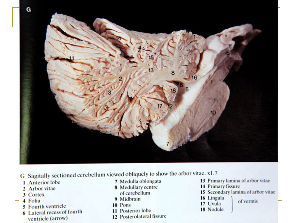
Cerebellum External Configurations
Located in posterior cranial fossa Tentorium cerebelli (cerebrum), 4th ventricle (brain stem) Communicate with other structure via superior, middle, and inferior cerebellar peduncle
, 4th. ventricle (brain stem) Communicate with other structure via. superior, middle, and inferior cerebellar peduncle.")
5
MRI at the level of the 4th ventricle

6
External features Consists of two cerebellar hemisphere united in the midline by the vermis

7
External features Three peduncles
Inferior cerebellar peduncle 下小腦脚 -connect with medulla and with spinal cord, contain both afferent and efferent fibers Middle cerebellar peduncle 中小腦脚-connect with pons, contain afferent fibers Superior cerebellar peduncle 上小腦脚-connect with midbrain, contain mostly efferent fibers

8
Cerebellum Longitudinal division Transverse division
Vermis, Paravermal Region, Cerebellar Hemisphere Transverse division Anterior Lobe Posterior Lobe Flocculonodular Lobe

9
Lobes Anterior lobe corpus of cerebellar Primary fissure
Posterior lobe Flocculonodular lobe Posterolateral fissure

10
Lobes Two deep fissures Three lobs Primary fissure
Posterosuperior fissure Three lobs Flocculonodular lobe 絨球小結葉 flocculus and nodule Anterior lobe Posterior lobe Corpus of cerebellar

11
External features Superior surface Tonsil of cerebellum 小腦扁桃体 two elevated masses on inferior surface of hemispheral portion just nearby foramen magnum Tonsil View from below

12
Functional divisions of cerebellar cortex
Cbm unfold

13
Cerebellum & Brainstem, Inferior Surface, Anterior View
External features Cerebellum & Brainstem, Inferior Surface, Anterior View

15
Cerebellum External Configurations
Subdivision of Flocculonodular Lobe Nodulus Flocculus Subdivision of Anterior Lobe Vermis Hemisphere Lingula Central Lobule Ala Central Lobule postcentral fissure Culmen Quadriangular Lobule

16
Cerebellum External Configurations
Subdivision of Posterior Lobe Vermis Hemisphere Declive Simple Lobule postcentral fissure Folium Superior Semilunar Lobule horizontal fissure Inferior Semilunar Lobule Tuber Gracile Lobule prepyramidal fissure Pyramis Biventer Lobule secondary fissure Uvula Tonsil

18
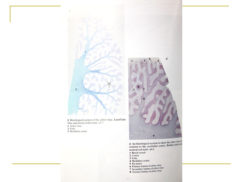
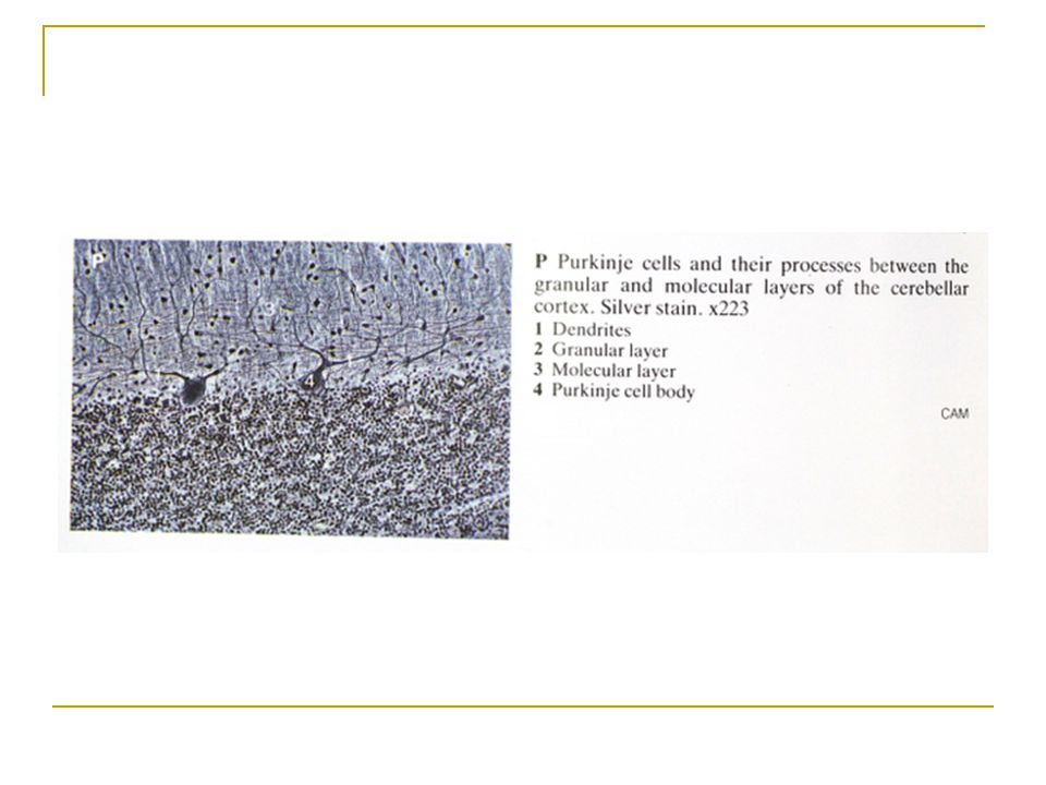
Cerebellum Internal Configurations
Cerebellar Cortex Molecular Layer Purkinje Cell Layer Granular Layer Corpus Medullare (Medullary Center) Deep Cerebellar Nuclei Fastigial Nuclei Nucleus Interpositus Emboliform Nucleus Globose Nucleus Dentate Nucleus
 Deep Cerebellar Nuclei. Fastigial Nuclei. Nucleus Interpositus. Emboliform Nucleus. Globose Nucleus. Dentate Nucleus.")
19
Internal structures White matter-medullary center 髓体 Gray matter
Cerebellar cortex Cerebellar nuclei Dentate nucleus 齒狀核 Fastigial nucleus 頂核 Interposed nucleus 中間核 Emboliform nucleus 栓狀核 Globose nucleus球狀核 White matter-medullary center 髓体

20
Deep Nuclei 1. fastigial nucleus 2. globose 3. emboliform 4. dentate

21
Internal structures Fastigial nucleus Cerebellar cortex
Globose nucleus Dentate nucleus Emboliform nucleus medullary center

26
Cerebellar Cortex Inputs Climbing fibers •from Inferior olive
Mossy fibers Output Purkinje neurons Interneurons Granule neurons Stellate neurons Basket neurons Golgi neurons Molecular Purkinje Granular NTA Fig

27
Cerebellum Internal Configurations
Cerebellar Cortex I. Molecular Layer Stellate Cell --- taurine (inhibitory) afferent: parallel fiber efferent: Purkinje cell dendrite Basket Cell ---- GABA (inhibitory) efferent: Purkinje cell soma Parallel Fiber granule cell axon Purkinje Cell Dendrite
 afferent: parallel fiber. efferent: Purkinje cell dendrite. Basket Cell ---- GABA (inhibitory) efferent: Purkinje cell soma. Parallel Fiber. granule cell axon. Purkinje Cell Dendrite.")
31
Cerebellum Internal Configurations
Cerebellar Cortex II. Purkinje Cell Layer Purkinje Cell -- 15,000,000 in number -- GABA (inhibitory) afferent from: parallel fiber climbing fiber stellate cell basket cell efferent to: deep cortical nuclei Bergman’s glial cell
 afferent from: parallel fiber. climbing fiber. stellate cell. basket cell. efferent to: deep cortical nuclei. Bergman’s glial cell.")
32
Cerebellum Internal Configurations
Cerebellar Cortex III. Granular Layer Granular Cell -- 50,000,000,000 in number -- glutamic acid (excitatory) afferent: mossy fiber efferent: Purkinje cell dendrite basket cell, stellate cell Golgi cell Golgi Cell -- GABA (inhibitory) afferent: parallel fiber, mossy fiber rosette efferent: granule cell dendrite
 afferent: mossy fiber. efferent: Purkinje cell dendrite. basket cell, stellate cell. Golgi cell. Golgi Cell. -- GABA (inhibitory) afferent: parallel fiber, mossy fiber rosette. efferent: granule cell dendrite.")
34
1. Purkinje cell 2. granule cell 3. basket cell 4. Golgi cell 5. stellate cell 6. climbing fiber 7. mossy fiber 8. parallel fiber 9. inferior olivary nucleus 10. deep cerebellar nuclei

35
Cerebellum Internal Configurations
Synaptic Glomerulus Afferent terminals on granular layer Mossy Fiber Rosette -- afferent fibers except inferior olivary input -- 2/3 of medullary center Granular Cell Dendrite -- main afferent input Golgi Cell Axon -- synapse on granule cell dendrite -- GABA (inhibitory) - Surrounded by Astrocyte Foot Process
 - Surrounded by Astrocyte Foot Process.")
36
Synaptic Glomerulus

37
Cerebellum Classifications
Classification by Phylogenetic and Ontogenic Development Archicerebellum Paleocerebllum Neocerebellum Classification by Afferent Connection Vestibulocerebellum Spinocerebellum Pontocerebellum Classification by Efferent Connection Vermis Paravermal Region Cerebellar Hemisphere

38
Archicerebellum (nodulus) (flocculus) Paleocerebellum Neocerebellum
 (flocculus) Paleocerebellum Neocerebellum")
39
Spinocerebellum Pontocerebellum Vestibulocerebellum

40
Cerebellum Connections
Afferent Connections (1): 1. Inferior Cerebellar Peduncle Restiform Body Posterior Spinocerebellar Tract Olivocerebellar tract Cuneocerebellar Tract Reticulocerebellar Tract Juxtarestiform Body Vestibulocerebellar Tract Primary Vestyibular Fiber
: 1. Inferior Cerebellar Peduncle. Restiform Body. Posterior Spinocerebellar Tract. Olivocerebellar tract. Cuneocerebellar Tract. Reticulocerebellar Tract. Juxtarestiform Body. Vestibulocerebellar Tract. Primary Vestyibular Fiber.")
41
Cerebellum Connections
Afferent Connections (2): 2. Middle Cerebellar Peduncle Pontocerebellar fiber Corticopontocerebellar Fiber Reticulocerebellar Fiber 3. Superior Cerebellar Peduncle Anterior Spinocerebellar Tract Cerulocerebellar fiber Raphecerebellar fiber Rubrocerebellar fiber Hypothalamocerebellar fiber
: 2. Middle Cerebellar Peduncle. Pontocerebellar fiber. Corticopontocerebellar Fiber. Reticulocerebellar Fiber. 3. Superior Cerebellar Peduncle. Anterior Spinocerebellar Tract. Cerulocerebellar fiber. Raphecerebellar fiber. Rubrocerebellar fiber. Hypothalamocerebellar fiber.")
42
Cerebellum Connections
Efferent Connections : 1. Superior Cerebellar Peduncle Cerebellothalamic fiber - from 3 deep nuclei to VPLo, VLc, CL Cerebellorubral fiber - from nucleus interpositus and dentate nucleus ascending portion of uncinate fasciculus of Russell 2. Inferior Cerebellar Peduncle Fastigiovestibular fiber descending portion of

43
Three functional divisions
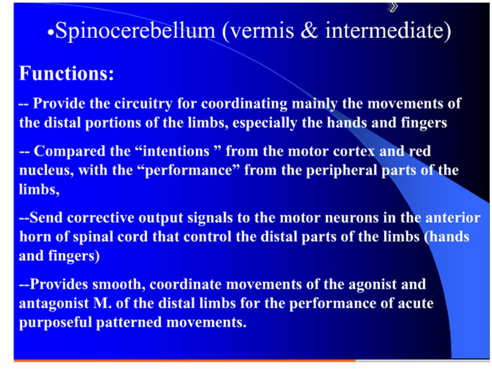
Vestibulocerebellum 前庭小腦 Archicerebellum 原小腦 Flocculonodular lobe Spinocerebellum 脊髓小腦 Paleocerebellum 舊小腦 Vermis and intermediate zone Cerebrocerebellum 大腦小腦 Neocerebellum 新小腦 Lateral zone Vermis Intermediate zone Lateral zone Flocculonodular lobe

44
Cerebellar divisions Spinocerebellum: Vermis Intermediate hem.
(Vermis + Intermed. Hem) Control of limbs and trunk Lateral hem. Cerebrocerebellum: Cerebrocerebellum (Lateral hemisphere) Planning of movement+ IVth vent Floculo-nodular lobe Vestibulo-cerebellum (Floculo-nodular lobe) Control of eye & head movements Balance NTA Fig. 13-1
 Control of limbs. and trunk. Lateral hem. Cerebrocerebellum: Cerebrocerebellum. (Lateral hemisphere) Planning of movement+ IVth vent. Floculo-nodular lobe. Vestibulo-cerebellum. (Floculo-nodular lobe) Control of eye & head movements. Balance. NTA Fig")
45
Connections and function of cerebellum
Vestibulocerebellum Connections Afferents: receive input from vestibular nuclei and primary vestibular Efferents: projects to the vestibular nucleus → (1) vestibulospinal tract → motor neurons of anterior horn for reflexively control of equilibrium (2) vestibulo-ocular tract → medial longitudinal fasciculus → CN nucleus 3, 4, 6 for EOM control. Function: involved in eye movements and maintain balance
 vestibulospinal tract → motor neurons of anterior horn for reflexively control of equilibrium. (2) vestibulo-ocular tract → medial longitudinal fasciculus → CN nucleus 3, 4, 6 for EOM control. Function: involved in eye movements and maintain balance.")
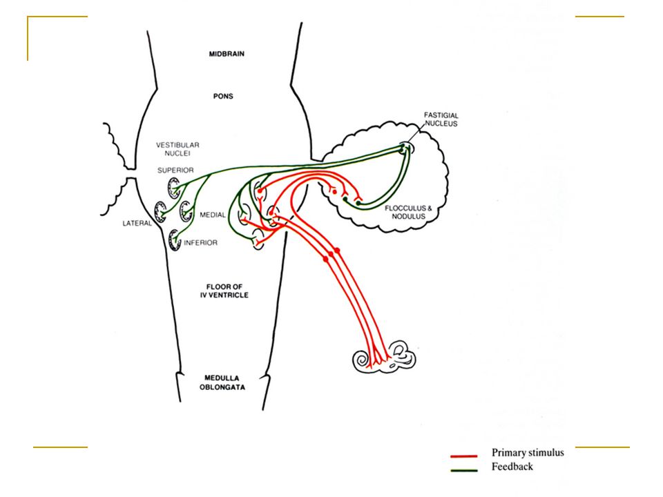
46
Main Connections of the Vestibulocerebellum
Vestibular Organ Floculonodular Lobe Vermis VESTIBULAR NUCLEUS vestibulospinal tract MLF FASTIGIAL NUCLEUS lower motor neuron ARCHICEREBELLUM LMN

48
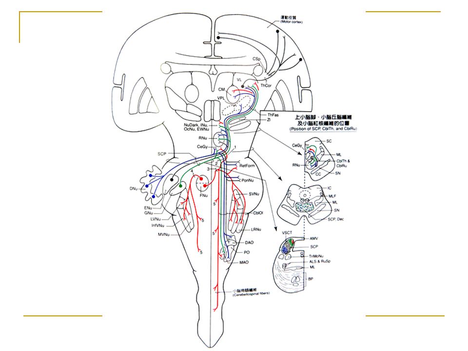
Main Connections of the Paleocerebellum
RED NUCLEUS NUCLEUS INTERPOSITUS rubrospinal tract Inferior Olivry Nucleus ANTERIOR LOBE PARAVERMAL ZONE lower motor neuron PALEOCEREBELLUM SPINAL CORD spinocerebellar tract

49
Spinocerebellar tracts
End mainly in the anterior lobe, the paramedian lobule, and the pyramis of the posterior lobe

51
Connections and function of cerebellum
Cerebrocerebellum Connection Afferents: receives input from the cerebral cortex via a relay in pontine nuclei Efferents: projects to dentate nucleus → VI → primary motor cortex → corticospinal tract → motor neurons of anterior horn Function: participates in planning movements

56
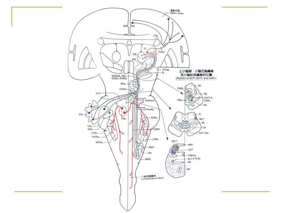
Main Connections of the Neocerebellum
CEREBRAL CORTEX THALAMUS DENTATE NUCLEUS pyramidal tract Pontine Nucleus POSTERIOR LOBE CEREBELLAR HEMISPHERE lower motor neuron NEOCEREBELLUM LMN

57
Pyramidal Tract and Associated Circuits
upper motor neuron UMN Cerebellum BASAL GANGLIA pyramidal tract lower motor neuron UMN

58
Cerebellum and Automatic Motor Control Lower Motor Neuron (LMN)
Motor Cortex CEREBELLUM Red Nucleus Vestibular Nucleus Reticular Formation Lower Motor Neuron (LMN) Proprioceptors
 Proprioceptors.")
59
Cerebellum---- connection
Corticonuclear Connections A zone fastigial nucleus medial vestibular nucleus B zone lateral vestibular nucleus C1, C3 zone --- emboliform nucleus C globose nucleus D parvocellular portion of dentate nucleus D magnocellular portion of dentate nucleus

60
1. vermis 2. paravermal region 3. cerebella hemisphere 4. nodulus 5. flocculus 6. fastigial nucleus 7. globose nucleus 8. emboliform nucleus 9. dentate nucleus 10. medial vestibular nucleus 11. lateral vestibular nucleus

61
Cerebellum Connections
Olivocerebellar Connections (1) Caudal portion of medial and dorsal accessory olivary nucleus vermis of cerebellar cortex (A and B) fastigial nucleus vestibular nucleus (2) Rostral portion of paravermal region (C1, C2, C3) nucleus interpositus (3) Principal Inferior Olivary Nucleus cerebellar hemisphere (D1, D2) dentate nucleus
 Caudal portion of. medial and dorsal accessory olivary nucleus vermis of cerebellar cortex (A and B) fastigial nucleus. vestibular nucleus. (2) Rostral portion of paravermal region (C1, C2, C3) nucleus interpositus. (3) Principal Inferior Olivary Nucleus cerebellar hemisphere (D1, D2) dentate nucleus.")
62
Olivo-cerebellar system
The inferior olivary complex Principal olive, medial and dorsal accessory olive, and medial lamina. 1.5 million cells Send climbing fibers to all cerebellar cortex in a specific topographic manner A Small area in the inferior olive is linked to a definite area of the cerebellar output

63
The inferior olivary complex
Receives projections from The spinal cord crossed ventral and dorsal spino-olivary tracts The brainstem (esp. red nuclei) Cerebellar nuclei Interpositus & dentate nuclei exert an inhibitory effect Pretectal nuclei Relaying optokinetic information Cerebrum Motor cortex (area 4), premotor cortex (area 6) Visual and Vestibular areas Zona incerta of the thalamus Relay for the projections from the motor cortex, prefrontal, cingulate, parietal and temporal area
 Cerebellar nuclei. Interpositus & dentate nuclei exert an inhibitory effect. Pretectal nuclei. Relaying optokinetic information. Cerebrum. Motor cortex (area 4), premotor cortex (area 6) Visual and Vestibular areas. Zona incerta of the thalamus. Relay for the projections from the motor cortex, prefrontal, cingulate, parietal and temporal area.")
64
caudal portion rostral portion
Principal Inferior Olivary Nucleus medial and dorsal accessory olivary nucleus

65
Functional divisions of cerebellar cortex
Spinocerebellum Vermis Intermediate hem Cerebrocerebellum Lateral hemisphere Vestibulo- cerebellum Floculo-nodular lobe

66
Functional divisions of cerebellar cortex --> Deep nuclei
Spinocerebellum Vermis Intermediate hemisphere Inter posed To lateral sysetms Cerebrocerebellum Lateral hemisphere Dentate Fastigial To medial sysetms Vestibulo- cerebellum To vestibular nuclei Eye mvt & balance To frontal motor areas Motor Planning +++ Motor execution

68
Reticular nuclei Lateral reticular nuclei (lateral to inferior olive)
Sent mossy fibers to bilateral cerebellum (ipsilateral dominant) via superior cerebellar peduncle. Receive excitatory reciprocal input from the cerebellar nuclei Receive collaterals from propiospinal neurons Relay motor information to cerebellum
 via superior cerebellar peduncle. Receive excitatory reciprocal input from the cerebellar nuclei. Receive collaterals from propiospinal neurons. Relay motor information to cerebellum.")
70
Organization of Motor Subsystems

78
Cerebellum Function Maintenance of Equilibrium
- balance, posture, eye movement Coordination of half-automatic movement of walking and posture maintenance - posture, gait Adjustment of Muscle Tone Motor Leaning – Motor Skills Cognitive Function

79
Balance

80
Motor Skill Pablo Casals

81
Cerebellum Clinical Syndromes
Ataxia: incoordination of movement - decomposition of movement - dysmetria, past-pointing - dysdiadochokinesia - rebound phenomenon of Holmes - gait ataxia, truncal ataxia, titubation Intention Tremor Hypotonia, Nystagmus Archicerebellar Lesion: medulloblastoma Paleocerebellar Lesion: gait disturbance Neocerebellar Lesion: hypotonia, ataxia, tremor

82
Posture Gait – Ataxia Tremor

83
Cerebellar Ataxia a b c d Ataxic gait and position:
Left cerebellar tumor a. Sways to the right in standing position b. Steady on the right leg c. Unsteady on the left leg d. ataxic gait d

84
Cerebellar Medulloblastoma Cerebellar tumors on vermis
- Truncal Ataxia - Frequent Falling The child in this picture: - would not try to stand unsupported - would not let go of the bed rail if she was stood on the floor.

85
Thank you for your attention

Similar presentations


 ◦ Mossy (Not Olive) ◦ Parallel Output ◦ Purkinje Cells M P G W Climbing.>")


