Download presentation
Presentation is loading. Please wait.

1
BY DR ABIODUN MARK AKANMODE.

2
What is happening here?

3
Post-maturity. There are few changes indicative of postmaturity (gestation beyond 42 weeks). Here are long fingernails that can be seen with post maturity.

4
Apgar score.

5
What is the pathology here

6
Meconium ileus. Meconium ileus is most often seen in the first few days of life in neonates with cystic fibrosis, but can rarely occur in infants with a normal pancreas. In cystic fibrosis, the abnormal pancreatic secretions lead to inspissated meconium that produces intestinal obstruction. The dilated coils of ileum are opened here to reveal the inspissated green meconium (which may also be tarry or gritty), while the unopened colon at the upper left and the appendix at the lower left beyond the ileocecal valve are not dilated, and little or no meconium is passed per rectum
, while the unopened colon at the upper left and the appendix at the lower left beyond the ileocecal valve are not dilated, and little or no meconium is passed per rectum.")
7
What is the pathology here?

8
Syndactyl Syndactyly represents fusion of two or more digits. It can be an isolated finding or part of syndromes that define patterns of anomalies

9
What is the pathology here?

10
Rocker bottom feet. This is the appearance of a "rocker bottom" foot with a prominent calcaneus and rounded bottom. Such an anomaly may suggest a chromosomal abnormality such as trisomy 18.

11
What is the pathology here?

13
Hydrops fetalis. Generalized edema from fluid collection in the soft tissues results in hydrops fetalis. There are many causes for fetal hydrops. The most common are "non-immune" types that include infections, congestive failure (from anemia or cardiac abnormalities), and congenital anomalies. Immune hydrops, from maternal antibody formed against fetal red blood cells, is not common when Rh immune globulin is employed in cases of potential Rh incompatibility.
, and congenital anomalies. Immune hydrops, from maternal antibody formed against fetal red blood cells, is not common when Rh immune globulin is employed in cases of potential Rh incompatibility..")
14
What is the pathology here?

15
Twin-twin transfusion syndrome. If there is a vascular connection across a monochorionic twin placenta, then a twin-twin transfusion syndrome can develop. In this condition, there is diminished blood flow to one twin (the "donor") and increased blood flow to the other twin (the "recipient"). The pale appearing donor is smaller and may die for lack of sufficient blood flow. More commonly, the larger plethoric recipient may die from congestive heart failure.
 and increased blood flow to the other twin (the recipient ). The pale appearing donor is smaller and may die for lack of sufficient blood flow. More commonly, the larger plethoric recipient may die from congestive heart failure..")
16
What is the pathology here?

17
IDENTIFY THE DIFFRENCE.

18
NECROTISING ENTEROCOLITIS. A complication of prematurity and low birth weight is neonatal necrotizing enterocolitis (NEC) in which ischemia results in focal to confluent areas of bowel necrosis, most often in the terminal ileum. Seen at autopsy here is a dark red appearance to the small intestine of a premature neonate.
 in which ischemia results in focal to confluent areas of bowel necrosis, most often in the terminal ileum. Seen at autopsy here is a dark red appearance to the small intestine of a premature neonate..")
19
WHAT IS THE PATHOLOGY HERE?

20
HYALINE MEMBRANE DISEASE. This is hyaline membrane disease due to prematurity and lack of surfactant production from type II pneumonocytes within the immature lung. Note the thick pink membranes lining the alveolar spaces.

21
SAMPLE QUESTION. The mother of this three-year-old male noticed a large mass in his abdomen. There were no other complaints. Physical examination and x-rays--including an intravenous pyelogram (IVP)--showed a large right renal mass. At surgery, a 550-gram right kidney was removed. There was no lymph node involvement, no extrarenal mass, and no involvement of the other kidney. Lung and bone x-rays showed no metastases. However, microscopic study of the surgical specimen showed infiltration by the tumor (Grade II neoplasm) which had been difficult to separate from the duodenum. The child was placed on chemotherapy. Four months after the surgery to remove the tumor, a nodule was found in the lungs. The chemotherapy was resumed but the patient died four weeks later due to complications of Gram-negative sepsis
--showed a large right renal mass. At surgery, a 550-gram right kidney was removed. There was no lymph node involvement, no extrarenal mass, and no involvement of the other kidney. Lung and bone x-rays showed no metastases. However, microscopic study of the surgical specimen showed infiltration by the tumor (Grade II neoplasm) which had been difficult to separate from the duodenum. The child was placed on chemotherapy. Four months after the surgery to remove the tumor, a nodule was found in the lungs. The chemotherapy was resumed but the patient died four weeks later due to complications of Gram-negative sepsis.")
23
Wilms' tumor This is a gross photograph of a bladder (1) to which are attached a normal kidney (2) and a kidney with Wilms' tumor (3). A large mass extends from the superior pole of the affected kidney. The renal capsule can be seen extending around this tumor.

26
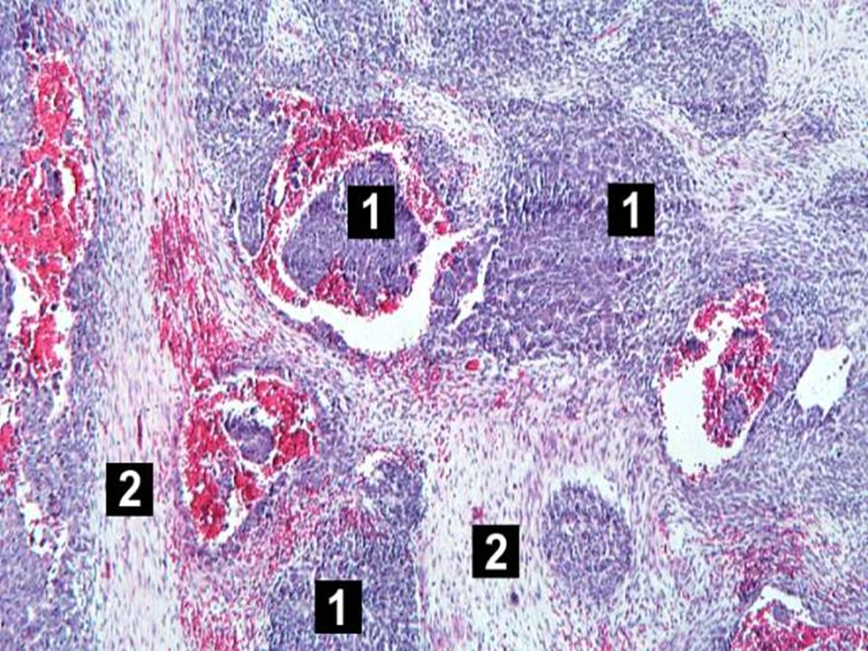
WT. This low-power photomicrograph of tumor shows the two cell types making up this neoplasm. The basophilic cellular component termed "blastema" (1) can be distinguished from less cellular eosinophilic areas with fibroblast-like cells (2).
 can be distinguished from less cellular eosinophilic areas with fibroblast-like cells (2)..")
28
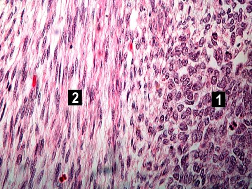
WT. This high-power photomicrograph shows the differences in cell morphology between the blastema (1) and the fibroblast type cells (2).
 and the fibroblast type cells (2)..")
29
What is the pathology here?

30
Neuroblastoma This is the gross appearance of a neuroblastoma arising in the right adrenal gland. It is the most common pediatric malignancy in infancy, and 75% of cases are diagnosed in children less than 4 years old. These tumors most often present as an abdominal or mediastinal mass.

32
Muchas gracias Al final.

Similar presentations


 Department of Pediatrics>")


