Download presentation
Presentation is loading. Please wait.

2
Mitosis Why do cells need to divide?

5
Recap… Cell theory… Cells are the basic structural and functional unit of life (Hooke) All living things (plants, animals, fungi, bacteria, protista) are made of cells (Schleiden, Schwann, Van Leewoehoek) Cells come from pre-existing cells (Virchow)
 All living things (plants, animals, fungi, bacteria, protista) are made of cells (Schleiden, Schwann, Van Leewoehoek) Cells come from pre-existing cells (Virchow)")
6
Overview Why do cells need to divide? Repair, growth, development Types of reproduction Sexual Reproduction Genetically different 2 parents Takes time to develop, better chance of survival More chance of mutation Process begins by making gametes…Uses MEIOSIS asexual Genetically identical One parent Many offspring very quickly Less chance of mutations (by MITOSIS)
.")
7
DNA Blueprint of life, nucleic acid Chromatin Granular genetic material, spread out in nucleus of non-dividing cells Chromosomes Condensed genetic material, in dividing cells Sister chromatids Identical copies of Chromosomes joined by a centromere (“centro-” middle)
")
10
Humans 46 chromosomes 46 sister chromatids One from your mom, one from your dad

11
Cell Cycle: Life of a Cell

12
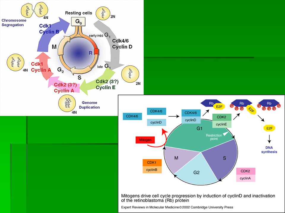
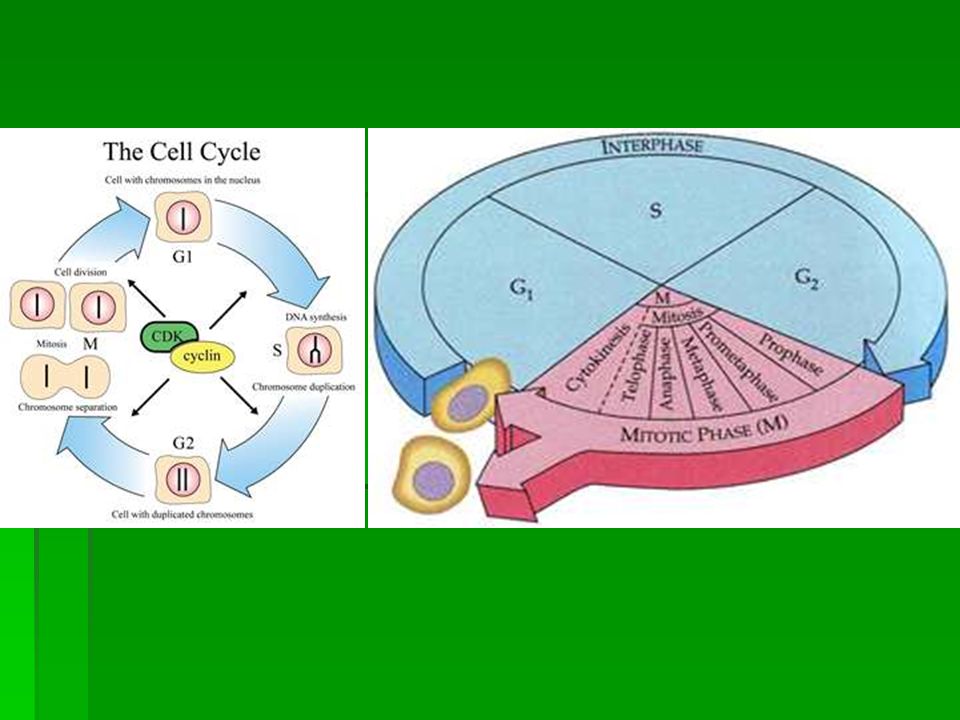
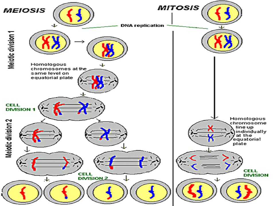
Cell Cycle Interphase 90 % of cell’s life, non dividing G1 phase Grows, makes organelles S phase DNA Synthesis…DNA replicates G2 phase Cell prepares to divide, makes sure it has all important organelles for division M phase When the cytoplasm and nucleus of the cell divides

13
Cell Cycle There are check points in G1, S, and G2 Make sure cell is ready to move onto the next phase (has all necessary organelles, copied DNA, etc.) Once the cell has past the G1 checkpoint, it will complete the cell cycle Some cells stay in the G1 phase all their life (muscle cell, brain cells)
 Once the cell has past the G1 checkpoint, it will complete the cell cycle Some cells stay in the G1 phase all their life (muscle cell, brain cells)")
14
Regulators of Cell Cycle Cyclins Protein that regulates the timing of the cell cycle in eukaryotic cells Levels of cyclins rise and fall throughout the cell cycle Cyclin-dependent Kinases (cdks) Enzymes that are activated when they bind with cyclin and they make the cell cycle continue
 Enzymes that are activated when they bind with cyclin and they make the cell cycle continue")
17
Regulators Internal Factors within the cell that control cell cycle Cyclin and CDKs Allow cell cycle to proceed only when certain processes have occurred Replication of chromosomes Chromosome Attachment to spindle before anaphase External Factors Outside the cell Growth factors molecules that bind to cell surface that signal cell to divide Similar cells have molecules that have opposite effect so that when it becomes to crowded, cells stop dividing

22
M-phase Consists of mitosis and cytokinesis Mitosis Process by which the nucleus of a cell divides One parent cell makes two identical daughter cells This is how organisms repair tissue and grow and develop Cytokinesis-division of the cytoplasm

24
Depending on cell type… Mitosis can take a few minutes or a few days Muscle cells (non-dividing) Nerve cells (non-dividing) Skin cells (divide all the time) Digestive Tract cells (divide all the time)
 Nerve cells (non-dividing) Skin cells (divide all the time) Digestive Tract cells (divide all the time)")
25
Life Span of Some Human Cells Cell typeLife spanCell division Lining of esophagus2-3 daysCan divide Lining of small intestine1-2 daysCan divide Lining of the large intestine 6 daysCan divide Red blood cellsLess than 120 daysCannot divide White blood cells10 hours to decadesMany do not divide Smooth muscleLong-livedCan divide Cardiac (heart) muscleLong-livedCannot divide Skeletal muscleLong-livedCannot divide Neurons (nerve) cellsLong-livedMost do not divide
 muscleLong-livedCannot divide Skeletal muscleLong-livedCannot divide Neurons (nerve) cellsLong-livedMost do not divide")
28
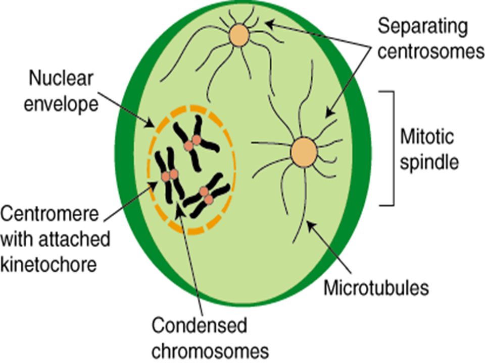
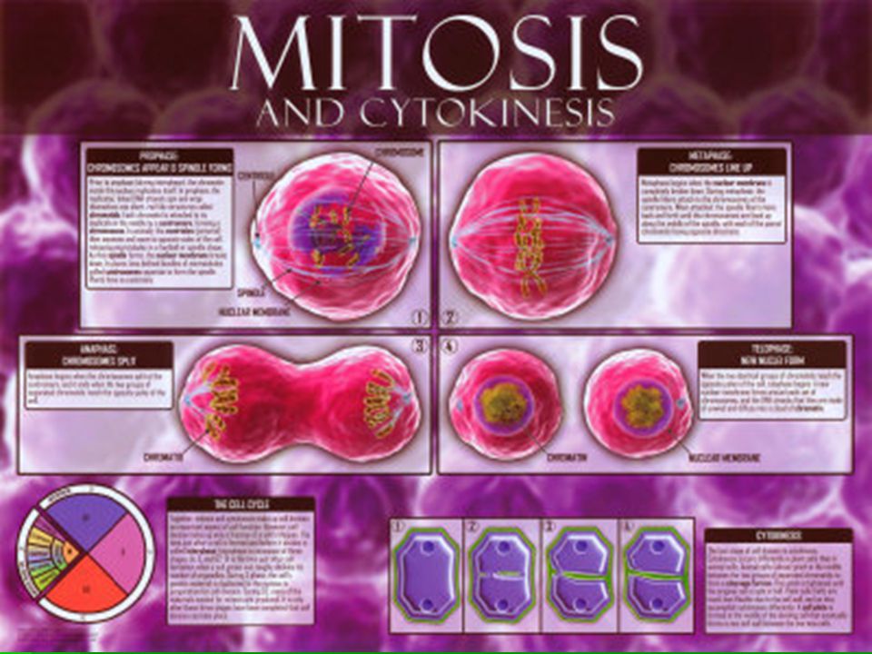
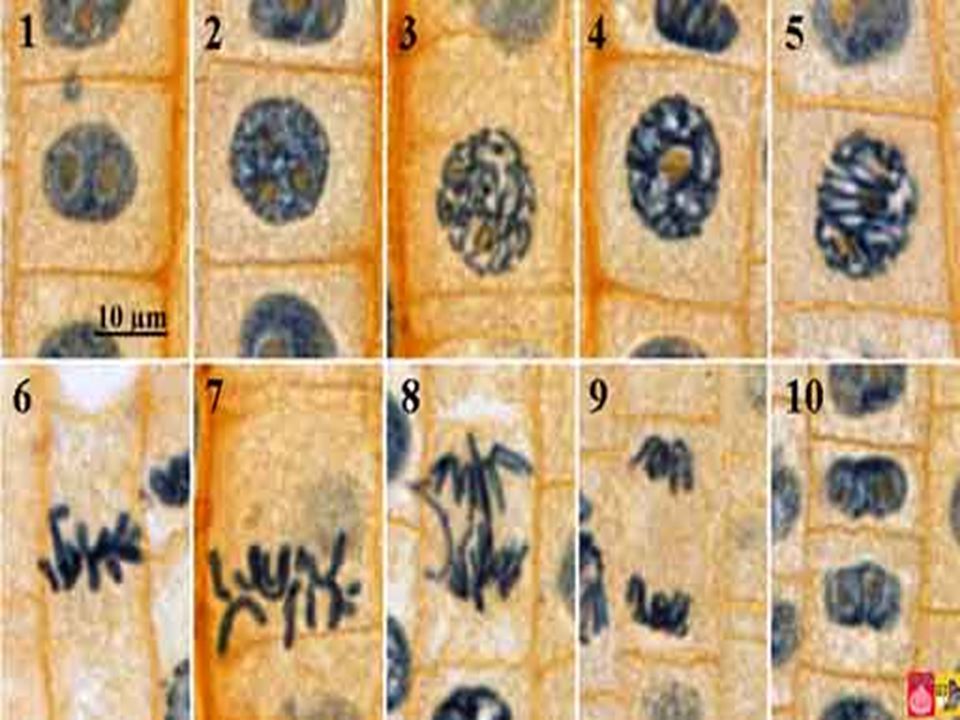
Prophase 50-60% of time Chromosomes become visible Centrioles develop in cytoplasm near nuclear envelope Centrioles separate and migrate to opposite ends of nuc. Env. Centrosome Region where Centrioles are found Organize the “spindle” Fan like microtubule structure that helps separate chromosomes Plants do NOT have Centrioles

31
End of prophase Chromosomes coil together tightly Nucleolus disappears Nuclear envelope breaks down

32
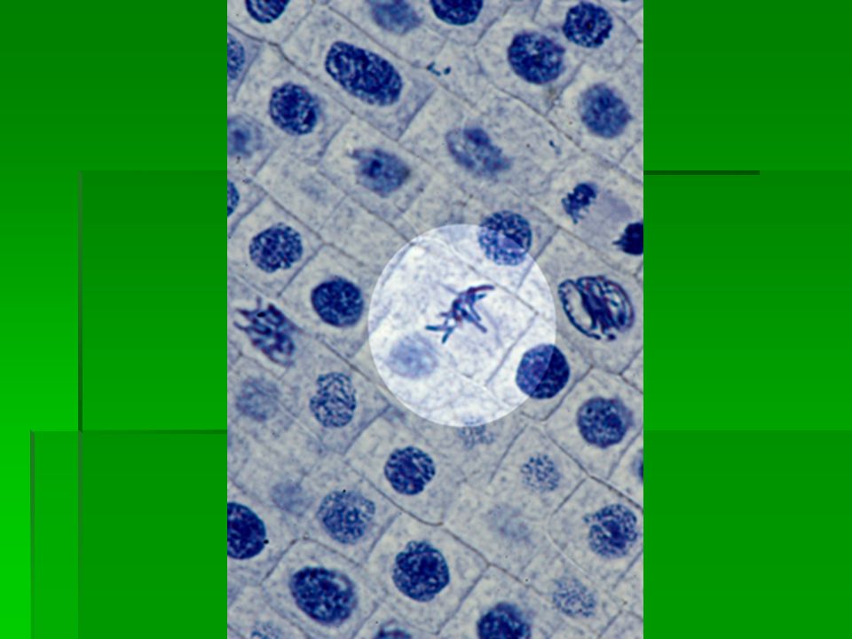
Metaphase Few minutes Chromosomes line up in middle (M in metaphase MIDDLE) Microtubules connect centromere of each chromosome to the 2 poles of spindle
 Microtubules connect centromere of each chromosome to the 2 poles of spindle")
36
Anaphase Centromeres joining sister chromatids separate and become individual chromosomes They are dragged by fibers to opposite poles Ends when chromosomes stop moving

40
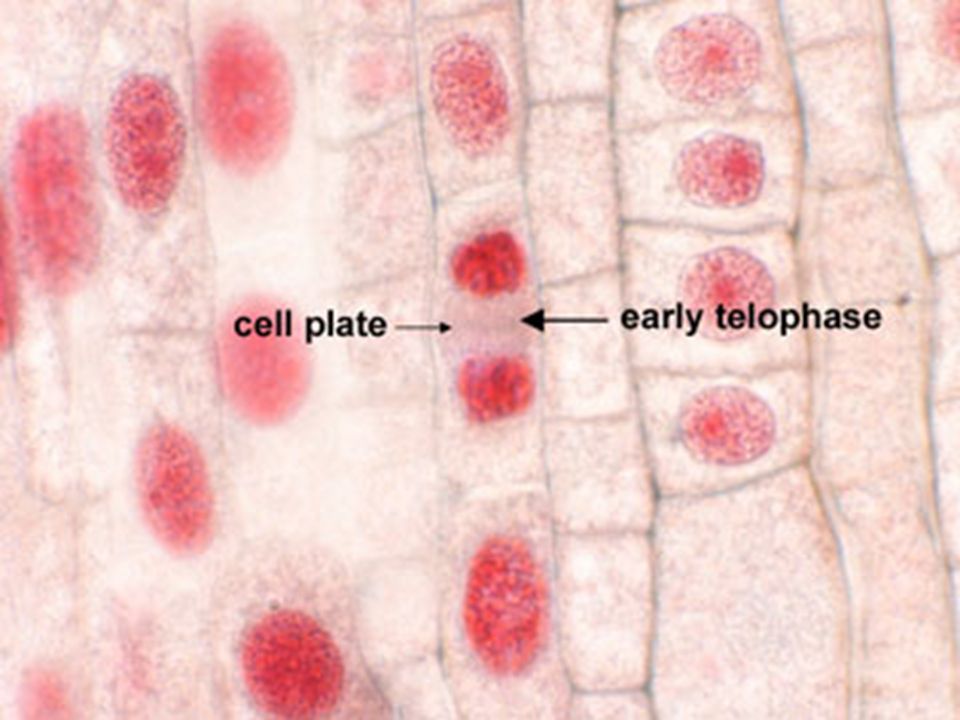
Telophase Opposite of prophase Condensed chromosomes disperse into tangle of material Nuclear envelope reforms Spindle breaks apart Nucleolus becomes visible At the end 2 identical nuclei in one cell

44
Cytokinesis Happens at the same time as Telophase Division of cytoplasm Animal Cells Cell membrane drawn inward until it pinches off and forms 2 id daughter cells Plant Cells Cell plate forms between nuclei Cell Plate develops into separate membrane Cell wall appears

51
MEIOSIS

52
MEIOSIS

53
Gregor Mendel 1822 Austrian monk University of Vienna In charge of the Garden

54
What Gregor Mendel Knew… Each organism must inherit a single copy of every gene from each of its “parents” Each of the organisms gametes must contain just one set genes When gametes are formed, there must be a process that separates the 2 sets of genes so each gamete gets one set

55
Karyotype A photograph of a person's chromosomes, arranged according to size

60
Chromosome Number Homologous chromosomes Chromosome that has a corresponding chromosome from the opposite-sex parent Fruit fly has 8 chromosomes 4 from mom 4 from dad

62
Before meiosis 1, each chromosome is replicate (s-phase of cell cycle) Tetrad STRUCTURE MADE WHEN EACH CHROMOSOME PAIRS UP WITH ITS HOMOLOGOUS CHROMOSOME 4 CHROMATIDS IN A TETRAD
 Tetrad STRUCTURE MADE WHEN EACH CHROMOSOME PAIRS UP WITH ITS HOMOLOGOUS CHROMOSOME 4 CHROMATIDS IN A TETRAD")
63
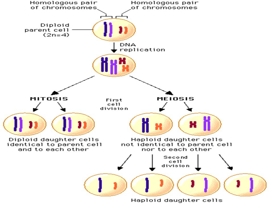
Diploid Di= two sets Cell that contains both sets of homologus chromosomes Cell contains 2 complete sets of chromosome 2 complete sets of genes Number of chrms in diploid cell represented by 2N For Drosophilia (fruit fly) 2N=8 Mendel said: Each adult cell contains two copies of each gene
 2N=8 Mendel said: Each adult cell contains two copies of each gene")
64
Haploid Means “one set” Refers to cells that contain only one set of chromosomes Gametes (sex cells) Represented by N Drosophilia fruit fly N=4
 Represented by N Drosophilia fruit fly N=4")
65
How are haploid (N) gametes made from diploid (2N) cells?
 gametes made from diploid (2N) cells")
66
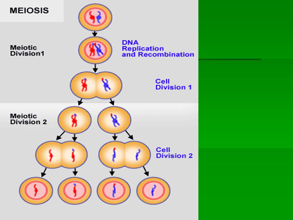
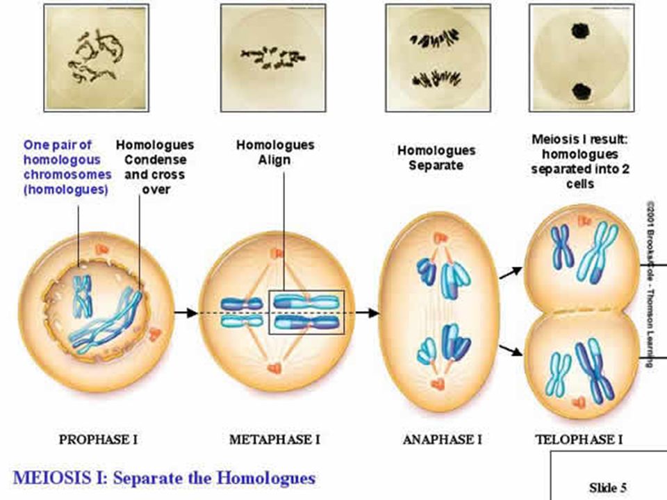
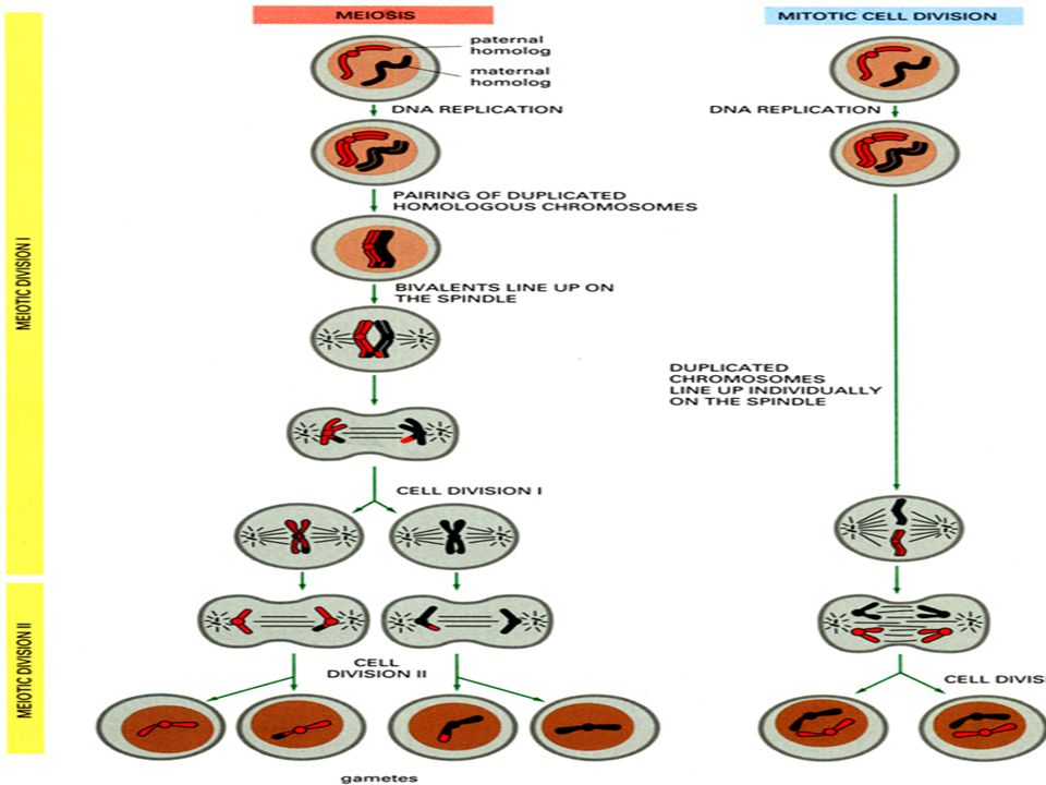
Meiosis Process of reduction division in which the number of chromosomes per cell is cut in half through the separation of homologous chromosomes in a diploid cell

67
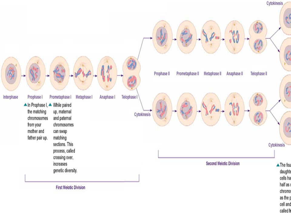
Meiosis 2222 distinct stages MMMMeiosis I AAAA diploid cell enters here MMMMeiosis II AAAAt the end of this, the diploid cell that entered meiosis has become 4 haploid cells

70
Meiosis I Before meiosis 1, each chromosome is replicate Tetrads line up like the sister chromatids did in mitosis What happened in mitosis? PMAT Tetrad STRUCTURE MADE WHEN EACH CHROMOSOME PAIRS UP WITH ITS HOMOLOGOUS CHROMOSOME 4 CHROMATIDS IN A TETRAD

71
Prophase 1 Each chromosome pairs with its homologous chromosome making a tetrad As they pair up in tetrads, chromosomes exchange portions of their chromatids in the process …. CROSSING OVER First way genetic variation in gametes is achieved

72
Crossing Over

74
Metaphase1 Homologous pairs line up in center of cell RANDOMLY…called Independent Assortment Lead s to genetic variation (in addition to crossing over) Anaphase 1 The spindles pull homologous chromosomes apart to opposite poles/ends Telophase 1 Nuclear membranes form and cell separates into two new cells
 Anaphase 1 The spindles pull homologous chromosomes apart to opposite poles/ends Telophase 1 Nuclear membranes form and cell separates into two new cells")
76
Now what do we have? 2 new daughter cells Are they identical to the parents? No Let’s say the parent started with 8 chromosomes Each daughter cell has 8 chromosomes but they are different because of crossing-over Each daughter cell has a set of chromosomes and alleles different from each other and different from the parent diploid cell

77
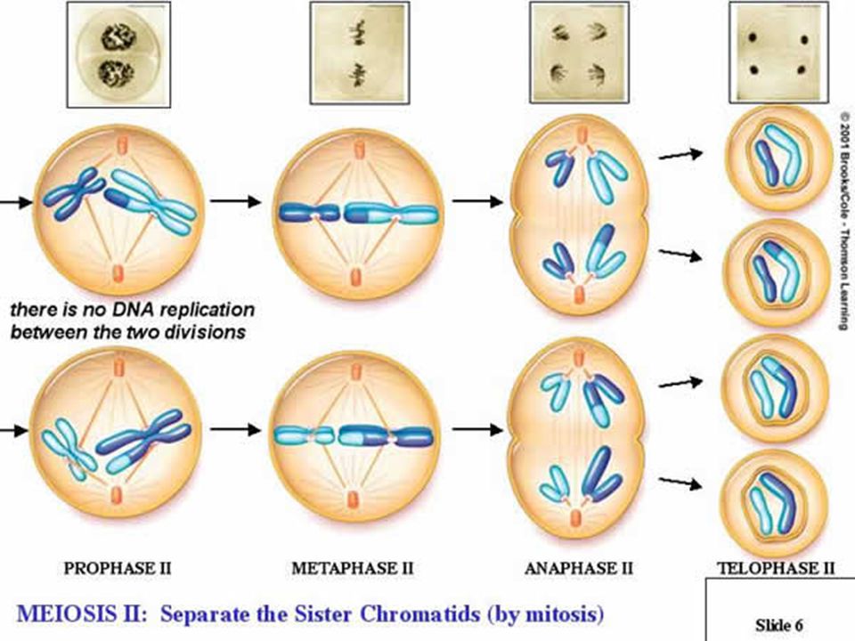
Meiosis II Unlike Mitosis, Neither cell goes through a round of chromosome replication

78
Prophase II Meiosis I resulted in 2 “seemingly” diploid cells Remember they are genetically different b/c of crossing over in prophase I We still need to cut this number in half to reach our goal of 4 haploid cells

79
Metaphase 2 Chromosomes line up in middle Anaphase 2 Sister chromatids separate and move to opposite poles Telophase 2 Meiosis II results in 4 haploid (N) daughter cells
 daughter cells")
83
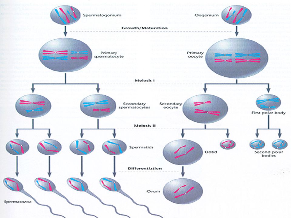
Gamete Formation Male Haploid gametes produced by meiosis are called spermatozoa Spermatogenesis begins at puberty and continues throughout one’s life Under hormone and environmental control Female Haploid gamete produced by meiosis is called an oocyte Cell divisions at the end of meiosis one and two are uneven so one cell gets most of the cytoplasm (the EGG) and the other three are called polar bodies (don’t participate in reproduction) IMPORTANT! Female gametes are stuck in Prophase 1 until puberty Complete Meiosis 1 every month and the secondary is released from ovary Female gametes only reach and complete meiosis 2 if they are fertilized

93
Mitosis vs. Meiosis Mitosis Results in the production of two genetically identical DIPLOID cells Daughter cells have sets of chromosomes identical to each other and to parent cell MITOSIS allows body to grow and replace other cells Asexual reproduction Meiosis Results in four genetically different HAPLOID cells MEIOSIS is how sexually reproducing organisms make gametes

95
Cancer Accounts for 1 in 4 deaths in developed countries More than 100 different forms of cancer Lung cancer=1 in 17 of all deaths in Britain in the 1990’s Most common cancer in men-lung Most common form of cancer in women- breast

96
Cancer A disease that is the result of uncontrolled mitosis Tumor-irregular mass of cells created by uncontrolled mitosis

97
Origin of Cancer Caused by changes in genes that control cell division Mutation- a change in any gene Not unusual Most mutated cells are either crippled in some way that results in their early death or they are destroyed by the body’s immune system Cancer cells bypass both these fates of mutated cells Oncogene- term for mutated gene that causes cancer Onkos in Greek means “mass” or “Bulk”

98
Cancer Although mutation may have occurred in one cell, it can be passed down to all of this one cells’ descendents By the time it is detected, a typical tumor consists of about a thousand million cells

99
Mutagen A factor that brings about any mutation Things can be described as mutagenic Carcinogen Anything agent that causes cancer These can described as carcinogenic Some mutagens are carcinogenic Factors that increase mutation rates (thus cancer) are as follows: 1.Ionizing Radiation 2.Chemical 3.Viral Infections 4.Hereditary predisposition
 are as follows: 1.Ionizing Radiation 2.Chemical 3.Viral Infections 4.Hereditary predisposition")
100
Ionizing Radiation X-rays, gamma rays, particles of a decaying radioactive element Creates the formation of damaging ions inside cells that break DNA strands UV light also breaks the DNA strands (but it does not cause the formation of ions) Depletion of the ozone layer is becoming a concern Leads to more UV radiation hitting Earth’s surface=increase risk to skin cancer
 Depletion of the ozone layer is becoming a concern Leads to more UV radiation hitting Earth’s surface=increase risk to skin cancer")
101
Chemicals Chemical compounds found in many consumer products 25% of all cancers in developed countries are caused by the carcinogens in tar of tobacco smoke Certain dyes (aniline) are well-known carcinogens
 are well-known carcinogens")
102
Viral Infections Viruses are genetic material and protein Cancer viruses are estimated to cause 15 to 20 percent of all cancers in humans Viruses that cause cancer usually carry oncogenes, or regulatory genes that can become oncogenes The tumor viruses change cells by integrating their genetic material with the host cell’s DNA via a permanent insertion mechanism This differs depending on whether the nucleic acid in the virus is DNA or RNA In DNA viruses, the genetic material can be directly inserted into the host's DNA RNA viruses must first transcribe RNA to DNA and then insert the genetic material into the host cell's DNA.

103
Viruses and Cancer DNA Viruses The Epstein-Barr virus has been linked to Burkitt's lymphoma Infects B cells and epithelial cells Causes mononucleosis, but can also cause a few different types of lymphoma and nasopharyngeal cancer The hepatitis B virus has been linked to liver cancer in people with chronic infections Human papilloma viruses have been linked to cervical cancer RNA Viruses Human T lymphotrophic virus type 1 (HTLV-I), a retrovirus, has been linked to T-cell leukemia The hepatitis C virus has been linked to liver cancer in people with chronic infections
, a retrovirus, has been linked to T-cell leukemia The hepatitis C virus has been linked to liver cancer in people with chronic infections")
104
Hereditary disposition Genetic link based on studying patterns in family members Disease itself is not inherited but susceptibility to the factors that can cause the disease are inherited Some forms of cancer are believed to be caused by the inheritance of one faulty gene Example: Retinoblastoma…caused by error on chromosome 13 Starts in both eyes during childhood and spreads to brain causing blindness and death if left untreated

105
Tumors Small groups of tumor cells are called primary growths 2 types Benign tumors Tumors that do not spread from site of origin They can compress and displace other tissues, causing discomfort and even death Warts, ovarian cysts, brain tumors Malignant (cancerous) tumors Dangerous Spread throughout the body, invading and destroying other tissues Interfere with normal functioning of the area they have started to grow Mutated cells break off the tumor and enter either the blood or lymph via vessel formation and spread all over the body creating secondary growths METASTASIS is the spread of cancer from the origin to other parts of the body most dangerous form of caner…can be very difficult to find secondary growths and remove them Both benign and malignant tumors involve a huge drain on the human body due to the high demand for nutrients that is created by the rapid and continual cell division
 tumors Dangerous Spread throughout the body, invading and destroying other tissues Interfere with normal functioning of the area they have started to grow Mutated cells break off the tumor and enter either the blood or lymph via vessel formation and spread all over the body creating secondary growths METASTASIS is the spread of cancer from the origin to other parts of the body most dangerous form of caner…can be very difficult to find secondary growths and remove them Both benign and malignant tumors involve a huge drain on the human body due to the high demand for nutrients that is created by the rapid and continual cell division")
109
Genes

111
Old meiosis slides

112
Gregor Mendel 1822 Austrian monk University of Vienna In charge of the Garden

113
What Gregor Mendel Knew… Each organism must inherit a single copy of every gene from each of its “parents” Each of the organisms gametes must contain just one set genes When gametes are formed, there must be a process that separates the 2 sets of genes so each gamete gets one set

114
Karyotype A photograph of a organism’s chromosomes, arranged according to size

119
Chromosome Number Homologous chromosomes Chromosome that has a corresponding chromosome from the opposite-sex parent Fruit fly has 8 chromosomes 4 from mom 4 from dad

120
Diploid Di= two sets Cell that contains both sets of homologus chromosomes Cell contains 2 complete sets of chromosome 2 complete sets of genes Number of chrms in diploid cell represented by 2N For Drosophilia (fruit fly) 2N=8 Mendel said: Each adult cell contains two copies of each gene
 2N=8 Mendel said: Each adult cell contains two copies of each gene")
121
Haploid Means “one set” Refers to cells that contain only one set of chromosomes Gametes (sex cells) Represented by N Drosophilia fruit fly N=4
 Represented by N Drosophilia fruit fly N=4")
122
How are haploid (N) gametes made from diploid (2N) cells?
 gametes made from diploid (2N) cells")
123
Meiosis Process of reduction division in which the number of chromosomes per cell is cut in half through the separation of homologous chromosomes in a diploid cell

124
Meiosis 2222 distinct stages MMMMeiosis I AAAA diploid cell enters here MMMMeiosis II AAAAt the end of this, the diploid cell that entered meiosis has become 4 haploid cells

127
Meiosis I Before meiosis 1, each chromosome is replicate Then they divide like in mitosis What happened in mitosis? PMAT Tetrad STRUCTURE MADE WHEN EACH CHROMOSOME PAIRS UP WITH ITS HOMOLOGOUS CHROMOSOME 4 CHROMATIDS IN A TETRAD

128
Prophase 1 Each chromosome pairs with its homologous chromosome making a tetrad As they pair up in tetrads, chromosomes exchange portions of their chromatids in the process …. CROSSING OVER

129
Crossing Over

131
Metaphase1 Spindle fibers attach to chromosomes Anaphase 1 The spindles pull homologous chromosomes apart to opposite poles/ends Telophase 1 Nuclear membranes form and cell separates into two new cells

133
Now what do we have? 2 new daughter cells Are they identical to the parents? No The parent has 4 chromosomes Each daughter cell has 4 chromosomes but they are different because of crossing-over Each daughter cell has a set of chromosomes and alleles different from each other and different from the parent diploid cell

134
Meiosis II Unlike Mitosis, Neither cell goes through a round of chromosome replication Each cell’s chromosome has 2 chromatids

135
Prophase II Meiosis I resulted in 2 “seemingly” diploid cells Remember they are genetically different b/c of crossing over in prophase I We still need to cut this number in half to reach our goal of 4 haploid cells

136
Metaphase 2 Chromosomes line up in middle Anaphase 2 Sister chromatids separate and move to opposite poles Telophase 2 Meiosis II results in 4 haploid (N) daughter cells 4 daughter cells contain haploid number of chromosomes, just 2 each
 daughter cells 4 daughter cells contain haploid number of chromosomes, just 2 each")
139
Gamete Formation Male Haploid gametes produced by meiosis are called sperm Female Haploid gamete produced by meiosis is called an egg Cell divisions at the end of meiosis one and two are uneven so one cell gets most of the cytoplasm (the EGG) and the other three are called polar bodies (don’t participate in reproduction)
 and the other three are called polar bodies (don’t participate in reproduction)")
145
Mitosis vs. Meiosis Mitosis Results in the production of two genetically identical DIPLOID cells Daughter cells have sets of chromosomes identical to each other and to parent cell MITOSIS allows body to grow and replace other cells Asexual reproduction Meiosis Results in four genetically different HAPLOID cells MEIOSIS is how sexually reproducing organisms make gametes

149
Genes

152
Microscope Lab Analysis Mitosis/Meiosis Microscope Lab Lab notebooks Title “Cell Division Microscope Lab” MUST sketch each stage and label the power Label slide name Stage of mitosis or meiosis Power of the objective used to observe cell Need to observe each stage of mitosis and meiosis

Similar presentations