Download presentation
Presentation is loading. Please wait.

1
Muscle System Ch. 10

2
Four functional groups of muscle:
1- prime movers (agonists): Muscle that bears the prime responsibility for effecting a movement 2- antagonist: Muscle that opposes the action of another muscle. 3- synergists: these muscles act as stabilizers to the prime movers. They may also contribute extra force to the prime mover as well. 4- fixator: muscle that immobilizes one or more bones (scapula)
: Muscle that bears the prime responsibility for effecting a movement. 2- antagonist: Muscle that opposes the action of another muscle. 3- synergists: these muscles act as stabilizers to the prime movers. They may also contribute extra force to the prime mover as well. 4- fixator: muscle that immobilizes one or more bones (scapula)")
3
Pennate - fascicle branch form central tendon diagonally
Muscle are classified according to their fascicular arrangement or attachment. Parallel - long axis of fascicle runs with longitudinal axis of muscle: sartorius muscle Pennate - fascicle branch form central tendon diagonally Unipennate - extensor muscles Bipennate - rectus femoris Multipennate - deltoid Convergent - fascicle converge toward a single tendon: pectoralis major Circular - fascicle are arranged in concentric rings Orbicularis oris and oculi

5
Types of muscle attachments: direct and indirect
Direct (tendon) - epimysium of muscle fused to periosteum of bone or perichondrium of cartilage. Indirect (aponeurosis) - muscle fascia extends beyond the muscle to bone or another muscle. Latissimus dorsi or galea aponeurotica
 - epimysium of muscle fused to periosteum of bone or perichondrium of cartilage. Indirect (aponeurosis) - muscle fascia extends beyond the muscle to bone or another muscle. Latissimus dorsi or galea aponeurotica.")
6
Muscle have points of attachment on the skeleton via tendons or aponeurosis
Origin and insertion Origin: end of the muscle attaches to the stationary bone (non-moveable) Insertion: end of muscle attaches to the moving bone Biceps (two heads) have two origins on the scapula Biceps inserts on the radius (crosses elbow joint)
 Insertion: end of muscle attaches to the moving bone. Biceps (two heads) have two origins on the scapula. Biceps inserts on the radius (crosses elbow joint)")
10
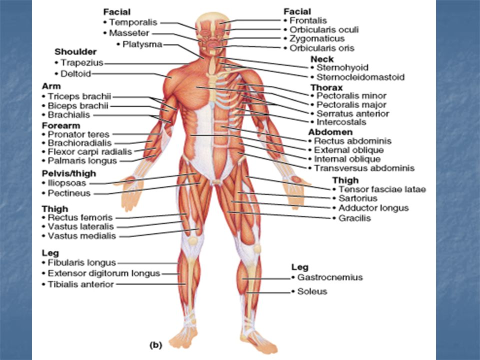
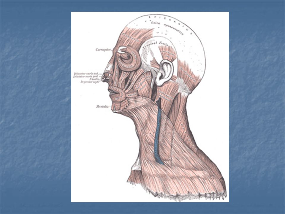
Muscle list 1- Frontalis: O: galea aponeurotica I: skin of eyebrows N: Facial nerve (CN VII) A: raises eyebrows 2- Orbicularis oculi: O: frontal bone I: tissues of eyelid N: Facial nerve A: closes eyes

11
3- Temporalis: O: temporal fossa I: coronoid process of mandible N: Trigeminal nerve (CN V) A: closes jaw (muscle of mastication) 4- Orbicularis oris O: indirectly from maxilla and mandible I: encircles mouth N: Facial nerve A: puckers lips (kissing and whistling)
")
13
O: subcutaneous skin over delto-pectoral region
5- Platysma: O: subcutaneous skin over delto-pectoral region I: skin widely over the mandible A: depress mandible and lower lip tenses the skin over the lower neck N: facial nerve 6- Scalenes: located deep in the neck anterior middle posterior

16
7- Sternohyoid: O: manubrium I: hyoid bone A: depresses hyoid and larynx 8- Digastric: consists of two bellies that are connected by an intermediate tendon 9- Sternocleidomastoid: O: manubrium and medial portion of clavicle I: mastoid process A: together they flex head forward; separately they rotate head opposite to muscle contracting N: Spinal accessory (CN XI)
")
17
10- Deltoid: O: lateral 1/3 of clavicle, acromion and spine of scapula I: deltoid tuberosity A: arm abduction N: axillary nerve 11: Pectoralis minor: Thin muscle located underneath pectoralis major O: ribs 3-5 I: coracoid process

19
12- Pectoralis major O: medial 1/3 of clavicle I: lateral lip of bicipital groove to the crest of the greater tubercle A: arm flexion, rotates arm medially N: lateral and median pectoral nerves 13- Serratus anterior O: ribs 1-8 I: vertebral border of scapula A: protract and hold scapula N: long thoracic nerve 14- Intercostals: accessory muscle to respiration

21
15- Rectus abdominis: O: pubic symphysis I: xiphoid process and costal cartilage A: flex lumbar spine N: intercostal nerves 16- Diaphragm: O: inferior surface of rib cage I: central tendon A: primary muscle of respiration N: phrenic nerve (C3,4,5)
")
25
17- Triceps brachii: O: long head: infraglenoid tubercle of scapula medial head: posterior humeral shaft lateral head: posterior humeral shaft I: olecranon process of ulna A: forearm extension N: radial nerve 18- Biceps brachii : O: short head: coracoid process long head: supraglenoid tubercle I: radial tuberosity A: forearm flexor N: musculocutaneous nerve

28
19- Brachialis: Forearm flexor (lifts ulna) 20- Brachioradialis: “drinking muscle” O: distal end of humerus I: styloid process of radius A: forearm flexor N: radial nerve 21- Flexor carpi radialis: A: wrist flexor N: median nerve

30
22- Palmaris longus: O: medial epicondyle of humerus I: palmar aponeurosis A: weak wrist flexor N: median nerve Absent in 15% of the population (More females than males) 23- Lumbricals: 4 worm shaped muscles A: flex fingers at MCP joint
 23- Lumbricals: 4 worm shaped muscles. A: flex fingers at MCP joint.")
31
24- Iliopsoas: combination iliacus and psoas muscle
A: primary hip and trunk flexor 25- Pectineus: Short flat muscle that adducts thigh O: pubis I: lesser trochanter 26- Tensor fasciae latae: O: ASIS I: Iliotibial tract (Gerdy’s tubercle) A: thigh abduction
 A: thigh abduction.")
32
27- Sartorius: “tailor’s muscle”
O: anterior superior iliac spine (ASIS) I: upper medial surface of body of tibia A: Flexes, abducts and laterally rotates thigh N: Obturator nerve 28: Adductor longus Common thigh adductor 29: Gracilis: O: pubis I: tibia A: adducts thigh
 I: upper medial surface of body of tibia. A: Flexes, abducts and laterally rotates thigh. N: Obturator nerve. 28: Adductor longus. Common thigh adductor. 29: Gracilis: O: pubis. I: tibia. A: adducts thigh.")
34
Quadriceps: 30: Rectus femoris O: ASIS I: patella by way of quadriceps tendon and tibial tuberosity by way of patellar ligament A: hip flexor and knee extension N: femoral nerve 31: Vastus lateralis medius knee extension intermedius

36
32: Tibialis anterior: O: lateral condyle of tibia I: medial cuneiform and 1st MTP. A: dorsiflexion of foot N: deep fibular nerve 33: Flexor hallucis longus: flexes great toe “push off” while taking a step

39
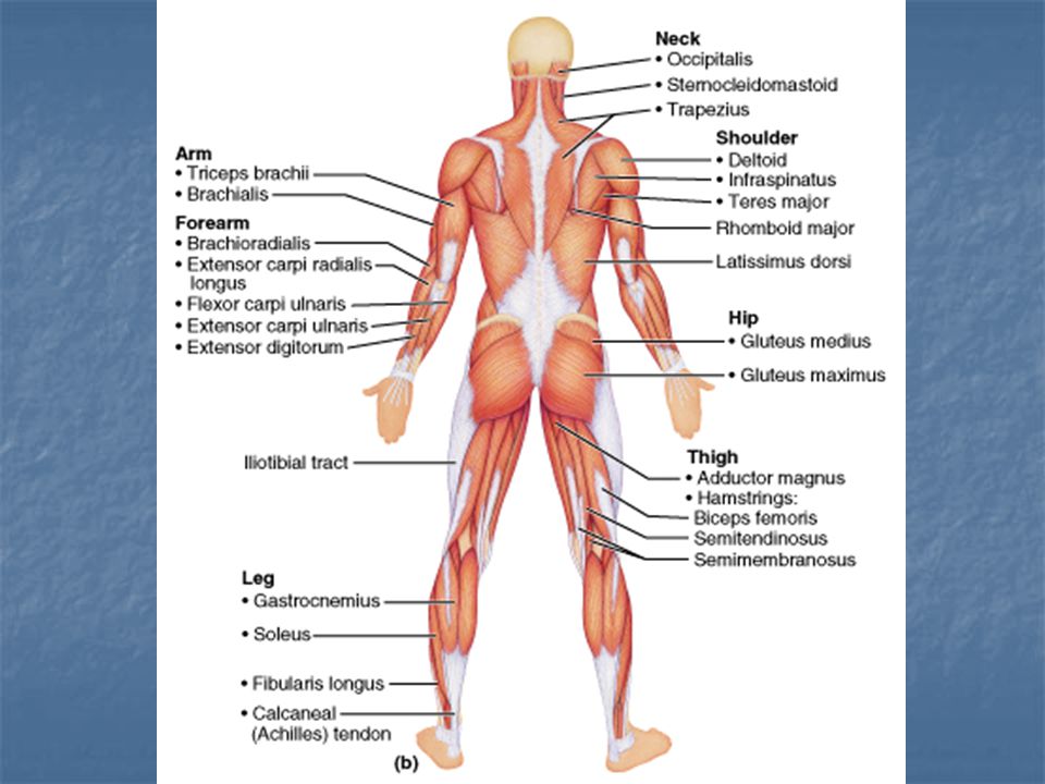
Posterior 1- Occipitalis: O: occipital bone I: galea aponeurotica A: pulls scalp posteriorly I: facial nerve 2- Trapezius O: Occipital bone, vertebra of cervical and thoracic spinous processes I: acromion, spine of scapula and lateral 1/3 of clavicle A: extends head and raises scapula. “shrugging shoulders” N: Spinal Accessory nerve (CNXI)
")
41
Rotator cuff of shoulder: Holds humerus tight to glenoid fossa (S.I.T.S.)
3- Supraspinatus: O: supraspinous fossa of scapula I: greater tubercle of humerus A: assists in abduction 4- Infraspinatus O: infraspinous fossa of scapula A: rotates humerus laterally

42
5- Subscapularis O: subscapular fossa I: lesser tubercle of humerus A: medial rotation of humerus 6- Teres minor: A: Arm adductor

43
7- Teres major: A: posteromedial extension of humerus 8- Rhomboid major and minor O: spinous processes of C7-T1 (minor) I: medial border of scapula A: retract scapula “standing up straight” N: dorsal scapular nerve

46
9- Erector Spinae: Iliocostalis Longissimus Spinalis A: prime muscles for back extension 10- Latissimus dorsi O: Thoracolumbar fascia (aponeurosis) - T6-T12 and L1-L5 and iliac crest I: intertubercular groove of humerus A: arm extension and adductor N: thoracodorsal nerve
 - T6-T12 and L1-L5 and iliac crest. I: intertubercular groove of humerus. A: arm extension and adductor. N: thoracodorsal nerve.")
48
11- Extensor carpi ulnaris:
Extension of the wrist 12- Extensor carpi radialis Extends and abducts wrist 13- Flexor carpi ulnaris Powerful wrist flexor N: ulnar nerve 14- Extensor digitorum Prime mover of finger extension

50
15- Gluteus medius A: abducts and medial rotates thigh N: superior gluteal nerve 16- Gluteus maximus O: dorsal ilium, sacrum and coccyx I: gluteal tuberosity of femur A: powerful thigh extender N: inferior gluteal nerve

52
Hamstrings (crosses knee joint- popliteal)
17- Biceps femoris: O: ischial tuberosity I: head of fibula and lateral condyle of tibia A: thigh extension and knee flexion N: Sciatic nerve 18- Semitendinosus: A: extends thigh and knee flexion 19- Semimembranosus A: extends thigh and flexes knee

55
20- Gastrocnemius O: medial and lateral condyles of femur I: posterior calcaneus (achilles tendon) A: plantar flexes foot (raise up on toes) N: Tibial nerve 21- Soleus A: plantar flexes foot and raise up on toes while seated
 N: Tibial nerve. 21- Soleus. A: plantar flexes foot and raise up on toes while seated.")
Similar presentations


>")