Download presentation
Presentation is loading. Please wait.

1
TACHYCARDIAS Dr Kirsten Cohen
ANTIARRYTHMIC DRUGS TACHYCARDIAS Dr Kirsten Cohen

2
Normal Sinus Rhythm Heart rhythm is determined by SA node = Cardiac Pacemaker Called sinus rhythm Specialised pacemaker cells spontaneously generate APs APs spread through the conducting pathways Normal sinus rate beats/min

3
Conducting System SAN AP triggers atrial depolarisation
AVN - Only pathway for AP to enter ventricles Conducts slowly: Complete atrial systole before ventricular systole Conducts rapidly through His Bundles & Purkinje – Ventricular depolarization & contraction

4
Conducting System Permits rapid organized depolarization of ventricular myocytes Necessary for the efficient generation of pressure during systole Atrial activation complete 0.09s after SAN firing Delay at AVN Septum activated 0.16s Whole ventricle activated by 0.23s

5
Cardiac Action Potential
Phase 4: RMP AP depolarizes cells to threshold -70mV Phase 0: Rapid depolarization Caused by a transient opening of fast Na channels Increases inward directed depolarizing Na+ currents Generate "fast-response" APs

6
Cardiac Action Potential
Phase 1: Initial repolarization Open K channel: transient outward hyperpolarizing K+ current Large increase in slow inward gCa++ occurs at the same time L-type CaCh open -40mV Repolarization delayed Phase 2: Plateau phase Plateau phase prolongs AP duration vs APs in nerves and skeletal muscle

7
Cardiac Action Potential
Phase 3: Repolarization K channels open Inactivation of Ca++ channels Action potential in non-pacemaker cells is primarily determined by relative changes in fast Na+, slow Ca++ and K+ conductances and currents

8
Refractory Periods Once an AP is initiated, there is a period (phase 0,1,2, part 3) that a new AP cannot be initiated. Effective or Absolute refractory period (ERP or ARP) Stimulation of cell by adjacent cell depolarizing does not produce new propagated APs Prevents compounded APs from occurring & limits frequency of depolarization and HR
 Stimulation of cell by adjacent cell depolarizing does not produce new propagated APs. Prevents compounded APs from occurring & limits frequency of depolarization and HR.")
9
SAN Pacemaker Potential
Fully repolarized -60mv No stable RMP Phase 4: Spontaneous depolarization or pacemaker potential Slow, inward Na+ channels open - "funny" currents Cause the membrane potential to begin to spontaneously depolarize During Ph4 there is also a slow decline in the outward movement of K+

10
SAN Pacemaker Potential
-50mV T-type CaCh open Ca in: further depolarizes -40 mV L-type CaCh open More Ca in: further depol AP threshold -35mV Phase 0: Depolarization Primarily caused by Ca++ conductance through the L-type Ca++ channels Movement of Ca++ through these is slow so the rate of depolarization (Phase 0 slope) is slower than in other cardiac cells
 is slower than in other cardiac cells.")
11
SAN Pacemaker Potential
Phase 3: Repolarization K+ channels open Increase the outward hyperpolarizing K+ currents At the same time the L-type Ca++ channels close gCa++ decreases Inward depolarizing Ca++ currents diminish Repolarization

12
Regulation of Cardiac APs
SNS - Increased with concurrent inhibition vagal tone: NA binds to B1 Rec Increases cAMP Increases Ca and Na in Decreases K out Increases slope phase 0 Non-Nodal tissue: More rapid depolarisation More forceful contraction Pacemaker current (If) enhanced Increase slope phase 4 Pacemaker potential more rapidly reaches threshold Rate increased
 enhanced Increase slope phase 4. Pacemaker potential more rapidly reaches threshold. Rate increased.")
13
Regulation of Cardiac APs
PSNS (Vagal N) Ach binds M2 rec Increases gK+ Decreases inward Ca & Na Non-Nodal tissue: More rapid depolarisation More forceful contraction Pacemaker current (If) suppressed Decreases pacemaker rate Decrease slope of Phase 4 Hyperpolarizes in Phase 4 Longer time to reach threshold voltage
 Ach binds M2 rec. Increases gK+ Decreases inward Ca & Na. Non-Nodal tissue: More rapid depolarisation. More forceful contraction. Pacemaker current (If) suppressed. Decreases pacemaker rate. Decrease slope of Phase 4. Hyperpolarizes in Phase 4. Longer time to reach threshold voltage.")
14
What is an Arrhythmia ? Irregular rhythm Abnormal Rate
Conduction abnormality

15
What causes an arrhythmia?
Changes in automaticity of the PM Ectopic foci causing abnormal APs Reentry tachycardias Block of conduction pathways Abnormal conduction pathways (WPW) Electrolyte disturbances and DRUGS Hypoxic/Ischaemic tissue can undergo spontaneous depolarisation and become an ectopic pacemaker
 Electrolyte disturbances and DRUGS. Hypoxic/Ischaemic tissue can undergo spontaneous depolarisation and become an ectopic pacemaker.")
16
Re-Entry Mechanism Branch 2 has a unidirectional block
Impulses can travel retrograde (3 to 2) but not orthograde. An AP will travel down the branch 1, into the common distal path (br 3), then travel retrograde through the unidirectional block in branch 2. When the AP exits the block, if it finds the tissue excitable, it will continue by traveling down (reenter) the branch 1. If it finds the tissue unexcitable (ERP) the AP will die. Tming is critical –AP exiting the block must find excitable tissue to propagate. If it can re-excite the tissue, a circular pathway of high frequency impulses (tachyarrhythmia) will become the source of APs that spread throughout a region of the heart (ventricle) or the entire heart.
 but not orthograde. An AP will travel down the branch 1, into the common distal path (br 3), then travel retrograde through the unidirectional block in branch 2. When the AP exits the block, if it finds the tissue excitable, it will continue by traveling down (reenter) the branch 1. If it finds the tissue unexcitable (ERP) the AP will die. Tming is critical –AP exiting the block must find excitable tissue to propagate. If it can re-excite the tissue, a circular pathway of high frequency impulses (tachyarrhythmia) will become the source of APs that spread throughout a region of the heart (ventricle) or the entire heart.")
17
Rationale for Antiarrhythmic Drugs
Restore normal rhythm, rate and conduction or prevent more dangerous arrhythmias Alter conduction velocity (SAN or AVN) Alter slope 0 depolarisation or refractoriness Alter excitability of cardiac cells by changing duration of ERP (usually via changing APD) ERPinc – Interrupts tachy caused by reentry APDinc – Can precipitate torsades 3. Suppress abnormal automaticity
 Alter slope 0 depolarisation or refractoriness. Alter excitability of cardiac cells by changing duration of ERP (usually via changing APD) ERPinc – Interrupts tachy caused by reentry. APDinc – Can precipitate torsades. 3. Suppress abnormal automaticity.")
18
Vaughan-Williams Classification
Mechanism Example I Na channel blockers Membrane Stabilisers Lignocaine II Beta Blockers Metoprolol III K channel blockers Amiodarone IV Ca channel blockers Verapamil Other Digoxin. Adenosine. MgSO4. Atropine Links AA drugs to a MOA Not all drugs are included – Some commonly used Some drugs overlap classes - Sotalol

19
Class I A Agents Block open ACTIVATED Na channels
Slow phase 0 depolarisation - upstroke of AP Lengthen APD and ERP. Prolong QRS duration on ECG Anticholinergic S/E. Also blocks K Ch. Greater affinity for rapidly firing channels Disopyramide: Prevent rec VT. - Inotrope Quinidine: SVT and VT. Torsades Procainamide

20
Class I B Agents Block INACTIVATED Na channels
Slow phase 0 depolarisation- Slows upstroke of AP Shorten APD and ERP Ratio ERP/APD is increased Greater affinity for ischaemic tissue that has more inactivated channels, little effect on normal cells – dissociates quickly (0.5sec) Lignocaine: VT in heart with normal EF Phenytoin
 Lignocaine: VT in heart with normal EF. Phenytoin.")
21
LIGNOCAINE - Cardiac arrest: 1-1.5 mg/kg to max 3mg/kg
- Stable wide complex tachycardia: Start lower 0.5 Especially in presence of ischaemia Not if poor cardiac function (Poor EF) Watch for signs of toxicity New algorithm only in cardiac arrest Infusion within 10 min of effect mg/min
 Watch for signs of toxicity. New algorithm only in cardiac arrest. Infusion within 10 min of effect mg/min.")
22
Class I C Agents Block Na channels. Most potent Na channel block
Dissociate very slowly (10-20 sec) Strongly depress conduction in myocardium Slow phase 0 depolarisation - upstroke of AP No effect on APD No effect on QRS Flecainide: Prophylaxis in paroxysmal AF Propafenone
 Strongly depress conduction in myocardium. Slow phase 0 depolarisation - upstroke of AP. No effect on APD. No effect on QRS. Flecainide: Prophylaxis in paroxysmal AF. Propafenone.")
23
Class II Agents Beta Blockers - Block B1 receptors in the heart
Decrease Sympathetic activity Non-Nodal Tissue: Increase APD and ERP SA and AVN: Decrease SR Decrease conduction velocity (Block re-entry) Inhibit aberrant PM activity
 Inhibit aberrant PM activity.")
24
ATENOLOL Non-selective B-Blocker (B1 and B2)
Indications: Convert or Slow rate in SVTs 2nd line after Adenosine/Digoxin/Diltiazem IV atenolol 5 mg over 5 minutes Repeat to maximum 15 mg. 50 mg PO BID if IV works Contraindiactions: Asthma CCF. Poor EF. High degree heart block. Ca channel blockers. Cocaine use.

25
Class III Agents Anti-Fibrillatory agents. Block K channels
Prolong repolarisation Prolong APD and ERP Useful in Re-Entry tachycardias AMIODARONE (also Class IA, II BB) SOTALOL (also Class II BB)
 SOTALOL (also Class II BB)")
26
AMIODARONE Most tachyarrhythmias OK if impaired LV function
Rate control and converts rhythm Cardiac arrest: 300 mg IV push (max 2.2g/24hrs) Stable VT: 150 mg IV repeat 10 min or infusion 360 mg IV over 6 hrs (1mg/min) Maintenance infusion: 540 mg over 18 hrs (0.5mg/min) Side Effects: Hypotension. Negative Inotropy. Prolonged QT. Photosensitivity. Thyroid disorders. Pulmonary alveolitis. Neuropathy.
 Stable VT: 150 mg IV repeat 10 min or infusion 360 mg IV over 6 hrs (1mg/min) Maintenance infusion: 540 mg over 18 hrs (0.5mg/min) Side Effects: Hypotension. Negative Inotropy. Prolonged QT. Photosensitivity. Thyroid disorders. Pulmonary alveolitis. Neuropathy.")
27
Class IV Agents Calcium Channel Blockers Bind to L-type Ca channels
Vascular SmM, Cardiac nodal & non-nodal cells Decrease firing rate of aberrant PM sites Decrease conduction velocity Prolong repolarisation Especially active at the AVN VERAPAMIL DILTIAZEM

28
VERAPAMIL Narrow complex tachycardias Terminates PSVT/SVT
Rate control in AFib/Aflutter NOT WPW or VT or high degree block NOT with BBlockers Negative Inotropy Vasodilation – Hypotension Dose: 5mg IV bolus. Rpt 15 min max 30 mg Diltiazem less adverse effects

29
What does Adenosine Do? Purine nucleoside
Acts on A1 adenosine receptors Opens Ach sensitive K channels Inhibits Ca in current – Suppresses Ca dependent AP (Nodal) Increases K out current – Hyperpolarisation Inhibits AVN > SAN Increases AVN refractory period
 Increases K out current – Hyperpolarisation. Inhibits AVN > SAN. Increases AVN refractory period.")
30
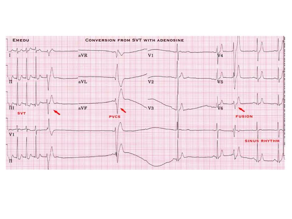
ADENOSINE Interrupts re-entry and aberrant pathways through AVN – Diagnosis and Treament Drug for narrow complex PSVT SVT reliant on AV node pathway NOT atrial flutter or fibrillation or VT Contraindications: VT – Hypotension and deterioration High degree AV block Poison or drug induced tachycardia Bronchospasm but short DOA

31
ADENOSINE Carotid massage and vagal maneuvers first
Rapid IV push 6mg – 12 mg – 12 mg Flush with 20ml N/S Record rhythm strip FLUSHING CHEST PAIN ASYSTOLE/BRADY VENTRICULAR ECTOPY

33
What does Digoxin Do? Reduces ventricular response to SVTs
Cardiac glycoside Blocks Na/K ATPase pump in heart Less ECF Na for Na/Ca pump Increased IC Ca Inotropic: Increases force of contraction AVN increased refractoriness Decreases conduction through AVN and SAN Negative chronotrope: Slows HR Reduces ventricular response to SVTs

34
DIGOXIN Contraindications: WPW. SSS.
Elderly or renal failure – reduce dose or TOXICITY 0.25 to 0.5 mg IV; then 0.25 mg IV every 4 to 6 hours to maximum of 1 mg 0.125 to 0.25 mg per day IV or orally

36
Take-Home Message Anti-arrhythmics are also pro-arrhythmics
Dangerous side effects If patient is unstable rather cardiovert Enlist expert help Stick to drugs you know Limited choice in SA anyway

Similar presentations


>")


