Download presentation
Presentation is loading. Please wait.

1
The Appendicular Skeleton

2
The Appendicular Skeleton
These consist of the limbs and their girdles. Their major function is to carry out movement.

3
The Appendicular Skeleton
These consist of the limbs and their girdles. Their major function is to carry out movement.

4
The Appendicular Skeleton
The pectoral girdle consists of the clavicle and scapula. 1) Only the clavicle attaches to the axial skeleton (sternal end) 2) The scapula is designed for flexibility
 Only the clavicle attaches to the axial skeleton (sternal end) 2) The scapula is designed for flexibility")
5
Figure 7.24a The pectoral girdle and clavicle.
Acromio- clavicular joint Clavicle Scapula (a) Articulated pectoral girdle
 Articulated pectoral girdle.")
6
Figure 7.24b The pectoral girdle and clavicle.
Sternal (medial) end Posterior Anterior Acromial (lateral) end (b) Right clavicle, superior view
 end. Posterior. Anterior. Acromial (lateral) end. (b) Right clavicle, superior view.")
7
The Appendicular Skeleton
The scapula has three borders. Superior border Medial border which lies toward the vertebral column The lateral border which has the glenoid cavity

8
(a) Right scapula, anterior aspect
Figure 7.25a The scapula. Acromion Suprascapular notch Superior border Coracoid process Superior angle Glenoid cavity Subscapular fossa Lateral border Medial border Inferior angle (a) Right scapula, anterior aspect
 Right scapula, anterior aspect.")
9
(b) Right scapula, posterior aspect
Figure 7.25b The scapula. Coracoid process Suprascapular notch Superior angle Acromion Supraspinous fossa Glenoid cavity at lateral angle Spine Infraspinous fossa Medial border Lateral border (b) Right scapula, posterior aspect
 Right scapula, posterior aspect.")
10
(c) Right scapula, lateral aspect Inferior angle
Figure 7.25c The scapula. Supraspinous fossa Supraglenoid tubercle Acromion Coracoid process Glenoid cavity Spine Supraspinous fossa Infraspinous fossa Infraglenoid tubercle Infraspinous fossa Subscapular fossa Subscapular fossa Posterior Anterior (c) Right scapula, lateral aspect Inferior angle
 Right scapula, lateral aspect. Inferior angle.")
11
The Appendicular Skeleton
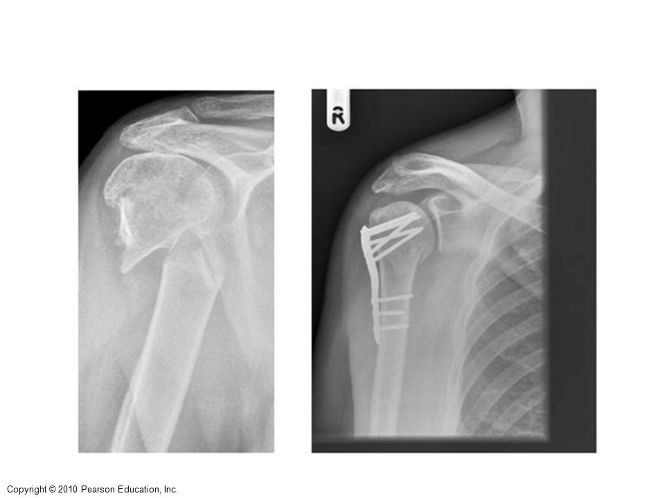
The humerus articulates with the scapula at the shoulder and the ulna and radius distally. The proximal head has the greater and lesser tubercles and anatomical neck which is where the rotator cuff muscles attach.

12
Greater Head of tubercle humerus Lesser Anatomical tubercle neck
Figure 7.26a The humerus of the right arm and detailed views of articulation at the elbow. Greater tubercle Head of humerus Lesser tubercle Anatomical neck Inter- tubercular sulcus Deltoid tuberosity Lateral supracondylar ridge Coronoid fossa Radial fossa Medial epicondyle Capitulum Trochlea (a) Anterior view
 Anterior view.")
13
The Appendicular Skeleton
Just distally is the surgical neck, the most frequently fractured portion of the humerus.

15
The Appendicular Skeleton
The deltoid tuberosity on the lateral side is for the attachment of the deltoid muscle Distally there are two condyles, the medial trochlea which articulates with the ulna and the lateral capitulum which articulates with the radius.

16
The Appendicular Skeleton

17
(c) Anterior view at the elbow region
Figure 7.26c The humerus of the right arm and detailed views of articulation at the elbow. Humerus Coronoid fossa Medial epicondyle Capitulum Trochlea Head of radius Coronoid process of ulna Radial tuberosity Radial notch Radius Ulna (c) Anterior view at the elbow region
 Anterior view at the elbow region.")
18
Telling Left from Right
Orient the bone so that the rounded head is superior (up) and pointing medially. Look for the deep olecranon fossa on the posterior side.
 and pointing medially. Look for the deep olecranon fossa on the posterior side.")
19
The Appendicular Skeleton
What is the medial epicondyle famous for?

20
The Appendicular Skeleton
What is the medial epicondyle famous for? The Funny Bone

21
The Funny Bone The ulnar nerve is the largest unprotected nerve in the human body unprotected by muscle or bone), so injury is common.
, so injury is common.")
22
The Funny Bone This nerve is directly connected to the little finger, and the adjacent half of the ring finger, supplying the palmar side of these fingers, including both front and back of the tips.

23
The Funny Bone The clawed hand can be a result of ulnar nerve damage.

24
The Appendicular Skeleton
The ulna and radial bones form the distal lower limb. The ulna is medial and the radius is lateral.

25
The Appendicular Skeleton
The olecranon process (elbow) and the coronoid processes are the major land marks on the proximal portion of the ulna. The ulna plays no major role in wrist movement. Its only action is extension and flexion of the lower limb.
 and the coronoid processes are the major land marks on the proximal portion of the ulna. The ulna plays no major role in wrist movement. Its only action is extension and flexion of the lower limb.")
27
Figure 7.27c Radius and ulna of the right forearm.
Olecranon process View Trochlear notch Coronoid process Radial notch (c) Proximal portion of ulna, lateral view
 Proximal portion of ulna, lateral view.")
28
The Appendicular Skeleton
The radius is shaped like the head of a nail. Its head is concave. Its distal end is highlighted by the styloid process. The radius allows for pronation and supination of the wrist.

30
Figure 7.27d Radius and ulna of the right forearm.
Ulnar notch of radius Articulation for lunate Articulation for scaphoid Styloid process View Head of ulna Styloid process (d) Distal ends of the radius and ulna at the wrist
 Distal ends of the radius and. ulna at the wrist.")
31
Telling Left from Right
Place the ulna so that the trochlear notch faces you, if the radial notch faces left, it is the right ulna. Place the radius so the distal styloid process is lateral. The radial tuberosity is to the right. It is the right radius.

32
The Hand The “hand” is composed of 8 carpals and 5 metacarpals. Distally are the phalanges, these begin at the knuckles.

33
Figure 7.28 Bones of the left hand.
Phalanges • Distal • Middle • Proximal Metacarpals • Head • Shaft Sesamoid bones • Base Carpals Carpals Carpals • Trapezium • Hamate • Trapezium • Trapezoid • Capitate • Trapezoid • Scaphoid • Pisiform • Scaphoid • Triquetrum Radius • Lunate Ulna Radius (a) Anterior view of left hand (b) Posterior view of left hand
 Anterior view of left hand. (b) Posterior view of left hand.")
34
The Hand Carpal tunnel syndrome is pain, tingling, and other problems in your hand because of pressure on the median in your wrist. It is a common repetitive injury.

39
The Appendicular Skeleton The Pelvic Girdle
The pelvic girdle attaches the lower limbs to the axial skeleton. The hip is also known as the os coxae It is made up of three separate bones: Ischium Ilium & Pubis

40
Base of sacrum Iliac crest Sacroiliac joint Iliac fossa Anterior
Figure Articulated pelvis showing the two hip (coxal) bones (which together form the pelvic girdle), the sacrum, and the coccyx. Base of sacrum Iliac crest Sacroiliac joint Iliac fossa Anterior superior iliac spine Sacral promontory Coxal bone (os coxae or hip bone) Anterior inferior iliac spine llium Sacrum Pubic bone Pelvic brim Coccyx Acetabulum Pubic tubercle Ischium Pubic crest Pubic symphysis Pubic arch
 bones (which together form the pelvic girdle), the sacrum, and the coccyx. Base of sacrum. Iliac crest. Sacroiliac. joint. Iliac fossa. Anterior. superior. iliac spine. Sacral. promontory. Coxal. bone. (os coxae. or hip. bone) Anterior inferior. iliac spine. llium. Sacrum. Pubic. bone. Pelvic brim. Coccyx. Acetabulum. Pubic tubercle. Ischium. Pubic crest. Pubic symphysis. Pubic arch.")
41
The Appendicular Skeleton The Pelvic Girdle
During infancy and child hood, these three bones are separate and fuse to one large irregular bone in adulthood.

42
The Appendicular Skeleton The Pelvic Girdle
Important Land Marks on the Ilium include: Acetabulum which is a socket that receives the head of the femur The ala or wing like projection of the ilium The greater sciatic notch where the sciatic nerve passes The gluteal lines which are the point of attachment for the gluteal muscles

43
The Appendicular Skeleton The Pelvic Girdle
Important Land Marks on the Ischium include: Ischial spine which projects medially into the pelvic cavity and is where the sacrospinous ligament attaches Ischial Tuberosity bears our weight when we sit, also a point of attachment for the ham string muscles Lesser sciatic notch where a number of blood vessels & nerves to the genitals pass

44
The Appendicular Skeleton The Pelvic Girdle
Important Land Marks of the Pubis include: Superior and inferior rami Obturator foramen which is a large empty circle Pubic symphysis which is where both pubic bones attach.

45
Figure 7.30c Bones of the bony pelvis.
Anterior gluteal line Ilium Posterior gluteal line Anterior superior iliac spine Posterior superior iliac spine Anterior inferior iliac spine Posterior inferior iliac spine Inferior gluteal line Acetabulum Greater sciatic notch Ischial body Pubic body Ischial spine Lesser sciatic notch Pubic tubercle Ischium Inferior ramus of pubis Ischial tuberosity Ischial ramus Obturator foramen (c) Lateral view, right hip bone
 Lateral view, right hip bone.")
46
Dimples of Venus These are indentations sometimes visible on the human lower back, just superior to the gluteal cleft. They are directly superficial to the two sacroiliac joints, the sites where the sacrum attaches to the ilium of the pelvis.

47
Table 7.4 Comparison of the Male and Female Pelves (1 of 3)
")
48
The femur is the longest and strongest bone of the body.

49
The Femur The femur is the longest and strongest bone of the body. Its identified by having a large and have a distinct rounded head

50
The Femur The femur has a distinct neck separating the head from the rest of the bone. The neck is the most common area of fracture in the elderly.

51
The Femur Important Features: The fovea capitis is the attachment point for the ligament between the head of the femur and the acetabulum The greater and lesser trochanter are the attachment point for the thigh muscles The gluteal tuberosity, linea apsera and supracondylar lines are sites of the “ham string attachment”

52
Medial and lateral condyles articulate with the tibia.
The Femur Medial and lateral condyles articulate with the tibia. Medial and lateral epicondyles Patellar surface articulates with the patella Intercondylar fossa is the attachment point for the cruciate ligaments

53
Figure 7.31b Bones of the right knee and thigh.
Neck Fovea capitis Greater trochanter Head Inter- trochanteric crest Lesser trochanter Intertrochanteric line Gluteal tuberosity Linea aspera Medial and lateral supra- condylar lines Lateral condyle Intercondylar fossa Lateral epicondyle Medial condyle Lateral epicondyle Adductor tubercle Patellar surface Medial epicondyle Anterior view Posterior view (b) Femur (thigh bone)
 Femur (thigh bone)")
54
Telling Left from Right
First orient the bones so that the rounded head is superior (up) and pointing medially (toward the body's midline). Look for the patellar surface, which is anterior. Note how the articulating surfaces of the condyles extends far back on the posterior side (since the knee bends back but not forward).
 and pointing medially (toward the body s midline). Look for the patellar surface, which is anterior. Note how the articulating surfaces of the condyles extends far back on the posterior side (since the knee bends back but not forward).")
55
The Patella The patella is a triangular, sesamoid bone enclosed in the quadriceps tendon. It helps to improve leverage of the thigh muscles on the tibia.

56
Figure 7.31a Bones of the right knee and thigh.
Apex Anterior Facet for lateral condyle of femur Facet for medial condyle of femur Surface for patellar ligament Posterior (a) Patella (kneecap)
 Patella (kneecap)")
57
Dislocation of the Patella
Kneecap (patella) dislocation is often seen in women. It usually occurs after a sudden change in direction when your leg is planted. This puts your kneecap under stress.
 dislocation is often seen in women. It usually occurs after a sudden change in direction when your leg is planted. This puts your kneecap under stress.")
58
Dislocation of the Patella
Dislocation may also occur as a direct result of injury. When the kneecap is dislocated, it can slip sideways and around to the outside of the knee.

60
The Tibia and Fibula The tibia is medial and the fibula is lateral. Only the tibia is weight bearing.

61
The tibia is a large, heavy bone and thus potentially confused with the femur or humerus.
Note that its superior end is rather flat-topped and lacks any sort of a rounded head.

62
Two large proximal condyles which articulate with the femur
Intercondylar eminence is the attachment for the cruciate ligaments Tibial tuberosity is the attachment point for the patella tendon Medial Malleolus articulates with the talus(“ankle”

63
Figure 7.32a The tibia and fibula of the right leg.
Lateral condyle Intercondylar eminence Head Medial condyle Proximal tibiofibular joint Tibial tuberosity Interosseous membrane Anterior border Fibula Tibia Distal tibiofibular joint Articular surface Lateral malleolus Medial malleolus (a) Anterior view
 Anterior view.")
64
Figure 7.32b The tibia and fibula of the right leg.
Articular surface of medial condyle Articular surface of lateral condyle Medial condyle Head of fibula Interosseous membrane Tibia Fibula Articular surface Medial malleolus Lateral malleolus (b) Posterior view
 Posterior view.")
65
Figure 7.32c The tibia and fibula of the right leg.
Lateral condyle Tibial tuberosity (c) Anterior view, proximal tibia
 Anterior view, proximal tibia.")
66
The Fibula Articulates with the tibia proximally and the talus distally Major land mark is the lateral malleolus

67
Injuries to the Tibia and Fibula
Pott’s Fracture is a common injury involving the fibula, tibia or both it’s a “broken ankle”

68
Injuries to the Tibia and Fibula
A shin splint is inflammation and pain along the inner part of the lower leg. It involves the tibia (shin bone).
.")
69
Injuries to the Tibia and Fibula
Shin splints occur when the tissue that connects muscles to the lining of the tibia becomes irritated and inflamed.

70
Risk factors for a shin splint include:
Improper stretching or failure to warm up before exercising Activities that involve repeated pounding of the legs on hard surfaces, such as running, basketball, or tennis Increasing intensity of exercise or mileage of running without proper preparation and conditioning Worn-out or ill-fitting footwear Improper running technique or problems with the way the foot hits the ground when running A strength imbalance between two opposing muscle groups in the leg Flattened foot arches Running on a slope

71
Figure 7.33a Bones of the right foot.
Phalanges Distal Middle Proximal 1 2 3 4 5 Metatarsals Medial cuneiform Intermediate cuneiform Lateral cuneiform Navicular Cuboid Tarsals Talus Trochlea of talus Calcaneus (a) Superior view
 Superior view.")
72
Figure 7.34 Arches of the foot.
Medial longitudinal arch Transverse arch Lateral longitudinal arch (a) Lateral aspect of right foot (b) X ray, medial aspect of right foot
 Lateral aspect of right foot. (b) X ray, medial aspect of. right foot.")
73
Problems with the Foot Each of your feet has 26 bones, 33 joints, and more than 100 tendons, muscles, and ligaments. No wonder a lot of things can go wrong.

74
Problems with the Foot Here are a few common problems: Bunions - hard, painful bumps on the big toe joint

75
Problems with the Foot It have several causes, including:
arthritis, a hereditary condition, an injury, or ill-fitting shoes

76
Problems with the Foot Corns and Callouses - thickened skin from friction or pressure Usually caused by poorly fitting shoes or abnormal gait.

77
Problems with the Foot Fallen arches - also called flat feet Usually caused by failure of the arch of the foot to develop.

78
Flip Flops and the Foot

Similar presentations


>")


