Download presentation
Presentation is loading. Please wait.

1
Anatomy and Physiology Chapter 14

2
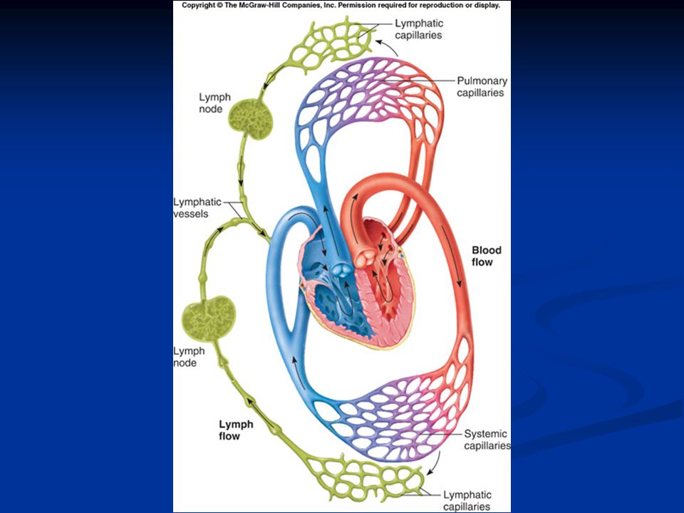
Introduction The lymphatic system is comprised of a network of vessels, cells, organs, and glands that produce and transport body fluids. Lymphatic vessels collect and carry away excess fluid from interstitial spaces The organs of the lymphatic system help defend against disease. Lymphatic capillaries are tiny, closed-ended tubes that extend into interstitial spaces. They receive fluid through their thin walls and once inside, tissue fluid is called lymph. The walls of lymphatic vessels are like veins but with thinner walls. Larger lymphatic vessels pass through lymph nodes and merge to form lymphatic trunks.

5
Lymphatic Trunks and Collecting Ducts
The lymphatic trunks drain lymph from the body and are named for the regions they drain. These trunks join one of two collecting ducts - either the thoracic duct or right lymphatic duct. The fluid is eventually returned to the subclavian vein.

6
Lymph Fluid Lymph is made up of water and dissolved substances that leave blood capillaries by filtration and diffusion. Most of the small molecules are returned to the capillaries by diffusion. Lymph then transports small foreign particles (bacteria, viruses, etc.) to lymph nodes. Forces that move blood in veins are the forces that propel lymph through lymphatic vessels.
 to lymph nodes. Forces that move blood in veins are the forces that propel lymph through lymphatic vessels.")
7
Lymph Nodes Lymph nodes are located along lymphatic pathways. They are bean-shaped, with two important parts: The hilum – area where blood vessels and nerves join a node The medulla – inner area where macrophages and T-cells are. They are covered with connective tissue that extends inside the node and divides it into nodules and spaces called sinuses. These contain both lymphocytes and macrophages which clean the lymph as it flows through the node. Lymph nodes are centers of lymphocyte production, which function in immune surveillance. The macrophages and lymphocytes within lymph nodes filter lymph and remove bacteria and cellular debris before lymph is returned to the blood. The lymph nodes generally occur in chains along the parts of the larger lymphatic vessels.

11
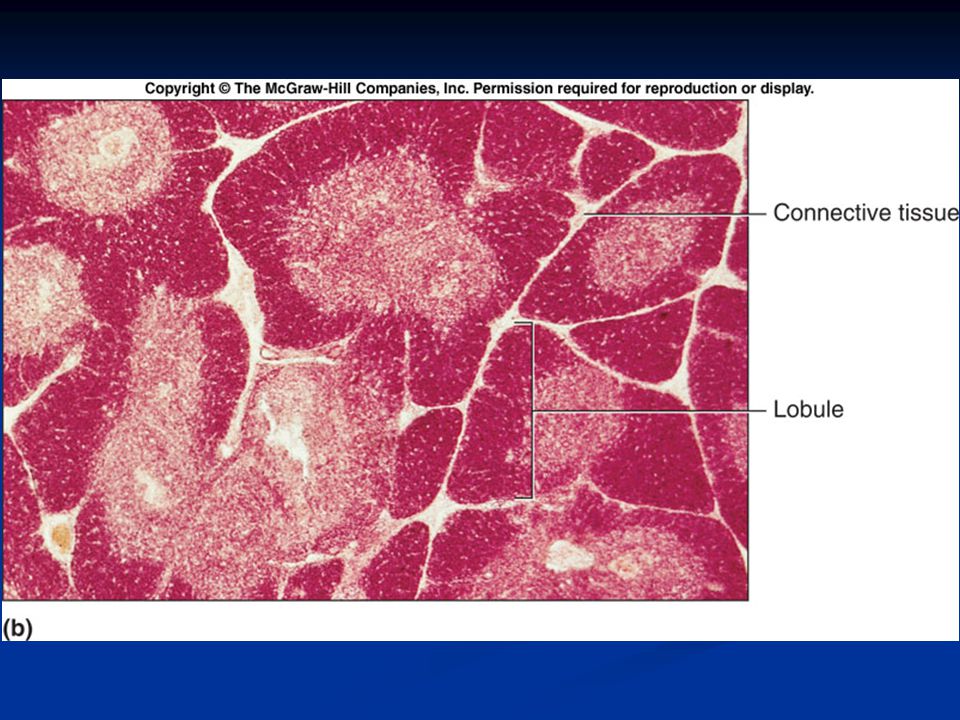
Thymus The thymus is a soft, bi-lobed organ located behind the sternum It shrinks in size during the lifetime (large in children, microscopic in the elderly). The thymus gland is divided into lobules, which are small clusters of thymus cells. Lobules contain lymphocytes, some of which mature into T lymphocytes (T cells) that leave the thymus to provide immunity. The thymus secretes the hormone thymosin, which influences the maturation of T lymphocytes once they leave the thymus.
. The thymus gland is divided into lobules, which are small clusters of thymus cells. Lobules contain lymphocytes, some of which mature into T lymphocytes (T cells) that leave the thymus to provide immunity. The thymus secretes the hormone thymosin, which influences the maturation of T lymphocytes once they leave the thymus.")
14
Spleen The spleen lies in the upper left abdominal cavity and is the body’s largest lymphatic organ. The spleen resembles a large lymph node except that it contains blood instead of lymph. Inside the spleen lies white pulp (containing many lymphocytes) and red pulp (containing red blood cells, macrophages, and lymphocytes). The spleen filters the blood and removes damaged blood cells and bacteria.
 and red pulp (containing red blood cells, macrophages, and lymphocytes). The spleen filters the blood and removes damaged blood cells and bacteria.")
16
Body Defenses Against Infection
Disease-causing agents, also called pathogens, can produce infections within the body. The body has two lines of defense against pathogens: Nonspecific defenses that guard against any pathogen Specific defenses (immunity) that mount a response against a very specific target. Specific defenses are carried out by lymphocytes that recognize a specific invader. Nonspecific and specific defenses work together to protect the body against infection.
 that mount a response against a very specific target. Specific defenses are carried out by lymphocytes that recognize a specific invader. Nonspecific and specific defenses work together to protect the body against infection.")
17
Innate (Nonspecific) Defenses
Species Resistance A species is resistant to diseases that affect other species because it has a unique environment. Pathogens from dogs tend not to infect humans. Doesn’t always work – H5N1 (Bird Flu) Mechanical Barriers The unbroken skin and mucous membranes of the body create mechanical barriers that prevent the entry of certain pathogens. Mechanical barriers represent the body’s first line of defense. Chemical Barriers Chemical barriers, such as the highly acidic and caustic environment of the stomach. Interferons are produced by cells when they are infected with viruses and induce nearby cells to produce antiviral enzymes that protect them from infection.
 Mechanical Barriers. The unbroken skin and mucous membranes of the body create mechanical barriers that prevent the entry of certain pathogens. Mechanical barriers represent the body’s first line of defense. Chemical Barriers. Chemical barriers, such as the highly acidic and caustic environment of the stomach. Interferons are produced by cells when they are infected with viruses and induce nearby cells to produce antiviral enzymes that protect them from infection.")
18
Fever Fever offers powerful protection against infection by interfering with the proper conditions that promote bacterial growth. During fever, the amount of iron in the blood is reduced Fewer nutrients are available to support the growth of pathogens. Phagocytic cells attack with greater vigor when the temperature rises. Inflammation Inflammation, a tissue response to a pathogen, is characterized by redness, swelling, heat, and pain. Major actions that occur during an inflammatory response include: Dilation of blood vessels Increase of blood volume in affected areas Invasion of white blood cells into the affected area Appearance of fibroblasts and their production of a sac around the area.

19
Adaptive (Specific) Defenses or Immunity
The response mounted by the body against specific, recognized foreign molecules. Antigens Before birth, the body makes an inventory of "self" proteins and other large molecules. Antigens are generally larger molecules that elicit an immune response. Lymphocyte Origins During fetal development, red bone marrow releases lymphocytes into circulation 70-80% become T lymphocytes (T cells) The remainder become B lymphocytes (B cells). Undifferentiated lymphocytes that reach the thymus become T cells and B cells are thought to mature in the bone marrow. Both B and T cells reside in lymphatic organs.
 The remainder become B lymphocytes (B cells). Undifferentiated lymphocytes that reach the thymus become T cells and B cells are thought to mature in the bone marrow. Both B and T cells reside in lymphatic organs.")
21
Lymphocyte Functions T cells attack foreign, antigen-bearing cells, such as bacteria, by direct cell-to-cell contact, providing cell-mediated immunity. T cells also secrete cytokines that enhance cellular response to antigens. T cells may also secrete toxins that kill target cells They can also produce growth-inhibiting factors or interferon to interfere with viruses and tumor cells. B cells attack pathogens by differentiating into plasma cells that secrete antibodies (immunoglobulins).
.")
22
There are three main types of T-Cells.
Helper T-Cells are cells that help activate B-cells to produce antibodies. They must come in contact with a cell that has already encountered the antigen. Macrophages contain Major Histocompatibility Complex (MHC) proteins that act as both ID badges AND antigen presenting proteins. If a Helper T-Cell comes in contact with a macrophage which is presenting an antigen, it becomes activated. Once activated, the Helper T-Cell then tells other B-cells to produce antibodies to the antigen. Cytotoxic T cells monitor the body's cells, recognizing and eliminating tumor cells and virus-infected cells. Cytotoxic T cells become activated when a antigen binds to its receptors. Memory T cells provide a no-delay response to any future exposure to the same antigen.
 proteins that act as both ID badges AND antigen presenting proteins. If a Helper T-Cell comes in contact with a macrophage which is presenting an antigen, it becomes activated. Once activated, the Helper T-Cell then tells other B-cells to produce antibodies to the antigen. Cytotoxic T cells monitor the body s cells, recognizing and eliminating tumor cells and virus-infected cells. Cytotoxic T cells become activated when a antigen binds to its receptors. Memory T cells provide a no-delay response to any future exposure to the same antigen.")
24
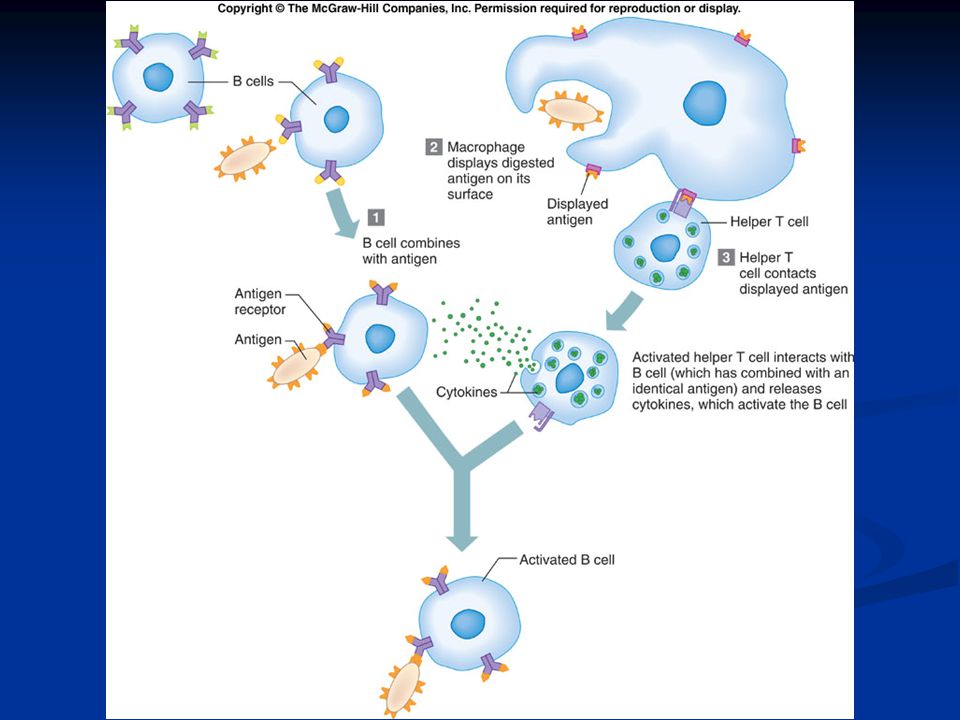
B Cells and the Humoral Immune Response
Most B cells need helper T cells for activation. The helper T cell releases cytokines that activate the B cell so that it can divide and form a clone. B cells may become activated and produce a clone of cells when its antigen receptor encounters its matching antigen. Some of the B cells become plasma cells, producing and secreting antibodies. Like T cells, some of the B cells become memory cells to respond to future encounters with the antigen.

26
Types of Antibodies There are five major types of antibodies (immunoglobulins) that constitute the gamma globulin fraction of the plasma. IgG is in tissue fluid and plasma and defends against bacterial cells, viruses, and toxins and activates complement. IgA is in exocrine gland secretions (breast milk, saliva, tears) and defends against bacteria and viruses. IgM is found in plasma and activates complement and reacts with blood cells during transfusions. IgD is found on the surface of most B lymphocytes and functions in B cell activation. IgE is found in exocrine gland secretions and promotes allergic reactions
 and defends against bacteria and viruses. IgM is found in plasma and activates complement and reacts with blood cells during transfusions. IgD is found on the surface of most B lymphocytes and functions in B cell activation. IgE is found in exocrine gland secretions and promotes allergic reactions.")
27
Antibody Actions Antibodies can react to antigens in three ways:
Direct attack Direct attack methods include agglutination, precipitation, and neutralization of antigens. Activation of complement This can produce inflammation or lysis in target cells or antigens. Stimulation of changes in areas that help prevent the spread of the pathogens.

29
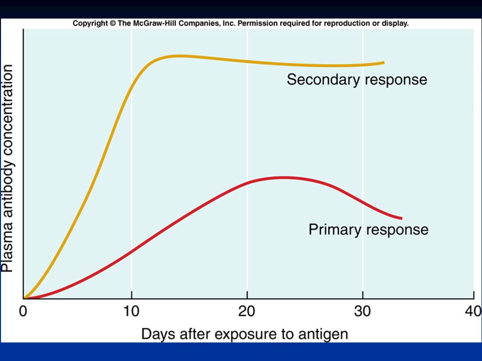
Immune Responses When B or T cells become activated the first time, their actions constitute a primary immune response This is when the cells acquire information about the pathogen and the antigens it contains. After this, some cells remain as memory cells. If the same antigen is encountered again, more numerous memory cells can mount a more rapid response, known as the secondary immune response. The ability to produce a secondary immune response may be long-lasting. Immunity is gained in an Active or Passive manner. Active immunity involves actual infection with pathogen. Allows body to produce its own antibodies. Passive immunity involves injection of actual antibodies. Short lived

31
Allergic Reactions Allergic reactions to allergens are excessive immune responses that may lead to tissue damage. Delayed-reaction allergy results from repeated exposure to substances that cause inflammatory reactions in the skin. Immediate-reaction allergy is an inherited ability to overproduce IgE. During allergic reactions, mast cells release histamine and leukotrienes, producing a variety of effects. Allergy mediators sometimes flood the body, resulting in anaphylactic shock, a severe form of immediate reaction allergy.

32
HIV HIV virus infects macrophages through special receptors on the cell surface Once virus is inside cell, it replicates and produces thousands of copies of itself. It then begins infecting the Helper T-Cells and they begin to die at a rapid rate. This affects B-Cell activation and thus antibody production. Eventually HIV begins binding to cytotoxic T-cells This lowers the body’s ability to patrol itself and prevent bacterial infections and cancers.

33
The End

Similar presentations


>")


 Immune system.>")


