Download presentation
Presentation is loading. Please wait.

1
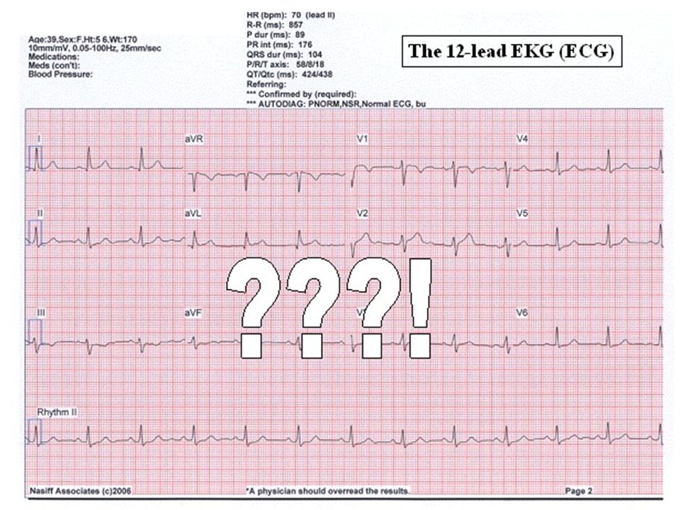
A brief introduction to the standard 12-lead ECG (EKG)
by James W. Grier Department of Biological Sciences North Dakota State University Fargo, ND Reference: ©2007, but may be copied and used without further permission

3
First, to make sure we know where the heart is …

4
Sensing the heart’s electrical activity via electrodes (contacts placed on the surface of the body)
")
5
Sensing the heart’s electrical activity via electrodes (contacts placed on the surface of the body)
Note: anatomical orientation is from the subject’s perspective:

6
The basic four limb electrodes:
right arm left arm electrical polarity: neutral or ground negative positive (manipulated by the EKG machine) right leg left leg
 right leg. left leg.")
7
Lead I (toward left) right arm left arm electrical polarity:
neutral or ground negative positive right leg left leg

8
Interpreting the view from an electrode
for any given viewing (positive) electrode: An approaching train of muscle fiber depolarizations (or repolarizations moving away) is seen as an upward trace on the recording (opposite movement = downward trace) Note: the normal average direction for the heart’s electrical activity is from the upper right, in the right atrium, to the lower left.
 electrode: An approaching train of muscle fiber depolarizations (or repolarizations moving away) is seen as an upward trace on the recording (opposite movement = downward trace) Note: the normal average direction for the heart’s electrical activity is from the upper right, in the right atrium, to the lower left.")
9
The main, typical waves of an EKG.
R T P Q S The main, typical waves of an EKG. (This particular tracing does not show a Q wave, a downward wave just before the R wave.)
")
10
P ATRIA: depol-pause-repol (atrial repolarization is obscured by ventricular depolarization)
")
11
QRS complex R T Q S VENTRICLES: depol-pause-repolarize

12
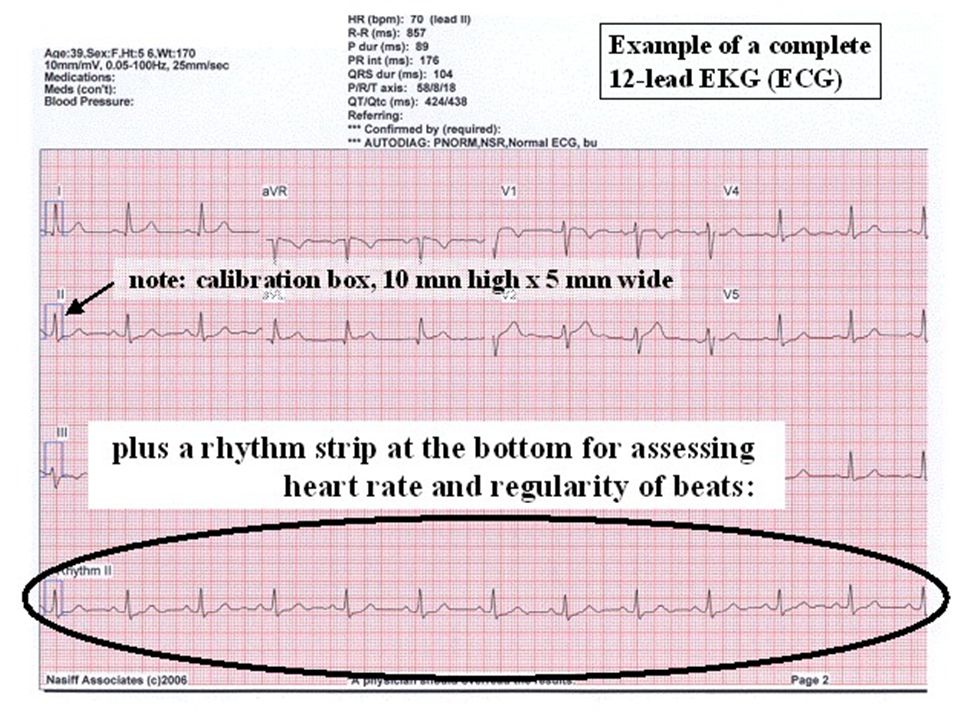
Standard calibration of EKG recordings
1 cm = 1 mV 1 mm = mV 1 mm = seconds 25 mm/second 5 mm = 0.20 seconds

13
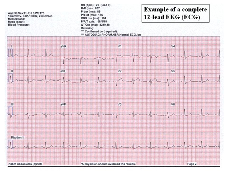
The appearance depends on the location of the electrode and what the heart’s electrical activity is doing (resting or active, normal vs various abnormalities, etc.). In addition to Lead I, here are the others …(following pages)
")
14
Lead II (toward left foot)
right arm left arm electrical polarity: neutral or ground negative positive right leg left leg

15
Lead III (down & rightward)
right arm left arm electrical polarity: neutral or ground negative positive right leg left leg

16
Leads I, II, & III together (“Einthoven’s triangle”)
right arm left arm electrical polarity: neutral or ground negative positive right leg left leg

17
Plus “augmented” leads, e.g., aVR
right arm left arm electrical polarity: neutral or ground negative positive right leg left leg

18
Frontal view of heart aVR aVL I Limb Leads III II aVF

19
Cross sectional view of heart
Chest leads

20
Summary: the 12 standard leads are :
Limb leads – I, from the right arm (-) toward the left arm (+) (taken together, these II, from the right arm toward the left leg three form the classic III, from the left arm toward the left leg "Einthoven's triangle") aVR, augmented lead toward the right (arm) (note: aVR is approx. aVL, augmented lead toward the left (arm) opposite of I and should aVF, augmented lead toward the foot essentially mirror the shape of I vertically) Chest leads – V1 through V6, starting over the right atrium with V1, and placed in a semi-circle of positions leftwards, to the left side of the left ventricle
 toward the left arm (+) (taken together, these. II, from the right arm toward the left leg three form the classic. III, from the left arm toward the left leg Einthoven s triangle ) aVR, augmented lead toward the right (arm) (note: aVR is approx. aVL, augmented lead toward the left (arm) opposite of I and should. aVF, augmented lead toward the foot essentially mirror the shape of I vertically) Chest leads – V1 through V6, starting over the right atrium with V1, and placed in. a semi-circle of positions leftwards, to the left side of the left ventricle.")
21
The normal progression of muscular contractions, hence, electrical activity, travels from the upper right part of the atria downward and leftwards to the ventricles, with the left ventricle being the strongest. Various combinations of limb leads and chest leads taken together provide a three-dimensional view into the electrical activity and workings of the heart for anyone who knows how to read an EKG. Abnormalities, such as heart attacks, arrhythmias, congenital problems, and a host of diseases and factors that affect the heart will cause sometimes major and sometimes subtle changes to the EKG patterns, which can be interpreted by a trained, experienced observer.

22
(plus the electrodes on the legs)
Positions of the electrodes: right arm left arm V1 V2 V3 V4 V6 V5 (plus the electrodes on the legs)
")
Similar presentations


>")


>")


>")
