Download presentation
Presentation is loading. Please wait.

1
Inflammasome activation by microbes and danger signals. Several NLRs can form multiprotein complexes called inflammasomes. Activation of the inflammasome results in activation of the cysteine protease caspase-1 and the resultant processing of pro-IL-1β, pro-IL-18, and pro-IL-33 into biologically active IL-1β, IL-18, and IL-33, respectively. Activation of the NLRC4 inflammasome following infection of macrophages with S. typhimurium, P. aeruginosa, S. flexneri, or L. pneumophila requires a functional type III or type IV secretion system. Bacterial-derived cytosolic flagellin augments caspase-1 activation following infection with L. pneumophila, S. typhimurium, and P. aeruginosa possibly through a Naip5-dependent pathway. Anthrax lethal toxin and MDP are capable of activating the NLRP1 inflammasome in a manner that may also require NOD2. A wide variety of stimuli including bacterial pore-forming toxins, ATP, DNA, bacterial RNA, and crystals such as silica, asbestos, uric acid, alum, and amyloid-β can activate the NLRP3 inflammasome. NLRP3 activating PAMPs and DAMPs induce a K+ efflux and the generation of mitochondrial- derived ROS that play a role in NLRP3 inflammasome activation by an unknown mechanism. Crystal induced lysosomal damage, and the resultant release of cathepsin B, are also postulated to play a role in NLRP3 inflammasome activation by an unknown mechanism.

3
Figure 1. Regulation of the inflammasomes by host factors and pathogen effectors. Inflammasomes are activated in a two-step process beginning with PRR-mediated induction of inflammasome components and pro-IL-1β production through NF-κB, followed by a second signal that activates the inflammasome and caspase-1 catalysis. This process can be regulated at multiple steps by host proteins that function as positive regulators (green) or inhibitors (orange) or targeted by pathogen effectors (red). Host COPs and POPs, the poxvirus proteins M13L and gp013L, and the anti- apoptotic factors Bcl-2 and Bcl-XL inhibit inflammasome assembly. Caspase-1 activation is inhibited by caspase-12, and multiple pathogen effectors, while murine caspase-11 and human caspase-5 are required for caspase-1 activation in response to certain stimuli. Type I IFN is required for AIM2 inflammasome activation in response to cytosolic DNA. The IL-1β and IL-18 pathways are also highly regulated. Endogenous IL-1 receptor antagonist (RA) prevents IL-1 signaling by binding to the IL-1 receptor, while the vaccinia virus proteins B15R and Molluscum contagiosum poxvirus MC53L and MC54L can bind and inhibit IL-1 and IL-18, respectively.
 or inhibitors (orange) or targeted by pathogen effectors (red). Host COPs and POPs, the poxvirus proteins M13L and gp013L, and the anti- apoptotic factors Bcl-2 and Bcl-XL inhibit inflammasome assembly. Caspase-1 activation is inhibited by caspase-12, and multiple pathogen effectors, while murine caspase-11 and human caspase-5 are required for caspase-1 activation in response to certain stimuli. Type I IFN is required for AIM2 inflammasome activation in response to cytosolic DNA. The IL-1β and IL-18 pathways are also highly regulated. Endogenous IL-1 receptor antagonist (RA) prevents IL-1 signaling by binding to the IL-1 receptor, while the vaccinia virus proteins B15R and Molluscum contagiosum poxvirus MC53L and MC54L can bind and inhibit IL-1 and IL-18, respectively..")
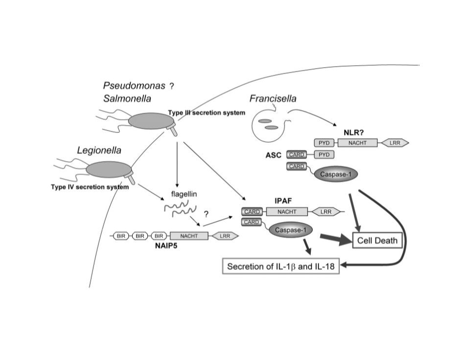
5
Figure 2. Microbial activation of the inflammasomes. Pathogenic microorganisms activate the inflammasomes through multiple agonists and pathways. S. typhimurium, L. pneumophila, and M. tuberculosis reside within the host cell phagosome and are capable of activating inflammasomes through secreted flagellin, effectors, or undefined NLRP3 agonists. F. tularensis and L. monocytogenes, which escape the phagosome activate AIM2 that senses cytosolic DNA. B. anthracis lethal toxin activates the NLRP1 inflammasome. C. albicans and hemozoin activate NLRP3 through SYK signaling. Viral-mediated inflammasome activation is heavily dependent on the detection of nucleic acids by NLRP3, AIM2, and RIG-I. Dotted lines indicate signaling through an unknown mechanism.

6
Macrophage/CD4+ CCR5+ Th1 lymphocytes cross-talk during MTB/HIV coinfection (1) CD4 CCR5 HIV MTB
 CD4 CCR5 HIV MTB")
7
Macrophage/CD4+ T lymphocytes cross-talk during MTB/HIV coinfection (2) Reinforced HIV replication and mycobacterial dissemination IL-1 /TNF- HIV MTB TLR-2
 Reinforced HIV replication and mycobacterial dissemination IL-1 /TNF- HIV MTB TLR-2")
9
La fagocitosi di corpi apoptotici ha sempre un esito anti-infiammatorio? …Dipende…

10
The four known inflammasomes. (a) The Nlrp3 inflammasome activates caspase-1 by recruiting ASC. (b) The Nlrp1 inflammasome has a FIIND and CARD domain in addition to its LRR, NACHT and Pyrin domains and can recruit caspase-5. (c) The Nlrc4 inflammasome has a CARD domain that can directly recruit pro–caspase-1. (d) AIM2 consists of a Pyrin and DNA- binding HIN domain that forms a complex with ASC and caspase-1. ASC, apoptosis-associated speck-like protein containing a CARD; FIIND, function to find domain; LRR, leucine-rich repeat; AIM2, absent from melanoma 2; MDP, muramyl dipeptide.
 The Nlrp1 inflammasome has a FIIND and CARD domain in addition to its LRR, NACHT and Pyrin domains and can recruit caspase-5. (c) The Nlrc4 inflammasome has a CARD domain that can directly recruit pro–caspase-1. (d) AIM2 consists of a Pyrin and DNA- binding HIN domain that forms a complex with ASC and caspase-1. ASC, apoptosis-associated speck-like protein containing a CARD; FIIND, function to find domain; LRR, leucine-rich repeat; AIM2, absent from melanoma 2; MDP, muramyl dipeptide..")
11
Differential IL-1β secretion pathways in monocytes and macrophages. Caspase-1 is constitutively active in monocytes, and these cells release mature IL-1β after a single stimulation with a TLR ligand. In contrast, macrophages need two signals for IL-1β secretion: one, such as a TLR-ligand, that induces IL-1β transcription, and a second signal that induces inflammasome activation.

12
IL-1β processing in acute and chronic stages of inflammation. Neutrophils are the major source for processing IL-1β via PR3 during acute inflammatory conditions. In chronic stages of inflammation when monocytes and macrophages play a more dominant role, caspase-1 and inflammasome activation become more important for the production of mature IL-1β.

13
Death and inflammation: how caspase-1 (ICE) activates IL-1β and IL-18 to induce innate and adaptive immunity and how this inflammation may be modulated. Apoptotic death induced by a variety of factors (such as ligation of Fas by its ligand) triggers a caspase cascade that can include ICE. Caspase-11 may be involved in the activation of ICE or may form a complex with it [16]. Activated ICE is then capable of cleaving the pro-forms of IL-1β and IL-18, which in turn trigger other proinflammatory cytokines. There is also evidence that activated ICE plays a role in the induction of apoptosis under some conditions (hence the question mark) [19]. During poxviral infection, CrmA can block the activity of ICE. A number of other factors may regulate or even abrogate the activation of innate and adaptive immune responses: ICE, or its substrates IL-1β and IL-18, may not be expressed by the dying cell; IL-1-receptor antagonist (IL-1RA) may modulate or block the effects of IL-1β; and macrophages that take up apoptotic cells through the phosphatidylserine (PS) receptor do not produce TNF-α in response to lipopolysaccharide (LPS) but may produce the anti-inflammatory cytokine, TGF-β[16][19]
 triggers a caspase cascade that can include ICE. Caspase-11 may be involved in the activation of ICE or may form a complex with it [16]. Activated ICE is then capable of cleaving the pro-forms of IL-1β and IL-18, which in turn trigger other proinflammatory cytokines. There is also evidence that activated ICE plays a role in the induction of apoptosis under some conditions (hence the question mark) [19]. During poxviral infection, CrmA can block the activity of ICE. A number of other factors may regulate or even abrogate the activation of innate and adaptive immune responses: ICE, or its substrates IL-1β and IL-18, may not be expressed by the dying cell; IL-1-receptor antagonist (IL-1RA) may modulate or block the effects of IL-1β; and macrophages that take up apoptotic cells through the phosphatidylserine (PS) receptor do not produce TNF-α in response to lipopolysaccharide (LPS) but may produce the anti-inflammatory cytokine, TGF-β[16][19].")
14
Figure 1 -γ-glutyamyl-meso-DAP (iE-DAP) and muramyl dipeptide (MDP), respectively, leading to recruitment of the adaptor proteins RICK and caspase recruitment domain 9 (CARD9). Subsequently, both TLRs and NOD1/NOD2 signaling pathways recruit TAK1, which mediates the activation of nuclear factor– kappa B (NF-κB) and mitogen-activated protein kinases (MAPKs), resulting in the transcriptional upregulation of proinflammatory genes. (c) Activation of NLRs by microbial or endogenous molecules in the cytosol results in the formation of caspase-1-activating inflammasomes. Activation of caspase-1 induces processing of the interleukin-1-beta (IL-1β) precursor and secretion of the mature cytokine. Abbreviations: ERK, extracellular signal–regulated protein kinase; IKK, I-kappa- B kinase; JNK, c-Jun N-terminal kinase; MKK, MAP kinase kinase; NEMO, NF-κB essential modulator.
 and mitogen-activated protein kinases (MAPKs), resulting in the transcriptional upregulation of proinflammatory genes. (c) Activation of NLRs by microbial or endogenous molecules in the cytosol results in the formation of caspase-1-activating inflammasomes. Activation of caspase-1 induces processing of the interleukin-1-beta (IL-1β) precursor and secretion of the mature cytokine. Abbreviations: ERK, extracellular signal–regulated protein kinase; IKK, I-kappa- B kinase; JNK, c-Jun N-terminal kinase; MKK, MAP kinase kinase; NEMO, NF-κB essential modulator..")
Similar presentations


 Lecture 3 8/9/2015.>")

. Toll-Like Receptor Signaling Toll receptor initially discovered in Drosophila as important receptor in dorso-ventral embryonic.>")


>")