Download presentation
Presentation is loading. Please wait.

1
Paediatric Lower Limb Deficiencies
Natasha Hankin March 2009

2
Outline Congenital vs. Acquired Limb Morphogenesis
Terminology and Classification Transverse Deficiencies Longitudinal Deficiencies Acquired Amputations

3
Congenital & Acquired deficiencies
John angel with severe Femur fibula ulna syndrome, Vanessa Smith with severe meningococcal sepsis

4
Aetiology Congenital : Genetic Vascular Intrauterine amputation
Maternal factors Acquired: Meningococcal Burns Trauma Vascular malformations Tumour Usually the cause of congenital limb deficiency is unknown. congenital Genetic: isolated or part of a multi-system syndrome or medical condition (e.g. Holt-Oram syndrome – hand heart syndrome) Current research suggests that many congenital anomalies have a genetic basis. Even when birth defects occur without a family history, DNA analysis may provide evidence of spontaneous genetic change Vascular: The type and severity of a vascular disruption depends on the gestational timing, the location of the involved vessel, the degree of tissue damage, and the adherence between tissues or organs. One of the major conditions with a vascular aetiology is amniotic band disruption syndrome Intrauterine amputation with or without amniotic bands causes about 10% of deficiencies seen Maternal factors (diabetes, factor V Leiden deficiency) Historically Thalidomide – proven to cause limb deficiency in children when mothers took it when pregnant Some acquired Focal neonatal Meningococcal Burns Trauma Vascular malformations Tumour
 Current research suggests that many congenital anomalies have a genetic basis. Even when birth defects occur without a family history, DNA analysis may provide evidence of spontaneous genetic change. Vascular: The type and severity of a vascular disruption depends on the gestational timing, the location of the involved vessel, the degree of tissue damage, and the adherence between tissues or organs. One of the major conditions with a vascular aetiology is amniotic band disruption syndrome. Intrauterine amputation with or without amniotic bands causes about 10% of deficiencies seen. Maternal factors (diabetes, factor V Leiden deficiency) Historically Thalidomide – proven to cause limb deficiency in children when mothers took it when pregnant. Some acquired. Focal neonatal. Meningococcal. Burns. Trauma. Vascular malformations. Tumour.")
5
Congenital Acquired No sense of loss Nothing new to adjust to
Prosthesis as an aid Family adjustment issues Profound sense of loss Period of readjustment How well they adjust affects acceptance of prosthetic limbs

6
Limb Bud development Key genes involved in growth and patterning of the limb buds Formation involves numerous genes, the actions of which are interlinked Limb bud development begins 4th week Key genes involved in growth and patterning of the limb buds & development of the extremities Formation of a limb involves numerous genes, the role of which often concerns predominantly one particular aspect of the limb, but the actions of which are interlinked Limb development begins during the fourth week along the flank of the embryo in predetermined regions, these are opposite cervicothoracic and lumbosacral segments

7
Limb Morphogenesis Thickening of lateral plate mesoderm signals the overlying ectoderm to thicken and form a ridge Apical ectodermal ridge (AER) controls proximal-distal limb Limb develops in a proximal-distal direction Initiation of growth involves thickening of lateral plate mesoderm that, in turn, signals the overlying ectoderm to thicken and form a ridge over the tip of the limb bud. This apical ectodermal ridge (AER) controls proximal-distal limb growth by keeping the mesenchyme cells beneath it in a rapidly proliferating state, i.e. progress zone. As mesoderm cells move away from this progress zone, they cease proliferating and begin to differentiate. Thus the limb develops in a proximal-distal direction from the limb girdle to the digits.
 controls proximal-distal limb. Limb develops in a proximal-distal direction. Initiation of growth involves thickening of lateral plate mesoderm that, in turn, signals the overlying ectoderm to thicken and form a ridge over the tip of the limb bud. This apical ectodermal ridge (AER) controls proximal-distal limb growth by keeping the mesenchyme cells beneath it in a rapidly proliferating state, i.e. progress zone. As mesoderm cells move away from this progress zone, they cease proliferating and begin to differentiate. Thus the limb develops in a proximal-distal direction from the limb girdle to the digits.")
8
Limb Morphogenesis (A) Limb begins with buds that form in mesoderm along the flank of the embryo. Growth of the UL precedes that of the LL by 1 to 2 days As the buds extend distally during the sixth week, the terminal portion flattens to form the hand and foot plates complete with digital rays (B) weeks: digits are present and the limbs have rotated to their normal position (C) 12 weeks: Cartilage first appears in proximal segments and ossification centres are present in the long bones
 8 weeks: digits are present and the limbs have rotated to their normal position. (C) 12 weeks: Cartilage first appears in proximal segments and ossification centres are present in the long bones.")
9
Congenital Limb Deficiencies
About 1 : 5-10,000 births May have complex genetics - important for geneticist to see family. In most cases cause unknown, low recurrence risk About 1 : 5-10,000 births May have complex genetics - important for geneticist to see family Much of the research has been completed on the fruit fly, the mouse and the chick. There has been progress in the understanding of the role different genes play in limb development, but large gaps still exist In most cases cause unknown, low recurrence risk

10
Congenital Limb Deficiencies
Most defects occur in period of limb morphogenesis Weeks 4-8 of gestation most critical time Sensitive period peaks 5th and 6th weeks Most defects occur in period of limb morphogenesis, when cells and tissues are rapidly proliferating and differentiating Weeks 4-8 of gestation most critical time The limb’s sensitive period reaches a peak during the fifth and sixth weeks after fertilization – often prior to mother being aware she is pregnant

11
Upper and Lower Limb buds rotated at 7 weeks but digits not separated
Final separation of the digits depends on additional cell death in the webbed area between each digit Upper and Lower Limb buds rotated at 7 weeks but digits not separated

12
Upper limb total deficiency

13
Terminology and Classification
ISO Classification 1989 is the accepted international standard Transverse limb developed normally to a particular level beyond which no skeletal elements exist Longitudinal Reduction or absence of an element/s within the long axis. There may be normal distal skeletal elements. Name the bones affected Partial / Total The International Organisation for Standardisation (ISO) has accepted this as the international standard (8548-1:1989), it is restricted to skeletal deficiencies, where the majority of such cases are due to failure of formation of parts. The deficiencies are described on the basis of anatomic and radiologic characteristics only. No attempt is made to classify in terms of embryology, aetiology, or epidemiology. Transverse The limb has developed normally to a particular level beyond which no skeletal elements exist, although there may be digital buds. Longitudinal There is a reduction or absence of an element or elements within the long axis of the limb, and in this case there may be normal skeletal elements distal to the affected bone or bones. To describe such a deficiency Name the bones affected in a proximo distal sequence by using the name as a noun. Any bone not named is present and of normal form. State whether each affected bone is totally or partially absent. In the case of partial deficiencies, the approximate fraction and the position of the absent part may be stated. Standard :1989
 has accepted this as the international standard (8548-1:1989), it is restricted to skeletal deficiencies, where the majority of such cases are due to failure of formation of parts. The deficiencies are described on the basis of anatomic and radiologic characteristics only. No attempt is made to classify in terms of embryology, aetiology, or epidemiology. Transverse. The limb has developed normally to a particular level beyond which no skeletal elements exist, although there may be digital buds. Longitudinal. There is a reduction or absence of an element or elements within the long axis of the limb, and in this case there may be normal skeletal elements distal to the affected bone or bones. To describe such a deficiency. Name the bones affected in a proximo distal sequence by using the name as a noun. Any bone not named is present and of normal form. State whether each affected bone is totally or partially absent. In the case of partial deficiencies, the approximate fraction and the position of the absent part may be stated. Standard :")
14
An example: What is in black is missing

15
Further Terminology Amelia: complete absence of the limbs
Hemimelia: absence of some portion of the limb Adactyly: absence of fingers Achiera: absence of a hand Apodia: absence of a foot

16
Conversion Amputation is never applicable in the Upper Limb
Paediatric deficiencies are often mixed and need to be considered in very functional terms : Shortening Unstable Terminal loss Conversion Amputation is never applicable in the Upper Limb Paediatric deficiencies are often mixed and need to be considered in very functional terms : Shortening : can it be lengthened? Unstable: stabilize with surgery, orthotics, prosthetics? Terminal loss : should there be a “ conversion amputation”? For children with congenital anomalies other than transverse deformity, surgical conversion of the limb to allow eventual prosthetic fitting often provides the most positive long term outcome. Each child would require separate assessment, but in general longitudinal absence of the fibula (complete) will requires a symes amputation. Longitudinal absence of the tibia (complete) will generally require knee disarticulation
 will requires a symes amputation. Longitudinal absence of the tibia (complete) will generally require knee disarticulation.")
17
Transverse Deficiencies
The limb has developed normally to a particular level beyond which no skeletal elements exist, although there may be digital buds Aetiology: Vascular disruption, Failure of formation, Constriction/ Amnionic Bands

18
Vascular Disruption may account for some transverse terminal losses
It is thought that premature vascular insufficiency results in a failure of normal mesodermal proliferation, thus not allowing full limb formation Vascular Disruption

19
Stump is usually smooth

20
Constriction Rings/ Amnionic Bands
The other major cause of transverse limb deficiencies is early amnion rupture with disruption of limb growth. Strands of amnion become entangled around limbs Fingers and toes are most often involved. Deep bands can cause distal oedema, nerve injury and impede growth. Amputation of digits or more proximal limb can also occur. Surgery for amnion disruption involves elective release and z-plasty of the bands

21
Longitudinal Deficiencies
Proximal Focal Femoral Deficiency (PFFD) Fibula Deficiency Tibial Deficiency Femur Fibula Ulna Syndrome Partial foot (lateral ray deficiency)
 Fibula Deficiency. Tibial Deficiency. Femur Fibula Ulna Syndrome. Partial foot (lateral ray deficiency)")
22
PFFD Profoundly short femur with bulbous thigh segment lying in external rotation & flexion flexed knee with cruciate insufficiency foot at level of opposite knee or just below most unilateral > 60% associated absence of fibula / other skeletal abnormality Proximal Focal Femoral Deficiency (PFFD) or Congenital deficiency of the femur
 or Congenital deficiency of the femur.")
23
PFFD

24
Proximal Femoral Focal Deficiency (PFFD)
Type A defect between femoral head & shaft with spontaneous restoration during growth Type B persistent discontinuity between hip joint & femur Type C femoral head never ossifies / dysplastic acetabulum Type D complete absence of the femoral head and acetabulum ISPO classification is longitudinal deficiency of the femur (partial, proximal) Radiograph shows failure of bone formation proximally - subtrochanteric pseudarthrosis to total absence of the proximal femur and acetabulum
 Radiograph shows failure of bone formation proximally - subtrochanteric pseudarthrosis to total absence of the proximal femur and acetabulum.")
25
PFFD Management options
Lengthening of femur Surgical procedures to provide hip stability & bony continuity Syme amputation / removal of foot + fusion of knee joint & prosthesis Van Nes rotationplasty non standard prostheses Need to consider: Inequality in leg length Malrotation Inadequate proximal musculature Instability of the proximal joints Lengthening of femur - Ilizarov (if 60% length of contralateral femur) Surgical procedures to provide hip stability & bony continuity Syme amputation / removal of foot + fusion of knee joint + prosthesis Van Nes rotationplasty (length 50% or < of the contralateral femur) non standard prostheses Van Nes rotationplasty In this procedure the tumour is removed while the neurovascular bundle and distal portion of the tibia and foot are maintained The tibia and foot are rotated 180 degrees, then attached to the remaining proximal femur so that the ankle is at the hight of the contralateral knee Benefits include have a functional “knee” joint, stable reconstruction, less energy consumption and potentially fewer future surgeries Major disadvantage is the appearance of the limb, especially for teenage girls for bilateral PFFD most functional without prostheses Important to differentiate from congenital short femur which has profoundly different functional potential and can be reconstructed. In congenital short femur the entire femur is abnormal rather than a proximal or focal defect.
 Surgical procedures to provide hip stability & bony continuity. Syme amputation / removal of foot + fusion of knee joint + prosthesis. Van Nes rotationplasty (length 50% or < of the contralateral femur) non standard prostheses. Van Nes rotationplasty. In this procedure the tumour is removed while the neurovascular bundle and distal portion of the tibia and foot are maintained. The tibia and foot are rotated 180 degrees, then attached to the remaining proximal femur so that the ankle is at the hight of the contralateral knee. Benefits include have a functional knee joint, stable reconstruction, less energy consumption and potentially fewer future surgeries. Major disadvantage is the appearance of the limb, especially for teenage girls. for bilateral PFFD most functional without prostheses. Important to differentiate from congenital short femur which has profoundly different functional potential and can be reconstructed. In congenital short femur the entire femur is abnormal rather than a proximal or focal defect.")
27
Severe Femur – Fibula - Ulna

28
Longitudinal Deficiency of Fibula
Shortening and anterior bowing of tibia absence of lateral metatarsal rays equinvalgus foot deformity cruciate ligament deficiency There can be hypoplasia with a portion of the fibula present or complete absence of the fibula Clinical features depend on severity of the deformity Shortening and anterior bowing of tibia, or it can be straight Involvement of other tissues and can have a dimple over the bow of the tibia absence of lateral metatarsal rays equinvalgus foot deformity cruciate ligament deficiency and valgus of the knee Possible to diagnoses at 16 weeks US

29
Fibula Deficiency Management Options
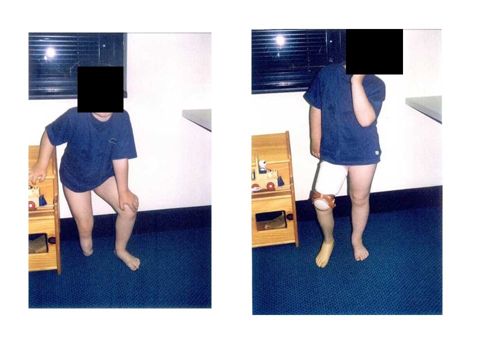
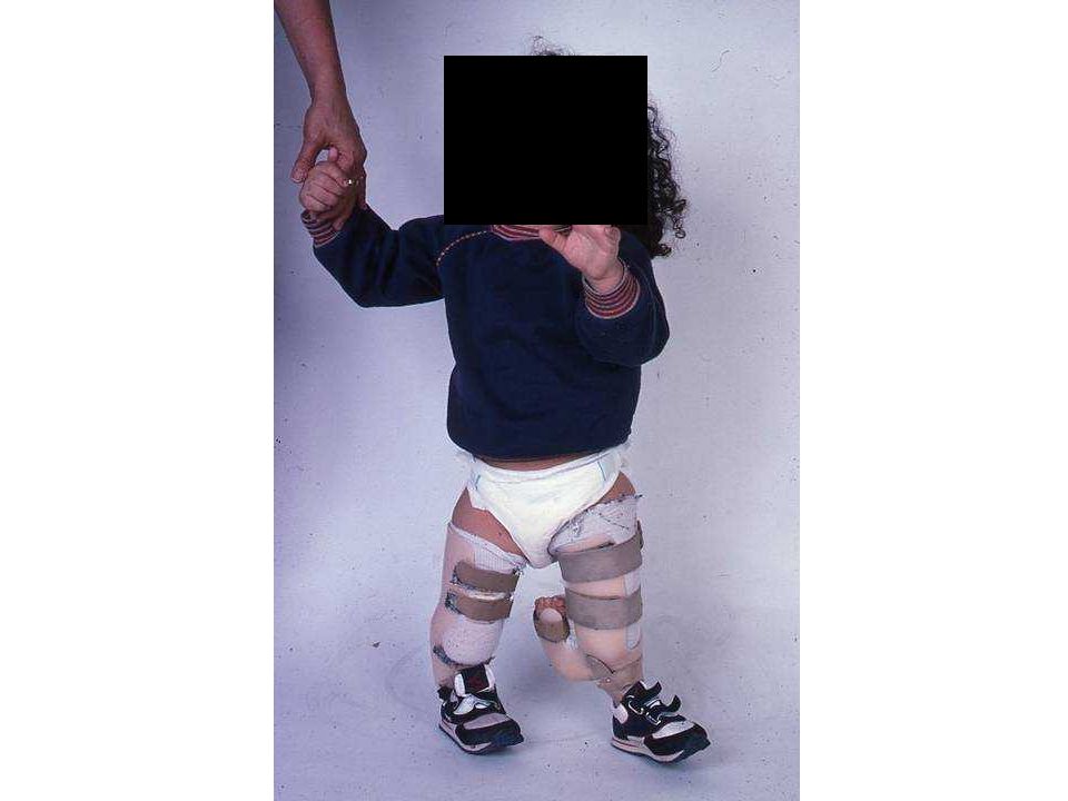
Extension prosthesis leg lengthening +/- ankle stabilisation conversion amputation through ankle & prosthetic restoration with supracondylar suspension for knee stability The major goal in the treatment of fibula deficiency, whether partial or complete, is maximising function. This means you need to achieve a plantargrade functional foot and appropriate length equalisation Some children may only need a shoe lift whereas other will require surgical intervention. Extension prosthesis – may have problems with cosmesis, especially in teenagers leg lengthening +/- ankle stabilization conversion amputation through ankle & prosthetic restoration with supracondylar suspension for knee stability

30
Improving ankle stability and leg length discrepancy

32
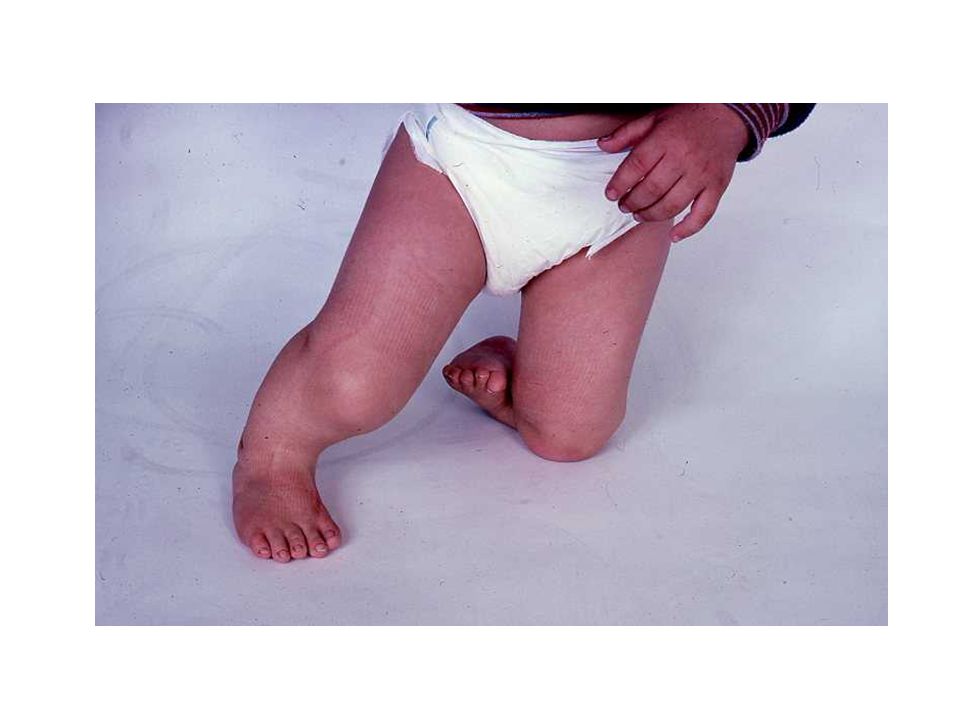
Bilateral Longitudinal Fibula deficiency and complete
deficiency of the 5th ray of the foot

35
Longitudinal Deficiency of Tibia
Complete or partial In complete absence: Short and relatively functionless leg Gross knee and ankle instability Equinovarus foot deformity No potential for development Congenital anomalies of the tibia are relatively rare and can be complete or partial Clinical findings vary depending on severity: Graded as type 1 to 4 In complete absence: Type 1 Severely short and relatively functionless leg Gross knee and ankle instability Equinovarus foot deformity No potential for development Type 1 is the most commonly occurring and most likely to be bilateral

37
Longitudinal Deficiency of Tibia
Management: Through knee amputation Ankle disarticulation Centralisation of fibula / reconstruction For patients with longitudinal deficiencies of the tibia, knee joint disarticulation is the typical treatment of choice, and patients generally function well with a prosthesis Amputation is optimally performed at about 12 months of age, although it is often deferred until the parents are convinced is and will be non functional Prosthetic fitting is appropriate when the patient is pulling to stand Only patients who are grade 4 (least severe) are candidates for attempted reconstruction of an ankle joint.
 are candidates for attempted reconstruction of an ankle joint.")
39
“Conversion” amputations
Aim for a weight bearing stump Enables better prosthesis use Joint disarticulation: Less risk of bony overgrowth as bones grow Maximises the residuals growth potential as leaves both growth plates intact

40
Acquired Amputations Lawn mower motor vehicle farm machinery burns
vascular catheterisations Landmines Tumours 75% of acquired amputations are due to trauma 25% due to other disease processes In children less than 10 years old power lawn mower injury is the most frequent cause of amputation. Acquired amputations show a 2:1 male to female predominance.

41
Tumours May require amputation or various strategies for limb salvage
The Van Nes Rotationplasty: distal femoral tumour May require amputation or various strategies for limb salvage The Van Nes Rotationplasty is used to salvage limb with a distal femoral tumour In this procedure the tumour is removed while the neurovascular bundle and distal portion of the tibia and foot are maintained The tibia and foot are rotated 180 degrees, then attached to the remaining proximal femur so that the ankle is at the hight of the contralateral knee Benefits include have a functional “knee” joint, stable reconstruction, less energy consumption and potentially fewer future surgeries Major disadvantage is the appearance of the limb, especially for teenage girls

42
Van Nes Rotationplasty
Tumour removed while the neurovascular bundle and distal portion of the tibia and foot are maintained Tibia and foot are rotated 180 degrees, attached to the remaining proximal femur The ankle is at the hight of the contralateral knee Benefits: functional “knee” joint Disadvantage: appearance of the limb In this procedure the tumour is removed while the neurovascular bundle and distal portion of the tibia and foot are maintained The tibia and foot are rotated 180 degrees, then attached to the remaining proximal femur so that the ankle is at the hight of the contralateral knee Benefits include have a functional “knee” joint, stable reconstruction, less energy consumption and potentially fewer future surgeries Major disadvantage is the appearance of the limb, especially for teenage girls

43
Tumour removed while the neurovascular bundle and distal portion of the tibia and foot are maintained Tibia and foot are rotated 180 degrees, attached to the remaining proximal femur The ankle is at the hight of the contralateral knee Benefits: functional “knee” joint Disadvantage: appearance of the limb

44
Questions?

Similar presentations


 Kelly Heikkila (0305975) Allison Pruys (0310660)>")


 Dr. Mazloumi MD Associate Professor Pediatric Orthopedic Surgeon.>")


