Download presentation
Presentation is loading. Please wait.

1
Labor and Birth Processes
Chapter 18

2
The 5 “Ps” of Labor: Passenger (fetus) Powers (uterine contractions)
Passage (the pelvis & maternal soft parts) Position (maternal) Psyche (maternal psychological status) These 5 components work together; they are all interrelated in their influence on the process of labor and birth.
 Position (maternal) Psyche (maternal psychological status) These 5 components work together; they are all interrelated in their influence on the process of labor and birth.")
3
PASSENGER (FETUS): Biological influences Mechanical influences
A pregnancy that terminates during the week gestation is likely to indicate a healthy fetus. Mechanical influences Fetal head Fetopelvic relationships Cardinal movements Biological influences: This statement is not necessarily true about postterm or preterm pregnancies. Fetal demise is often associated with an unripe uterus unresponsive for induction of labor. A fetus with adrenal hypoplasia is frequently associated with a prolonged gestation. Etc.

4
Fetal Head: ( a mechanical influence)
Bones: The head is the largest portion of the fetal body, & because it is a firm, noncompliant bony structure, it is the fetal component that is of most significance (from an obstetrical perspective). Sutures & Fontanelles: Between the bones of the fetal head are membranous spaces called sutures. The fontanelles are areas of the head where suture lines intersect. The ANTERIOR FONTANELLE: Is diamond shaped and is located at the intersection of the sagittal and coronal sutures. Does not close until the baby is about 18 months of age. Can be palpated during newborn assessment. The POSTERIOR FONTANELLE: Is triangle shaped and is located at the intersection of the sagittal and lambdoid sutures. Usually closes by 6-8 weeks after birth. Can be palpated on newborn assessment.
. Sutures & Fontanelles: Between the bones of the fetal head are membranous spaces called sutures. The fontanelles are areas of the head where suture lines intersect. The ANTERIOR FONTANELLE: Is diamond shaped and is located at the intersection of the sagittal and coronal sutures. Does not close until the baby is about 18 months of age. Can be palpated during newborn assessment. The POSTERIOR FONTANELLE: Is triangle shaped and is located at the intersection of the sagittal and lambdoid sutures. Usually closes by 6-8 weeks after birth. Can be palpated on newborn assessment.")
5
Landmarks: Head is divided into designated areas (1) the sinciput or brow portion; (2) the vertex, or top of the head between the 2 fontanelles; (3) the occiput or back of the head over the occipital bone. Diameters: During birth it is desirable that the smallest diameter of the fetal head move through the maternal bony pelvis. The diameter tht presents through the pelvis depends on the amount of flexion or extension of the head (attitude). LANDMARKS: Other landmarks are the bregma (anterior fontanelle), the glabella (the bridge of the nose, and thye mentum (chin). DIAMETERS: Suboccipitobregmatic diameter = the measurement from the lower edge of the occipital bone to the bregma (anterior fontanelle). Presents thru the pelvis when the fetal head is well flexed and averages 9.5 cm at term. Occipitofrontal diameter = extends from the occipital protuberance at the back of the head to the bridge of the nose. Presents thru the pelvis when the head is straight in a military attitude and averages 11.75cm at term. Occipitomental diameter = the measurement from just above the posterior fontanelle to the mentum (chin). It presents thru the pelvis when the head is extended back and averages 13.5 cm at term. Submentobregmatic diameter = is the measurement from the junction of the neck and lower jaw to the bregma (ant. Fontanelle). It is small at an average 9.5 cm, but DOES NOT promote an easy delivery since it occurs when the head is hyperextended back with a face presentation.
. LANDMARKS: Other landmarks are the bregma (anterior fontanelle), the glabella (the bridge of the nose, and thye mentum (chin). DIAMETERS: Suboccipitobregmatic diameter = the measurement from the lower edge of the occipital bone to the bregma (anterior fontanelle). Presents thru the pelvis when the fetal head is well flexed and averages 9.5 cm at term. Occipitofrontal diameter = extends from the occipital protuberance at the back of the head to the bridge of the nose. Presents thru the pelvis when the head is straight in a military attitude and averages 11.75cm at term. Occipitomental diameter = the measurement from just above the posterior fontanelle to the mentum (chin). It presents thru the pelvis when the head is extended back and averages 13.5 cm at term. Submentobregmatic diameter = is the measurement from the junction of the neck and lower jaw to the bregma (ant. Fontanelle). It is small at an average 9.5 cm, but DOES NOT promote an easy delivery since it occurs when the head is hyperextended back with a face presentation.")
18
Fetopelvic Relationships:
Fetal Lie: refers to the relationship of the long axis of the fetus, as related to the spinal column, to the long axis of the mother. (vertical lie = most common). Fetal Attitude: refers to the relationship of the fetal parts to one another. Fetus is described as being in a state of flexion or extension. VERTICAL LIE = the fetal spine is parallel to that of the mother. LONGITUDINAL LIE = the fetus could be either a cephalic or a breech presentation. TRANSVERSE LIE = horizontal. The fetal spine is perpendicular to the mother’s. OBLIQUE LIE = the fetal spine is at a slight angle from a true horizontal lie. The fetus in a transverse or an uncorrectable oblique lie is usually delivered by Cesarean birth, since the position would so adversly impede labor and could result in a catastrophic rupture of the uterus. FLEXION = the fetal back is rounded, the head is forward on the chest, and the arms and legs are folded against the body. Ideal for delivery, as it enables the smaller diameters of the fetal head to traverse the pelvis. EXTENSION = head extended or at least not well flexed. A Military attitude. A common variation, and can increase length of labor since the diameters of the head are larger than those with a well-flexed head. If it is so extended that the presenting part is the brow, or is completely extended as with a face presentation, labor is adversely affected by the fetal attitudes and it is more likely that a C/S may be needed.
. Fetal Attitude: refers to the relationship of the fetal parts to one another. Fetus is described as being in a state of flexion or extension. VERTICAL LIE = the fetal spine is parallel to that of the mother. LONGITUDINAL LIE = the fetus could be either a cephalic or a breech presentation. TRANSVERSE LIE = horizontal. The fetal spine is perpendicular to the mother’s. OBLIQUE LIE = the fetal spine is at a slight angle from a true horizontal lie. The fetus in a transverse or an uncorrectable oblique lie is usually delivered by Cesarean birth, since the position would so adversly impede labor and could result in a catastrophic rupture of the uterus. FLEXION = the fetal back is rounded, the head is forward on the chest, and the arms and legs are folded against the body. Ideal for delivery, as it enables the smaller diameters of the fetal head to traverse the pelvis. EXTENSION = head extended or at least not well flexed. A Military attitude. A common variation, and can increase length of labor since the diameters of the head are larger than those with a well-flexed head. If it is so extended that the presenting part is the brow, or is completely extended as with a face presentation, labor is adversely affected by the fetal attitudes and it is more likely that a C/S may be needed.")
19
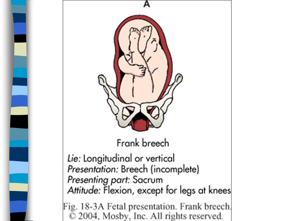
Fetal Presentation: The part of the fetal body that enters (or presents to) the maternal pelvis. Most common = cephalic presentation (head first). Fetal Position: refers to the relationship of an assigned area of the presenting part (often called the fetal denominator) to the maternal pelvis. Determine the fetal denominator. Mentally divide the maternal pelvis into 4 quadrants (R&L anterior, R&L posterior). Assign a standard abbreviation indicating the fetal position based on findings of vaginal exam. A noncephalic presentation can be either a breech or a shoulder presentation. A shoulder presentation occurs when the fetus is in a transverse lie. POSITION: When the fetal presentation is cephalic, the occiput is most often used as the fetal denominator to determine position (if the head is extended, the brow or chin is used). When the fetal presentation is breech, the fetal denominator is the sacrum; when the presentation is the shoulder, the fetal denominator is the fetal scapula. STEP 3: If the preseentation is cephalic, the fetal denominator is the occiput, and the occiput is located in the right anterior quadrant of the maternal pelvis; the abbreviation assigned is ROA (right occiput anterior). If the presentation is breech, the fetal denominator is the sacrum, and the sacrum is located in the left posterior quadrant of the maternal pelvis; the abbreviation assigned is LSP (left sacrum posterior). Most often, the fetus is in a mildly malpositioned placement. Nursing interventions such as repositioning te woman in labor, helping her ambulate, encouraging rocking, or providing ways to reduce pain (& thus reduce catecholamines which can interfere with labor) can positively change the influence of the passenger’s position on labor. Refer to handout of the most common fetal positions.
 to the maternal pelvis. Determine the fetal denominator. Mentally divide the maternal pelvis into 4 quadrants (R&L anterior, R&L posterior). Assign a standard abbreviation indicating the fetal position based on findings of vaginal exam. A noncephalic presentation can be either a breech or a shoulder presentation. A shoulder presentation occurs when the fetus is in a transverse lie. POSITION: When the fetal presentation is cephalic, the occiput is most often used as the fetal denominator to determine position (if the head is extended, the brow or chin is used). When the fetal presentation is breech, the fetal denominator is the sacrum; when the presentation is the shoulder, the fetal denominator is the fetal scapula. STEP 3: If the preseentation is cephalic, the fetal denominator is the occiput, and the occiput is located in the right anterior quadrant of the maternal pelvis; the abbreviation assigned is ROA (right occiput anterior). If the presentation is breech, the fetal denominator is the sacrum, and the sacrum is located in the left posterior quadrant of the maternal pelvis; the abbreviation assigned is LSP (left sacrum posterior). Most often, the fetus is in a mildly malpositioned placement. Nursing interventions such as repositioning te woman in labor, helping her ambulate, encouraging rocking, or providing ways to reduce pain (& thus reduce catecholamines which can interfere with labor) can positively change the influence of the passenger’s position on labor. Refer to handout of the most common fetal positions.")
20
Synclitism & Asynclitism: Asynclitic refers to a fetal head that is not parallel to the anteroposterior plane of the pelvis. The head is synclitic when the sagittal suture lies midway between the symphysis pubis and the sacral promontory. More often a woman will have the fetus engage in what is termed “posterior asynclitism” or with the sagittal suture (anteroposterior diameter) closer to the symphysis pubis than to the sacrum. Uterine contractions will tend to force the head both downward and laterally to correct this to a more normal synclitic position. Anterior asynclitism (also called Nagele obliquity) is associated with a lax abdominal wall, and the sagittal suture is closer to the sacrum than to the symphysis pubis. A fetus in this position often indicates that a labor may be longer and that interventions such as mild abdominal binding may be needed to encourage fetal repositioning.
 closer to the symphysis pubis than to the sacrum. Uterine contractions will tend to force the head both downward and laterally to correct this to a more normal synclitic position. Anterior asynclitism (also called Nagele obliquity) is associated with a lax abdominal wall, and the sagittal suture is closer to the sacrum than to the symphysis pubis. A fetus in this position often indicates that a labor may be longer and that interventions such as mild abdominal binding may be needed to encourage fetal repositioning.")
23
Cardinal Movements: Also called the “mechanisms of labor”.
A series of adaptations the fetus makes as it moves through the maternal bony pelvis during the process of lavor & birth. Influenced by the size and position of the fetus, the powers of labor, the size and shape of the maternal pelvis, and the mother’s position.

24
8 Cardinal Movements: (in an anterior occiput position)
Engagement Descent Flexion Internal rotation Extension Restitution External rotation of the shoulders Expulsion

25
Engagement: the mechanism by which the fetus nestles into the pelvis.
Also referred to as “dropping” or “lightening”. A fetus is engaged when the biparietal diameter of the fetal head reached the level of the maternal ischial spines; known as zero station. Leopold’s maneuvers: the head is more difficult to move and less of the head is able to be palpated abdominally after engagement. ENGAGEMENT: For a primipara, often occurs approx. 2 weeks before labor begins. Woman may report that it’s easier to breathe, since the pressure on the diaphragm and the lungs is decreased. May complaine of increased need to urinate or an increased pelvic heaviness. For a multiparous woman, engagement may not occur until labor begins.

34
Descent: describes the process that the fetal head undergoes as it begins its journey through the pelvis. Pressure from uterine ctx, hydrostatic forces, abdominal muscles, and gravity promote descent of the fetus through the pelvic inlet and midplane. Descent is continuous from the time of engagement until birth. Assessed by measurements called stations. Ranges from –3 to +3 station. STATIONS = based on an imaginary scale that uses the ischial spines of the maternal pelvis as its reference point. Ischial spines are located at the narrowest diameter of the pelvis. Halfway through the pelvic passage is zero station. If the presenting part is above zero station, it is assigned a negative number. If it is below zero station, a positive number is assigned. Use the “RULE OF THIRDS” to calculate station: The space above and below the ischial spines is divided into 3 levels. The presenting part is designated –3 if unengaged and +3 if fully crowning. Crowning = a term used to describe the visualization of the biparietal diameter of the fetal head at the vaginal introitus.

35
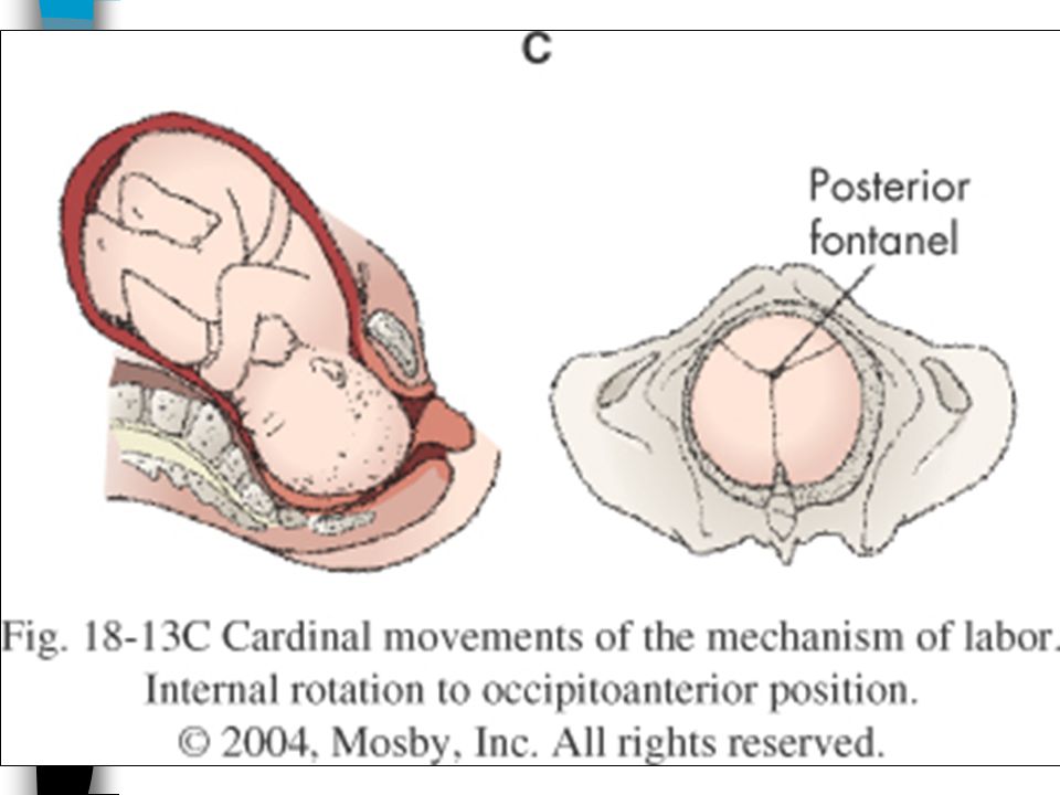
Flexion: the process of the fetal head’s nodding forward toward the fetal chest and occurs as a result of descent, the thickening of the uterine fundus, & increased resistance of the soft tissues. Engagement, descent and flexion tend to occur simultaneously. Internal Rotation: most commonly the fetus rotates internally from the occiput transverse position assumed at engagement into the pelvis to an occiput anterior position while continuously descending. INTERNAL ROTATION: the shape of the pelvis and the pelvic musculature encourage internal rotation to the occiput anterior position. Most often takes place in the 2nd stage of labor and is facilitated by the mother’s position and expulsion efforts.

36
Extension: enables the head to be born when the fetus is in a cephalic position. Results from the downward forces of the uterine contractions and the resistance of the pelvic floor muscles. Begins after the head has crowned and is complete when the head passes under the symphysis pubis and the occiput, anterior fontanelle, brow, face, and chin pass over the sacrum & coccyx and are born over the perineum.

37
Restitution: results in a realignment of the fetal head with the body, after the head is born.
It is common that as the head internally rotates to an anterior position before its birth, the shoulders may enter the pelvis in the oblique diameter. This allows the head to turn, but as a result, the neck twists. Restitution occurs when the head is free of pelvic resistance, allowing the head to turn back until it is again at right angles to the shoulders.

38
External Rotation: After the head is born & restitution occurs, the shoulders externally rotate so that they are in the anteroposterior diameter of the pelvis. This is the largest diameter of the outlet, it easily allows the birth of the broad shoulders. Shoulders are born by first delivering the anterior shoulder from under the symphysis pubis and then the posterior shoulder from over the perineum.

39
Expulsion: the last cardinal movement; consists of the birth of the entire body.
The body usually follows easily after the birth of the head and shoulders. The time of birth is often documented at the moment of expulsion.

40
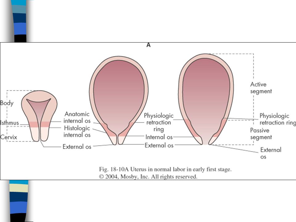
PASSAGE: “P” # 2 Major pelvic bones include the innominate bones (formed by the fusion of the ilium, ischium, and pubis around the acetabulum), the sacrum, and the coccyx. DIVISIONS: Pelvis is arbitrarily divided into halves – the false pelvis and the true pelvis. False pelvis: wide broad area btw. the iliac crests & has no major clinical significance for L&D. The passage consists of both the hard passage, or bony pelvis, and the soft passage, or maternal soft tissue structures.
, the sacrum, and the coccyx. DIVISIONS: Pelvis is arbitrarily divided into halves – the false pelvis and the true pelvis. False pelvis: wide broad area btw. the iliac crests & has no major clinical significance for L&D. The passage consists of both the hard passage, or bony pelvis, and the soft passage, or maternal soft tissue structures.")
41
True Pelvis: the actual bony passage that the fetus must traverse during labor and birth. Shape is a curved axis, not a straight passage , d/t the diameters & planes of the pelvis. PLANES: 3 common planes of the pelvis are the inlet (the pelvic brim), midpelvis, and outlet. A pelvis with an adequate inlet & midplane rarely if ever has reduced diameters for the outlet. The coccyx also has slight mobility, which increases the available space in the outlet. True Pelvis: This means that the fetus must first move down and then up over the sacrum as it descends and has implications for the positioning of women during the expulsion stage of labor.
, midpelvis, and outlet. A pelvis with an adequate inlet & midplane rarely if ever has reduced diameters for the outlet. The coccyx also has slight mobility, which increases the available space in the outlet. True Pelvis: This means that the fetus must first move down and then up over the sacrum as it descends and has implications for the positioning of women during the expulsion stage of labor.")
42
PRENATAL ASSESSMENT OF PELVIS:
Clinical pelvimetry reassures both the health care provider & the woman about the normalcy of the pelvis. When any variation exists in the pelvic structures, it can be discussed & anticipatory guidance given (ex- how to cope with back aches, back labor, etc.) Rarely an abnormal pelvis such as true android, guidance may include the planning for a C/S.
 Rarely an abnormal pelvis such as true android, guidance may include the planning for a C/S.")
43
SOFT PASSAGE THROUGH MATERNAL SOFT TISSUE STRUCTURES:
Soft tissues of the cervix, vagina, and perineum must stretch to allow passage of the fetus through the axis of the birth canal. Progesterone & relaxin help facilitate the softening & increase the elasticity of muscles & ligaments. RIPENING of soft tissues occurs as a result of: Braxton Hicks contractions Engagement of the fetal head (which serves as a wedge against the cervix). The fibromuscular vaginal passage becomes even more elastic throughout pregnancy. Hormonal changes cause increased vascularity and a thickening and lengthening of the vaginal walls. These changes allow the vagina to accomidate as the fetus moves through it. The muscles of the perineum also soften and becoje more stretchable.
. The fibromuscular vaginal passage becomes even more elastic throughout pregnancy. Hormonal changes cause increased vascularity and a thickening and lengthening of the vaginal walls. These changes allow the vagina to accomidate as the fetus moves through it. The muscles of the perineum also soften and becoje more stretchable.")
44
POWERS: “P” # 3 Uterine labor ctx. of the myometrium.
Ctx.phase consists of a descending gradient: The wave begins in the fundus (greatest # myometrial cells). Then moves downward through the corpus of the uterus. Intensity of ctx.diminishes from fundus to cervix. Retraction phase. Throughout labor, the upper uterine segment is more active, contracting more intensely and for a longer time than the lower uterine segment. The second part of the contraction is the retraction phase: After the muscle has contracted, it retracts as it relaxes by pulling up the cervix and lower uterine segment. The upper uterine segment becomes thicker in time, while the more passive lower segment becomes thinner. The synchronous nature of contractions is necessary for efficient dilatation and effacement of the cervix. Women who are dehydrated frequently experience preterm labor that can be stopped by being hydrated. Normal uterine ctx. are like waves, composed of an increment (the building up or ascending protion), an acme (the peak), and a decrement (the coming down or descending protion). Refer to handout
. Then moves downward through the corpus of the uterus. Intensity of ctx.diminishes from fundus to cervix. Retraction phase. Throughout labor, the upper uterine segment is more active, contracting more intensely and for a longer time than the lower uterine segment. The second part of the contraction is the retraction phase: After the muscle has contracted, it retracts as it relaxes by pulling up the cervix and lower uterine segment. The upper uterine segment becomes thicker in time, while the more passive lower segment becomes thinner. The synchronous nature of contractions is necessary for efficient dilatation and effacement of the cervix. Women who are dehydrated frequently experience preterm labor that can be stopped by being hydrated. Normal uterine ctx. are like waves, composed of an increment (the building up or ascending protion), an acme (the peak), and a decrement (the coming down or descending protion). Refer to handout.")
45
EFFACEMENT & DILATATION: The purpose of uterine ctx.
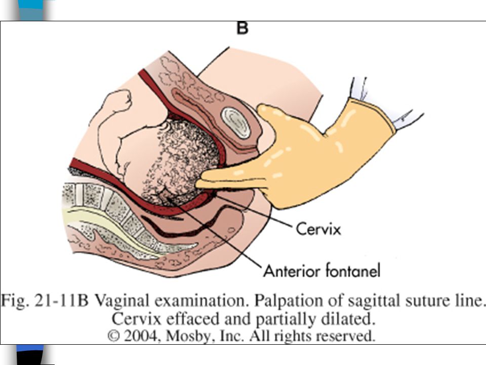
Accomplish the effacement and dilation of the cervix. Facilitate the descent & rotation of the fetus through the passages. Facilitate the separation & expulsion of the placenta. Control bleeding after delivery by compressing blood vessels. Effacement= the thinning or shortening of the cervix. During labor, effacement is accomplished by the upward retraction of the lower uterine segment as the upper uterine segment becomes thicker. Some effacement must take place before dilatation is possible. By the time the process of effacement is complete, the cervix is completely falt or paper thin, having been pulled up into the lower uterine segment. It assessed by palpation on vaginal exam, and is recorded in percentage ranging 0-100%.

46
Dilatation and effacement take place concurrently throughout labor.
Dilatation = the gradual opening of th cervix and is a continued extension of the contraction-retraction process already described. Dilatation and effacement take place concurrently throughout labor. Dilatation is assessed by vaginal examination, and is recorded in centimeters from 0-10 cm. Refer to handout.

47
Hydrostatic Force = another power that facilitates the process of labor and birth.
Includes the pressure of the fetus within the amniotic sac. As ctx. occur, the membranes and amniotic fluid facilitates dilation and effacement. Since the lower uterine segment and cervix are regions of lesser resistance, the additional pressure of the amniotic sac is of great importance in promoting the birth process.

48
Maternal pushing, or bearing down effort.
Abdominal Force = the final power for labor & birth. Intra-abdominal force. This power is reserved for the 2nd stage of labor, after effacement & dilation are complete. Maternal pushing, or bearing down effort. In the expulsion stage, the ctx.change in character, & many women begin to experience an involuntary urge to push. Pushing involves use of abd.muscles by contracting them to provide an auxiliary force to the uterine ctx, which increases intra-abdominal pressure to force the fetus through the vagina. For many years it was suggested that prolonged breath holding is the way in which a woman should push. Evidence has called into question both the efficacy and potential harm of pushing with a closed glottis (also called a Valsalva maneuver). Women have been observed to push effectively by either controlled exhalations or brief breath holding when they are not instructued to do otherwise. This is termed open glottis, exhale, or gentle pushing, and it avoids the risks of prolonged breath holding.
. Women have been observed to push effectively by either controlled exhalations or brief breath holding when they are not instructued to do otherwise. This is termed open glottis, exhale, or gentle pushing, and it avoids the risks of prolonged breath holding.")
49
POSITION: “P” # 4 In the last half of the 20th century, the position used most frequently for labor in the US has supine in a hospital bed. The most common position for birth has been a lithotomy position. Limited ambulation of laboring women resulted from use of continuous fetal monitoring, routine use of IV hydration, epidural anesthesia and use of analgesia. These positions evolved because of technology and the birth attendant’s comfort. The natural childbirth movement, gained popularity in the late 1960’s and 1970’s, which advocated birth without pain med. This movement has also increased the acceptance of women’s mobility during labor. The dorsal position in labor can have a number of detrimental physiologic effects. Refer to handout on positions, advantages and disadvantages.

50
PSYCHOLOGY OF BIRTH: “P” # 5
The progress of labor and birth can be adversely affected maternal fear and tension. Norepinephrine and epinephrine may stimulate both alpha and beta receptors of the myometrium and interfere with the rhythmic nature of labor. Anxiety can also increase pain perception and lead to an increased need for analgesia & anesthesia.

53
by Information Technology
Photo Album by Information Technology

Similar presentations


>")


