Download presentation
Presentation is loading. Please wait.

1
Objectives Chapter 7 Cell Structure

4
Microscopes are used to make cells look bigger The Compound Microscope The first person to discover microscopic life was a Dutch man cal led Antonie van Leeuwenhoek. In 1665 Robert Hooke first used the word 'Cell'. Cells are measured in micrometers The symbol µm is used. There are 1000 micro meters in 1mm. An animal cell is abou t 25µm in length.

5
Microscopes A compound microscope has two lenses: an eye piece lens and an objective lens Total magnification = eye piece lens X objective lens e.g. 10 X 4 = 40

6
Plant Cells These cells are usually rectangular in shape. They have a large central vacuole and have green Chloroplasts. Chloroplasts are small factories that make food. They catch sunlight and mix it with carbon dioxide and water to turn it into sugar. Chlorophyll is the green pigment (chemical) in the chloroplast that catches sunlight. An example of a plant cell is the onion cell.
 in the chloroplast that catches sunlight. An example of a plant cell is the onion cell..")
7
Compulsory Experiment

8
Experiment Results Note: If iodine stain is not used it is very difficult to see the onion cells clearly. Only the cell wall and nucleus are visible in the cell you will not be able to see the other parts of the cell at x400

9
Animal Cell An animal cell has no cell wall and no chloroplasts. An example of an animal cell is the cheek cell. Methylene blue stains the nucleus of the cell.

10
Experiment to view a cheek cell under a light microscope 1. Rub the inside of your cheek with a clean Finger 2. Smear your finger on a clean dry glass slide. 3. Apply a few drops of methylene blue stain. 4. Leave for a few minutes and allow excess stain to drain down the sink. 5. Apply a cover slip carefully to avoid air bubbles. 6. View under low, medium and high power under the microscope.

11
Cell Ultra structure

12
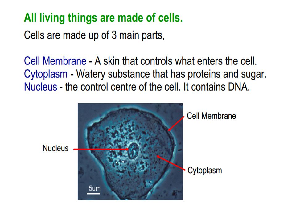
Nucleus The nucleus is the control centre of the cell. The nucleus has numerous small holes in its surface called nuclear pores. The nucleus contains strands of DNA (deoxyribonucleic acid). DNA is arranged into structures called Chromosomes. Every organism has a definite number of chromosomes. E.g. 46 chromosomes in humans. Genes are located randomly along chromosomes. Genes control features e.g. eye colour, production of enzymes etc. Genes are the units of inheritance.
. DNA is arranged into structures called Chromosomes. Every organism has a definite number of chromosomes. E.g. 46 chromosomes in humans. Genes are located randomly along chromosomes. Genes control features e.g. eye colour, production of enzymes etc. Genes are the units of inheritance..")
13
Nuclear Pores Nuclear pores allow a type of RNA (ribonucleic acid) called mRNA (messanger RNA) to pass in/out of the nucleus. Nucleolus The nucleolus is an area in the nucleus that stains very darkly and is responsible for making ribosomes. Cytoplasm Jelly like liquid that surrounds the nucleus in a cell. A number of organelles (small structures) e.g. chloroplasts, mitochondrion etc. are suspended in the cytoplasm.
 e.g. chloroplasts, mitochondrion etc. are suspended in the cytoplasm..")
14
Mitochondria (Singular is Mitochondrion) Mitochondria supply energy to the cell. They are the sites of respiration in the cell.

15
The DNA in mitochondria is inherited along the female line in the family from mother to daughter. Chloroplasts Chloroplasts are only found in plant cells. They are green structures in plants in which photosynthesis takes place. They are surrounded by a double membrane. They have membrane stacks, which contain the green pigment chlorophyll. They also have a loop of DNA. Chloroplasts are involved in photosynthesis in the cell.

16
Cell Wall The function of the cell wall is to support and strengthen the cells. Cell walls in plant cells are made of the polysaccharide cellulose. Cell walls are fully permeable (they allow all substances into/out of the cell). Ribosomes Ribosomes are very tiny, bead like structures found in cells. The are made of RNA (ribo nucleic acid) and protein. The function of ribosomes is to make proteins.
. Ribosomes Ribosomes are very tiny, bead like structures found in cells. The are made of RNA (ribo nucleic acid) and protein. The function of ribosomes is to make proteins..")
17
Generalised Plant and Animal Cells

18
Plant CellsAnimal Cells Have a cell wallDo not have a cell wall May have chloroplasts containing chlorophyll Do not have chloroplasts or chlorophyll Have a large vacuoleDo not have a large vacuole Differences between Plant & Animal Cells Prokaryotic & Eukaryotic Cells Living things (organisms) are divided into two categories depending on the structure and complexity of their cells. a)Prokaryotes b)Eukaryotes
Prokaryotes b)Eukaryotes.")
19
Prokaryotic cells do not have a nucleus or membrane enclosed organelles. Prokaryotic organisms are: Single celled Have a circular loop of DNA (not surrounded by a membrane and do not have a nucleus. Have small cells. Do not have a membrane and enclosed structures such as chloroplasts and mitochondria. Include bacteria.

20
Eukaryotic Cells have a nucleus and cell organelles, all of which are enclosed by membranes Eukaryotic organisms: Have a nucleus (i.e. DNA enclosed by a membrane) May have membrane-enclosed organelles such as mitochondria and chloroplasts. Have large cells. Include animal, plant and fungi Are more advanced than prokaryotes. Life originated with prokaryotic cells and has evolved into eukaryotic cells.
 May have membrane-enclosed organelles such as mitochondria and chloroplasts. Have large cells. Include animal, plant and fungi Are more advanced than prokaryotes. Life originated with prokaryotic cells and has evolved into eukaryotic cells..")
Similar presentations