Download presentation
Presentation is loading. Please wait.

1
Polymorphism & Restriction Fragment Length Polymorphism (RFLP)
")
2
Genome Variation Definition:
In 2 non-related individuals: 0.1% genome variation (1 in each 1500 bases) Polymorphism Mutation Clinical effect Clinically harmless (No effect on phenotype Potentially harmful (results in genetic disease incidence ≥ 0.1% rare: 0.1%
 Polymorphism. Mutation. Clinical effect. Clinically harmless (No effect on phenotype. Potentially harmful (results in genetic disease. incidence. ≥ 0.1% rare: 0.1%")
3
Polymorphism Polymorphism: is a variation in nucleotide sequence from one individual to another, occurring in the non-coding regions of DNA. RFLP: is a genetic variant that can be examined by cleaving the DNA into fragments (Restriction fragments) with a RE. The length of the restriction fragments is altered if the genetic variant alters the DNA so as to create or abolish a site of RE cleavage. It can be used to detect human genetic variations, e.g. in prospective parents or in fetal tissue 2 types of DNA variation result in RFLP: SNP VNTR Considered markers, which, in most cases, have NO known effect on the structure or rate of production of any particular protein.
 with a RE. The length of the restriction fragments is altered if the genetic variant alters the DNA so as to create or abolish a site of RE cleavage. It can be used to detect human genetic variations, e.g. in prospective parents or in fetal tissue. 2 types of DNA variation result in RFLP: SNP. VNTR. Considered markers, which, in most cases, have NO known effect on the structure or rate of production of any particular protein.")
4
Single Nucleotide Polymorphism (SNP; 90% of human genome variation)
")
5
Variable Number of Tandem Repeats (VNTR)
short sequences of DNA at scattered locations in the genome Repeated in tandem (one after another) The # of these units varies from person to person, but is unique for any given individual Serves as a molecular fingerprint
 The # of these units varies from person to person, but is unique for any given individual. Serves as a molecular fingerprint.")
6
Variable Number of Tandem Repeats (VNTR)
VNTR loci: sites in genome that are frequently showing VNTR, important in DNA fingerprinting analysis (e.g. in forensic & paternity identity)
")
7
RFLP of VNTR

8
Prenatal Diagnosis Methods available:
It is recommended if there is a history of severe genetic disease, (affected previous child or near relative) Its aim is to determine the presence of the disorder in a developing fetus Methods available: Visualization of the fetus e.g. by Ultrasound or fetoscopy: If the genetic abnormality → gross anatomic defects e.g. neural tube defect Amniotic fluid biochemical analysis: e.g. level of a fetoprotein (increase in open neural tube defects, & low in Down syndrome. Fetal cells in amniotic fluid or in chrorionic villi biopsy: Karyotyping: the morphology of the metaphase chromosomes (e.g trisomies, translocations →abnormal length of chromosome Fetal DNA analysis: (the most detailed genetic picture; DNA from WBC, Amniotic fluid, or Chorionic villi DNA has to be amplified first: In the past: cell culture Now: PCR
 Its aim is to determine the presence of the disorder in a developing fetus. Methods available: Visualization of the fetus e.g. by Ultrasound or fetoscopy: If the genetic abnormality → gross anatomic defects e.g. neural tube defect. Amniotic fluid biochemical analysis: e.g. level of a fetoprotein (increase in open neural tube defects, & low in Down syndrome. Fetal cells in amniotic fluid or in chrorionic villi biopsy: Karyotyping: the morphology of the metaphase chromosomes (e.g trisomies, translocations →abnormal length of chromosome. Fetal DNA analysis: (the most detailed genetic picture; DNA from WBC, Amniotic fluid, or Chorionic villi. DNA has to be amplified first: In the past: cell culture. Now: PCR.")
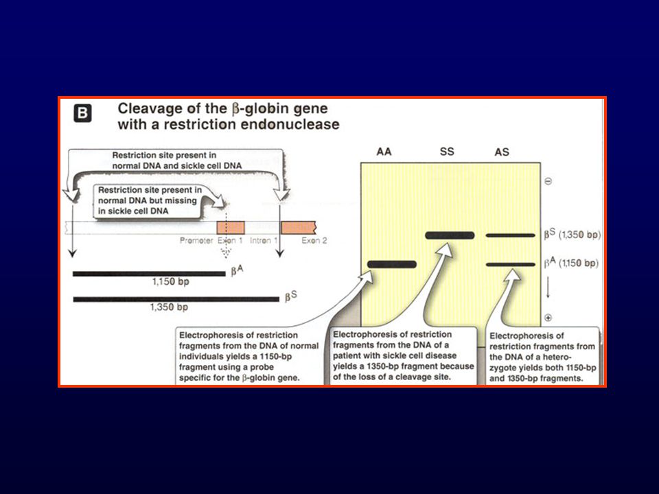
9
Examples of diseases diagnosed prenatally
Sickle cell anemia: Could be diagnosed prenatally by: I- Electrophoresis analysis of the amount & type of Hb obtained by hemolyzing fetal blood 1- risk in obtaining fetal blood 2- Late detection (2nd trimester) 2- Fetal DNA then do RFLP: Advantages: 1- Safe 2- early detection
 2- Fetal DNA then do RFLP: Advantages: 1- Safe. 2- early detection.")
12
RFLP in a family with a child affected by PKU

Similar presentations

 Molecular Medicine 2) Energy sources and environmental applications 3) Risk assessment 4) Bioarchaeology,>")


 The order of the base pairs in the sequence of every human varies In a single.>")


>")

 By Amr S. Moustafa, M.D.; Ph.D. Assistant Prof. & Consultant, Medical Biochemistry Dept. College of.>")


>")