Download presentation
Presentation is loading. Please wait.

1
Enterocele, the missed problem
By Dr. Khattab KAEO Prof. & Head of Obstetrics and Gynaecology Department Faculty of Medicine, Al-Azhar University, Damietta

2
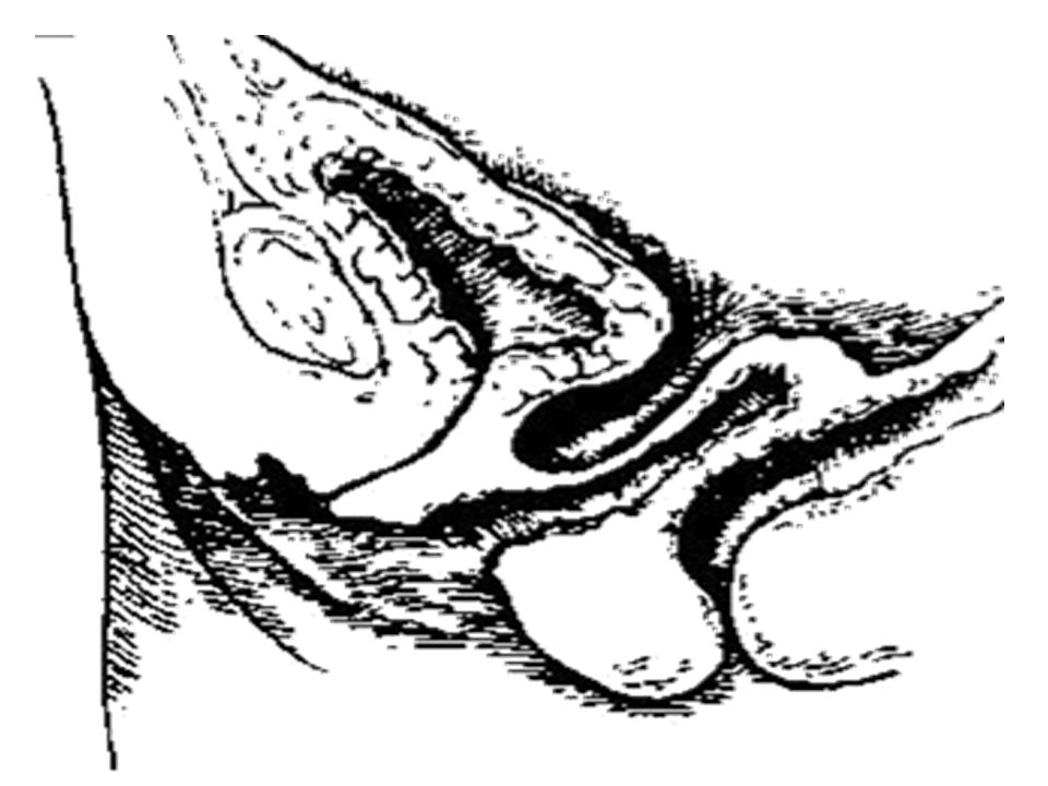
Vaginal axis

3
Vagina is not a straight tract.
Vaginal axis is 60-70 to the horizontal Vagina is not a straight tract.

4
It is angulated, with the perineal body supporting only the lower 3 cm, where the rectum and anal canal turns back sharply.

5
The upper vagina is almost horizontal
and the related part of the rectum lies on and parallel to the levator plate.

7
The upper fourth of the posterior vaginal wall is directly related to the posterior cul-de-sac.

8
The vagina is a potential space with its anterior and posterior walls are in contact with each others.

9
As intra-abdominal pressure increases, the pelvic diaphrag contracts & maintains position of the levator plate, and the horizontal vaginal axis. As a consequence, uterus, vagina and rectum are pushed against the levator plate and not through the genital hiatus

10
1- a break in the integrity of the utero sacral ligament complex,
Uterovaginal prolapse is attributed to: 1- a break in the integrity of the utero sacral ligament complex, 2- weakness of the pelvic floor musculature and 3- alteration of the normal vaginal axis.

11
Presence of congenital or developmental weakness of the supports;
Vault prolapse and recurrent prolapse have common aetiological factors: Presence of congenital or developmental weakness of the supports; - Omitting precipitating factors like chronic cough or constipation.

12
Inappropriate choice of operation (hysterectomy without repair);
- Failure to recognise or eradicate enterocele; - Poor surgical technique, particularly in the region around the cervix;

13
It should be emphasised that repairs are compensatory at best and to a great extent empirical.

14
Vault prolapse complicates hysterectomy in an incidence of 11
Vault prolapse complicates hysterectomy in an incidence of 11.6% if hysterectomy was performed for prolapse, and 1.8% when it was for other benign diseases.

15
Vault prolapse is a term variously applied to the following conditions: 1- Enterocele. 2- Collapse of the supports around the upper vagina & uterus. This is really represents what will be 3rd degree uterovaginal prolapse Nulliparous prolapse is commonly of this type. 3- Prolapse of the vaginal vault after hysterectomy.

16
Enterocele is hernia of the Douglas pouch.
The sac may or may not be filled with contents. Contents are loops of small bowel with elongated mesentery (beyond the usual 15 cm; this elongation is an important cause of recurrence!). Enterocele is usually progressive and can reach as far as the perineal body.
. Enterocele is usually progressive and can reach as far as the perineal body.")
17
There are at least 4 types of enterocele (identifiable with its location within the pelvis), each with a different aetiology). I- Congenital: The fetal sac of peritoneum between rectum and vagina fails to fuse, or it reopens. No associating rectocele or cystocele. The sac represents a split layer of the rectovaginal septum.

19
II- Pulsion: Increased intra-abdominal pressure creates the sac and often pushes the vault down with it. The vault, then, drags the anterior vaginal wall → cystocele, but no rectocele. This type is a sliding hernia along the anterior surface of the rectum and may eventually result in rectal prolapse and intussuception.

21
III- Traction: A large rectocele and a cystocele pull on the vault.

23
IV- Iatrogenic enterocele:
- Enterocele anterior to the vagina: It looks like a cystocele. Excess anterior peritoneum is not resected at the time of vaginal hysterectomy when there was difficulty identifying the vesico-uterine peritoneal fold, the peritoneal cavity was entered cranial to the bladder and close to the uterine fundus. - Most commonly iatrogenic enterocele results from a surgically-produced change in the vaginal axis (Burch, MMK,..). Ventral suspen-sion or ventrofixation of the uterus or vagina may render an unprotected cul-de-sac vulner-able to subsequent enterocele.
. Ventral suspen-sion or ventrofixation of the uterus or vagina may render an unprotected cul-de-sac vulner-able to subsequent enterocele.")
26
Lateral pudendal enterocele: This rare type results from sudden, short, massive increase in intra-abdominal pressure with rupture of the pelvic diaphragm, often lateral to the vagina. The sac lies lateral to the lateral vaginal wall and may extend to the vulva. The neck is small and intestinal obstruction may occur. During surgery, the hernia defect can be identified by tracing the bowel to its disappearance within the pelvic floor.

29
History Abdominal and back pains. These are much more likely to be due to primary back problems which may be referable to the abdomen. POP-related backache is diffuse, deep-seated, midline lumbo- sacral or sacral and unaccompanied by tender-ness. It is completely and immediately relieved by rest and is never experienced in bed or on rising in the morning. It is attributable to traction on the uterosacral & cardinal ligaments. Discomfort related to enterocele is caused by: Backache due to traction to the long mesentry against supports of the vault in the standing position. Backache is characteristically minimal in the morning, worsens as the day goes on, and peaks by evening. Lying down relieves it. Sensation of fullness in the pelvis. Dysparenia.

30
A proctoscope will trans-illuminate a rectocele but not an enterocele.
Examination Peristalsis may be seen through a thin wall. Diagnosis is suspected when fullness can be felt in the vault in the lithotomy posi., but most accurate when the patient is standing. Rectal examination is essential and shows that the rectum is pushed backwards by the swelling and not forming a part of it. A proctoscope will trans-illuminate a rectocele but not an enterocele.

31
The patient is examined systemically and locally in the supine, standing, squatting and straining positions, and some authors advocate in addition, the left lateral position using the Sims' speculum to differentiate a high rectocele from an enterocele (the speculum blade is held tightly in the posterior cul-de-sac, gently lifting the cervix, a finger in the rectum, then, outlines the upper limits of the rectocele. If an enterocele is present, it will be seen to roll down from the vaginal apex when the patient strains). High rectocele &enterocele often co-exist.

32
Combined PV and PR examination:
While the patient is in the upright position, the thumb is placed in the vagina & the index in the rectum. When the patient strains, a bowel-filled sac in the rectovaginal septum is felt. The thumb establishes whether there is a vault prolapse or cystocele. It elevates the vault, & the index is then introduced through the anus.

34
Treatment: Prophylactic:
Tightening of the uterosacral and cardinal ligaments, and their inclusion in the vault are important steps at hysterectomy.

35
Treatment: Prophylactic:
High peritoneal closure during vaginal hysterectomy is a useful technique. It comprises stitching through the surface of an uterosacral ligament above the posterior cut edge of peritoneum. In a circular direction posterior peritoneum - the other uterosacral ligament - the round ligament anterior to it - anterior peritoneum - the other round ligament are reefed.

36
Treatment: Prophylactic:
McCall culdoplasty: It comprises approximating the uterosacral ligaments using continuous sutures, so as to obliterate the peritoneum of the posterior cul-de-sac as high as possible.

37
Treatment: Prophylactic:
Recognizing a potential enterocele (deep cul-de-sac). An existing enterocele can be recognized with the bent finger. Packing a suspected sac with a gauze sponge helps to confirm and identify the excess peritoneum.
. An existing enterocele can be recognized with the bent finger. Packing a suspected sac with a gauze sponge helps to confirm and identify the excess peritoneum.")
38
Resecting redundant or excess peri-toneum
Resecting redundant or excess peri-toneum. If it is difficult to locate the anterior peritoneum, catheterize the patient and grasp the bladder wall with successive gentle bites on an unlocked hemostat (walk-up) until the cut edge of the peritoneum is reco-gnised and can be grasped with forceps. A full-length of absorbable 2-0 suture is used, held in a light hemo-stat for later identification.
 until the cut edge of the peritoneum is reco-gnised and can be grasped with forceps. A full-length of absorbable 2-0 suture is used, held in a light hemo-stat for later identification.")
39
Sacrospinous fixation at the time of vaginal hysterectomy is reco-mmended when the vault descends to the introitus during closure.

40
Principles of surgery for genital prolapse
1- Effective & sustained vault support; 2- Approximation of the utero-sacral ligaments (obliteration of the enterocele sac); 3- Repair of associating cystocele & rectocele. 4- Correction/prevention of urinary incontinence.
; 3- Repair of associating cystocele & rectocele. 4- Correction/prevention of urinary incontinence.")
41
Active treatment The sac is dissected to a point where excision of excess peritoneum extends across the anterior surface of the rectum. Any fat belongs on the rectal side of dis-section. The characteristic condensation of fat or the longitudinal muscle fibres in- dicate that the rectum has been reached. The anterior peritoneum is inspected and the redundant peritoneum is excised. The sac is closed purse-string by size 0 non-absorbable suture material in > one layer A coincident eversion of the vault is treated by colpopexy.

42
Vaginal repair of enterocele
Location of the ureter is critical. When the enterocele dissects its way down the rectovaginal septum and leaves the vaginal apex in its normal position, the ureters also remain in their usual post-hysterectomy location. But, when the vagina inverts, the ureters are brought down.

43
ureter ureter

44
Thank you

Similar presentations


>")


