Download presentation
Presentation is loading. Please wait.

1
Bacteria

5
Characteristics Prokaryotes
Microscopic (Eukaryotic cells are at least 10x bigger) Unicellular DNA is a single circular piece of DNA Asexual Reproduction Binary Fission Metabolism Aerobic Anaerobic
 Unicellular. DNA is a single circular piece of DNA. Asexual Reproduction. Binary Fission. Metabolism. Aerobic. Anaerobic.")
6
Genetic Exchange Conjugation –transfer DNA through contact
Transformation – acquire DNA from dead bacteria Transduction – DNA is transferred from one bacteria to another using a virus (genetic engineering)
")
7
http://highered. mcgraw-hill

10
Survival of the Fittest!!!
Bacteria have been around for 3.5 billion years!! How???? Cell Walls Capsules (surrounds cell wall) Asexual Reproduction, but can still acquire other genes Inhabit every place on Earth
 Asexual Reproduction, but can still acquire other genes. Inhabit every place on Earth.")
11
Super fast reproduction

12
allow them to withstand drought, high temps., lack of food, etc.
endospores

14
Bacteria are Classified according to Shape and arrangement of cells
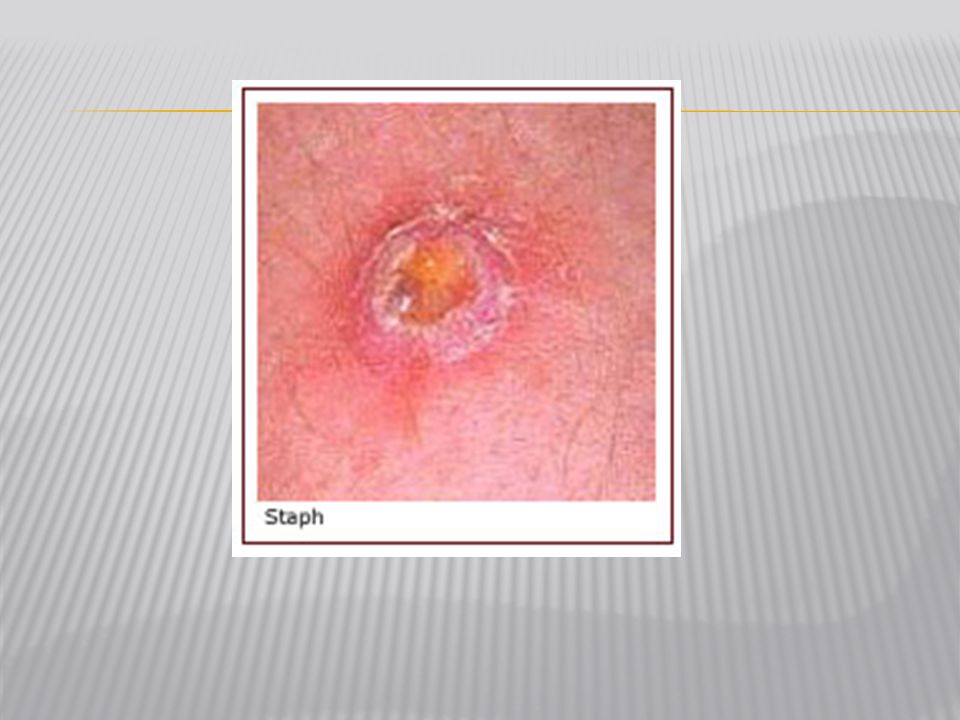
Shapes Coccus : Spheres Bacillus : Rods Spirillum : Spirals Arrangements Strept : Chains Staph : Clusters Diplo : Pairs

19
Gram Stain (pg. 529) Gram + simple walls, large amount of peptidoglycan Gram - less peptidoglycan, outer membrane contains lipopolysaccharides which are often toxic and provides additional protection more resistant to antibiotics Many antibiotics (penicillens) inhibit synthesis of cross links in peptidoglycan and prevent formation of a functional wall Gram negative Gram positive
 inhibit synthesis of cross links in peptidoglycan and prevent formation of a functional wall. Gram negative. Gram positive.")
20
Gram Positive Organisms
Aerobic, Gram-positive cocci Staphylococcus aureus (fig 1, 2, 3, 4) Staphylococcus epidermidis (fig 1) Staphylococcus sp. (Coagulase-negative)(fig 1) Streptococcus pneumoniae (Viridans group)(fig 1, 2, 3) Streptococcus agalactiae (group B)(fig 1) Streptococcus pyogenes (group A)(fig 1, 2) Enterococcus sp.(fig 1, 2, 3 ) Aerobic, Gram-positive rods Bacillus anthracis (fig 1, 2 ) Bacillus cereus (fig 1, 2) Bifidobacterium bifidum (fig 1) Lactobacillus sp. (fig 1, 2) Listeria monocytogenes (fig 1, 2) Nocardia sp.(fig 1, 2) Rhodococcus equi (coccobacillus)(fig 1) Erysipelothrix rhusiopathiae (fig 1) Corynebacterium diptheriae (fig 1, 2) Propionibacterium acnes (fig 1) Anaerobic, Gram-positive rods Actinomyces sp. (fig 1, 2) Clostridium botulinum (fig 1) Clostridium difficile (fig 1) Clostridium perfringens (fig 1, 2, 3) Clostridium tetani (fig 1, 2) Anaerobic, Gram-positive cocci Peptostreptococcus sp. (fig 1)
 Staphylococcus epidermidis (fig 1) Staphylococcus sp. (Coagulase-negative)(fig 1) Streptococcus pneumoniae (Viridans group)(fig 1, 2, 3) Streptococcus agalactiae (group B)(fig 1) Streptococcus pyogenes (group A)(fig 1, 2) Enterococcus sp.(fig 1, 2, 3 ) Aerobic, Gram-positive rods. Bacillus anthracis (fig 1, 2 ) Bacillus cereus (fig 1, 2) Bifidobacterium bifidum (fig 1) Lactobacillus sp. (fig 1, 2) Listeria monocytogenes (fig 1, 2) Nocardia sp.(fig 1, 2) Rhodococcus equi (coccobacillus)(fig 1) Erysipelothrix rhusiopathiae (fig 1) Corynebacterium diptheriae (fig 1, 2) Propionibacterium acnes (fig 1) Anaerobic, Gram-positive rods. Actinomyces sp. (fig 1, 2) Clostridium botulinum (fig 1) Clostridium difficile (fig 1) Clostridium perfringens (fig 1, 2, 3) Clostridium tetani (fig 1, 2) Anaerobic, Gram-positive cocci. Peptostreptococcus sp. (fig 1)")
21
Actinobacillus actinomycetemcomitans (fig 1)
Gram Negative Organisms Aerobic, Gram-negative cocci Neisseria gonorrhoeae (fig 1, 2, 3, 4) Neisseria meningitidis (fig 1; false color of the bacterium., 2) Moraxella catarrhalis (fig 1) Anaerobic, Gram-negative cocci Veillonella sp. (fig 1) Aerobic, Gram-negative rods Fastidious, Gram-negative rods Actinobacillus actinomycetemcomitans (fig 1) Acinetobacter baumannii(fig 1 really A. calcoaceticus) Bordetella pertussis (fig 1, 2) Brucella sp. (fig 1) Campylobacter sp.(fig 1) Capnocytophaga sp.(fig 1, 2) Cardiobacterium hominis (fig 1) Eikenella corrodens (fig 1) Francisella tularensis (fig 1,) Haemophilus ducreyi (fig 1, 2) Haemophilus influenzae (fig 1, 2) Helicobacter pylori (fig 1, 2, 3, 4) Kingella kingae (fig ) Legionella pneumophila (fig 1, 2, 3) Pasteurella multocida (fig 1) Enterobacteriaceae (glucose-fermenting Gram-negative rods) Citrobacter sp. (fig 1) Enterobacter sp. (fig 1) Escherichia coli (fig 1, 2) Klebsiella pneumoniae (fig 1, 2) Proteus sp. (fig 1) Salmonella enteriditis (fig 1) Salmonella typhi (fig 1) Serratia marcescens (fig 1, 2) Shigella sp. (fig 1) Yersinia enterocolitica (fig 1) Yersinia pestis (fig 1, 2) Oxidase-positive, glucose-fermenting Gram-negative rods Aeromonas sp. (fig 1) Plesiomonas shigelloides (fig 1) Vibrio cholerae (fig 1, 2) Vibrio parahaemolyticus (fig 1) Vibrio vulnificus (fig 1) Glucose-nonfermenting, Gram-negative rods Acinetobacter sp. (fig 1) Flavobacterium sp. (fig 1) Pseudomonas aeruginosa (fig 1, 2) Burkholderia cepacia (fig 1) Burkholderia pseudomallei (fig 1) Xanthomonas maltophilia or Stenotrophomonas maltophila(fig 1) Anaerobic, Gram-negative rods Bacteroides fragilis (fig 1) Bacteroides sp. (fig 1) Prevotella sp. (fig 1) Fusobacterium sp. (fig 1, 2) Gram-negative spiral Spirillum minus (minor)- (fig 1)
 Neisseria meningitidis (fig 1; false color of the bacterium., 2) Moraxella catarrhalis (fig 1) Anaerobic, Gram-negative cocci. Veillonella sp. (fig 1) Aerobic, Gram-negative rods. Fastidious, Gram-negative rods. Actinobacillus actinomycetemcomitans (fig 1) Acinetobacter baumannii(fig 1 really A. calcoaceticus) Bordetella pertussis (fig 1, 2) Brucella sp. (fig 1) Campylobacter sp.(fig 1) Capnocytophaga sp.(fig 1, 2) Cardiobacterium hominis (fig 1) Eikenella corrodens (fig 1) Francisella tularensis (fig 1,) Haemophilus ducreyi (fig 1, 2) Haemophilus influenzae (fig 1, 2) Helicobacter pylori (fig 1, 2, 3, 4) Kingella kingae (fig ) Legionella pneumophila (fig 1, 2, 3) Pasteurella multocida (fig 1) Enterobacteriaceae (glucose-fermenting Gram-negative rods) Citrobacter sp. (fig 1) Enterobacter sp. (fig 1) Escherichia coli (fig 1, 2) Klebsiella pneumoniae (fig 1, 2) Proteus sp. (fig 1) Salmonella enteriditis (fig 1) Salmonella typhi (fig 1) Serratia marcescens (fig 1, 2) Shigella sp. (fig 1) Yersinia enterocolitica (fig 1) Yersinia pestis (fig 1, 2) Oxidase-positive, glucose-fermenting Gram-negative rods. Aeromonas sp. (fig 1) Plesiomonas shigelloides (fig 1) Vibrio cholerae (fig 1, 2) Vibrio parahaemolyticus (fig 1) Vibrio vulnificus (fig 1) Glucose-nonfermenting, Gram-negative rods. Acinetobacter sp. (fig 1) Flavobacterium sp. (fig 1) Pseudomonas aeruginosa (fig 1, 2) Burkholderia cepacia (fig 1) Burkholderia pseudomallei (fig 1) Xanthomonas maltophilia or Stenotrophomonas maltophila(fig 1) Anaerobic, Gram-negative rods. Bacteroides fragilis (fig 1) Bacteroides sp. (fig 1) Prevotella sp. (fig 1) Fusobacterium sp. (fig 1, 2) Gram-negative spiral. Spirillum minus (minor)- (fig 1)")
23
Nutrition Autotrophic Heterotrophic Photosynthetic
Chemoautotrophic (nitrogen fixers) Heterotrophic Decomposer Parasitic (Treponema pallidum)
 Heterotrophic. Decomposer. Parasitic. (Treponema pallidum)")
24
Bacteria are used to produce medicines Insulin

25
First commerical use of genetic engineering: insulin

26
Important Recyclers in environment
Nitrogen cycle

27
Bacteria can produce chemicals
Acetone, Butanol

28
Bacteria are used to make food
Pickles, buttermilk, cheese, sauerkraut, olives, vinegar, sourdough bread, beer, wine

29
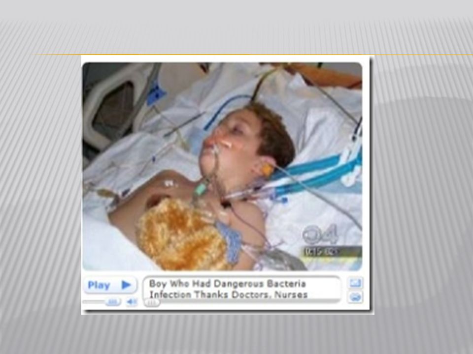
Bacteria cause disease
Produce toxins (Clostridium botulinum) Metabolize their host (Mycobacterium tuberculosis)
 Metabolize their host (Mycobacterium tuberculosis)")
35
History of Microbiology
1664: Robert Hooke - microscope 1684: Antoni van Leeuwenhoek - microorganisms 1798: Edward Jenner - smallpox vaccination 1864: Louis Pasteur - spontaneous generation 1884: Robert Koch - Koch’s postulates 1889: Martinus Beijerink - concept of virus 1929: Alexander Fleming - discovery of penicillin 1977: Carl Woese - discovery of Archaea 1981: First reports of AIDS 1983: Luc Montagnier - discovery of HIV 1995: Craig Venter - complete genome sequence

Similar presentations


 –found in marshes, swamps, hot sulfur springs, Great.>")

 All causes2,391,399 877.0 Diseases of.>")


 bacteria ◦ Live in oxygen free environment ◦ Produce.>")


 Autotrophs.>")
 No membrane bound organelles 3.5 billion years Unicellular Circular DNA Contain a cell wall Eukaryotic.>")
 –Single-celled –Single circular piece of DNA –May have pili (attachment)>")

 Crash Course: Bacteria Video.>")


