Download presentation
Presentation is loading. Please wait.

1
Imaging of Anal Fistula
Dr Sue Roach

2
Introduction Pre-operative confirmation of fistula complexity has been shown to facilitate surgical planning of sphincter saving techniques[1] and to reduce the incidence of unidentified sepsis, which is the leading cause of fistula recurrence [2].

3
Imaging Objectives Determine relationship of fistula to sphincter complex Identify any secondary fistulous tracks

4
Imaging Modalities Fistulography Endoanal ultrasound
Magnetic resonance

5
Fistulography Acute tracks may not have a patent lumen
Difficult to relate the track to the sphincter and levator ani Shown to be accurate in only 16% [3] Helpful for chronic fistulae with an external opening distant from the anus

6
Endoanal ultrasound Operator dependent
Highly accurate at identifying the internal opening [4] Depicts fewer secondary extensions than MR Difficulty differentiating active track from fibrosis

7
Magnetic Resonance Most accurate technique for evaluation of the primary track and any extensions [4]. More accurate predictor of patient outcome than surgical findings at EUA[5].
![Magnetic Resonance Most accurate technique for evaluation of the primary track and any extensions [4].](http://slideplayer.com/slide/3937986/13/images/7/Magnetic+Resonance+Most+accurate+technique+for+evaluation+of+the+primary+track+and+any+extensions+%5B4%5D..jpg "More accurate predictor of patient outcome than surgical findings at EUA[5].")
8
Prospective study 56 patients
Beets-Tan RGH, Beets GL, Gerritsen van der Hoop A. et al. Preoperative MR Imaging of Anal Fistulas: Does it Really Help the Surgeon? Radiology 2001; 218:75-84 Prospective study 56 patients MR prior to surgery but result witheld from surgeon until end of surgery while patient still anaesthetised Important additional information in 21%. Benefit greatest in crohns (40%), recurrent fistulas (24%), primary fistulas (8%)
, recurrent fistulas (24%), primary fistulas (8%)")
9
Prospective study 48 patients
Spencer JA, Chapple K, Wilson D et al. Outcome After Surgery for Perianal Fistula: Predictive Value of MR Imaging. AJR 1998; 171: Prospective study 48 patients MR and then surgical exploration blinded to MR MR categorised 41% complex. Surgery 38%. Only agreed in 8 cases 19 patients required further surgery. 13 of these considered complex on MR, 9 by surgery MR better at predicting outcome than surgery

10
Gadolinium? Post operative problems
Complex cases such as crohns disease[6]

11
Endoanal coil? Endocoils give superior anatomical resolution of fistula disease within the sphincter Resolution falls off rapidly outside the sphincter Complex tracks outside the sphincter are not well seen

15
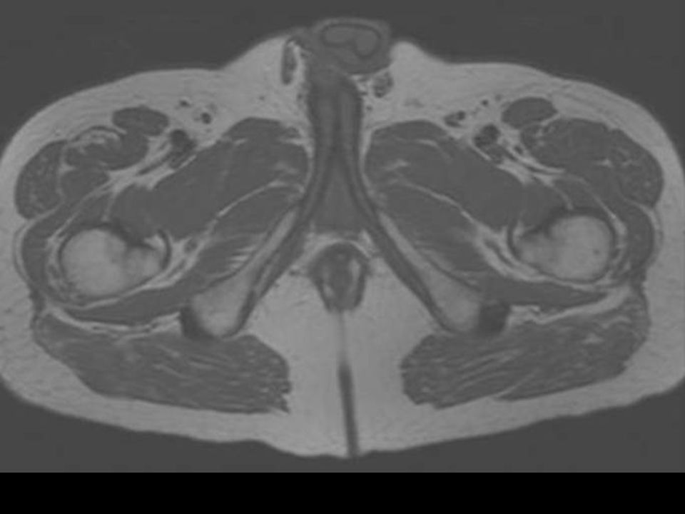
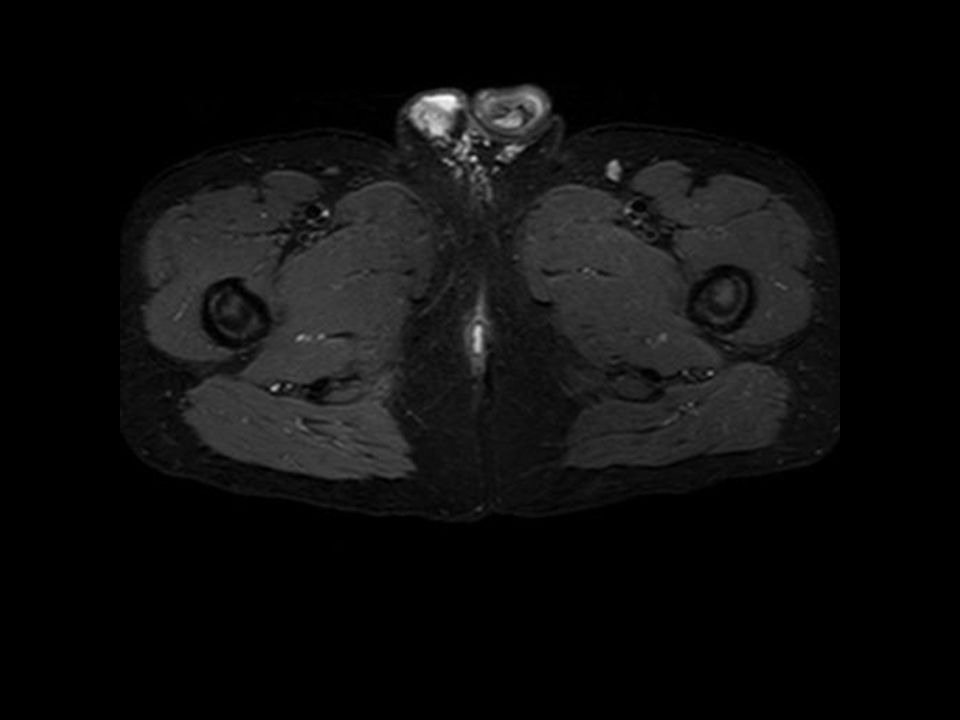
MR Technique Phased array pelvic coil
Axial and coronal imaging of the perineum T1 and short T1 inversion recovery (STIR) images obtained Additional saggital high resolution T2 images occasionally helpful IV gadolinium rarely administered
 images obtained. Additional saggital high resolution T2 images occasionally helpful. IV gadolinium rarely administered.")
19
Morris J, Spencer JA, Ambrose S
Morris J, Spencer JA, Ambrose S. MR Imaging Classification of Perianal Fistulas and Its implications for Patient Management. Radiographics 2000; 20:

20
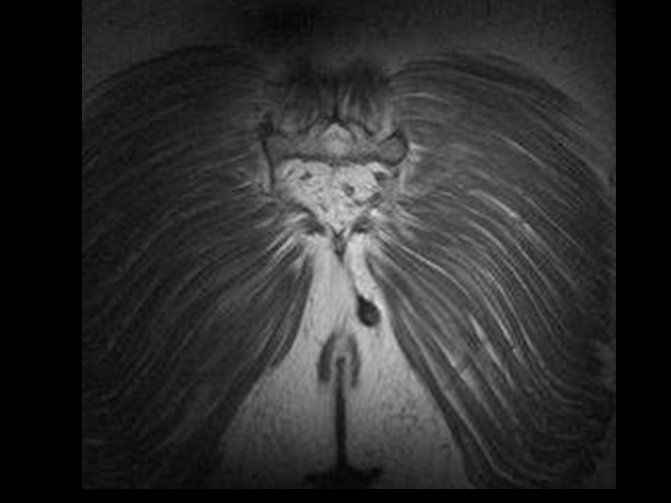
Grade 1 Simple Intersphincteric Fistula

21
Grade 2 Intersphincteric track with secondary track or abscess

22
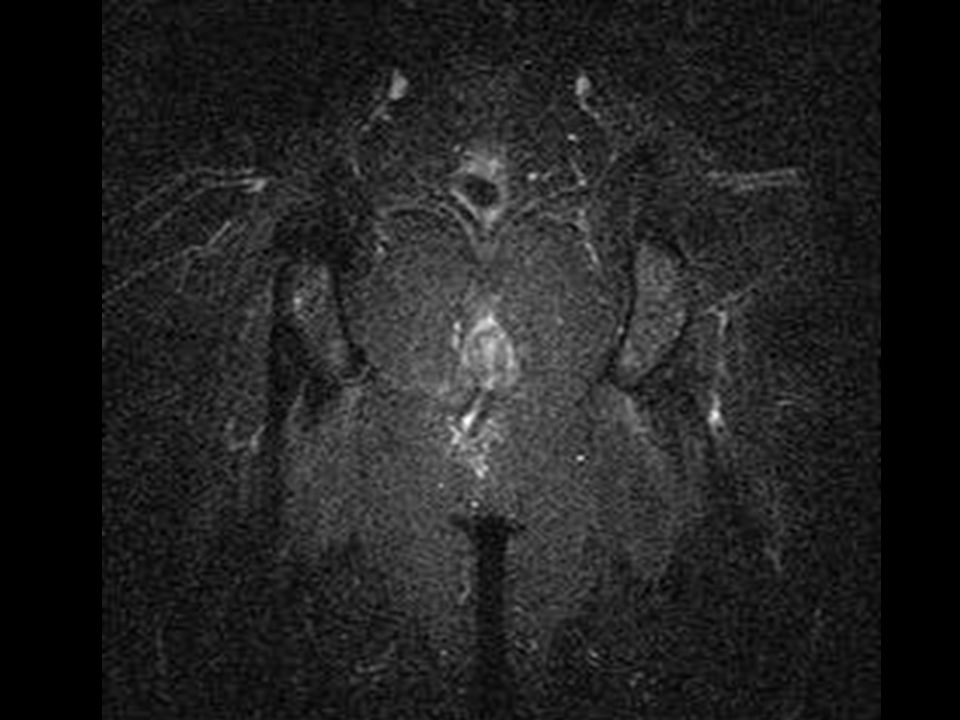
Grade 3 Trans-sphincteric Fistula

23
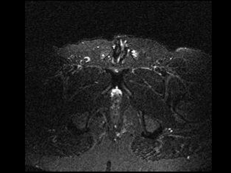
Grade 4 Trans-sphincteric Fistula With Abscess or Secondary Track

24
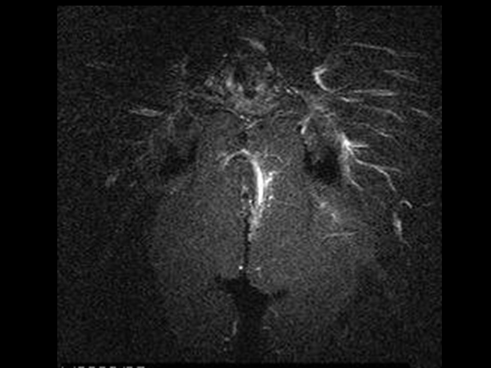
Grade 5 Supralevator and Translevator Disease

25
Aims To establish the common MR patterns of idiopathic peri-anal fistulation in Hope Hospital patients.

26
Methods Retrospective review
24 consecutive MR scans performed for idiopathic anal fistulation Scans performed on a 1 Tesla MR scanner with phased array pelvic coil technique

27
Results % of patients

28
Discussion Majority (50%) of patients with idiopathic peri-anal fistulation have uncomplicated disease 25% have trans-sphincteric fistulae complicated by secondary tracks or ischiorectal abscess Supra-levator or trans-levator disease is relatively rare in this patient group (8%).
.")
29
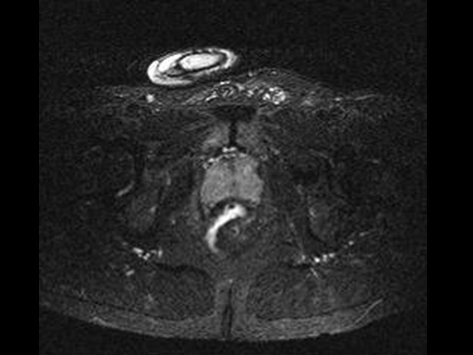
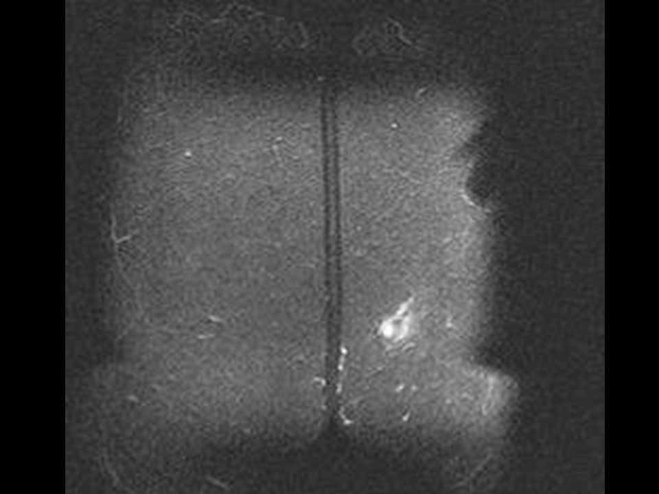
Grade 1- Intersphincteric fistula

38
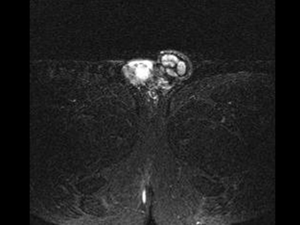
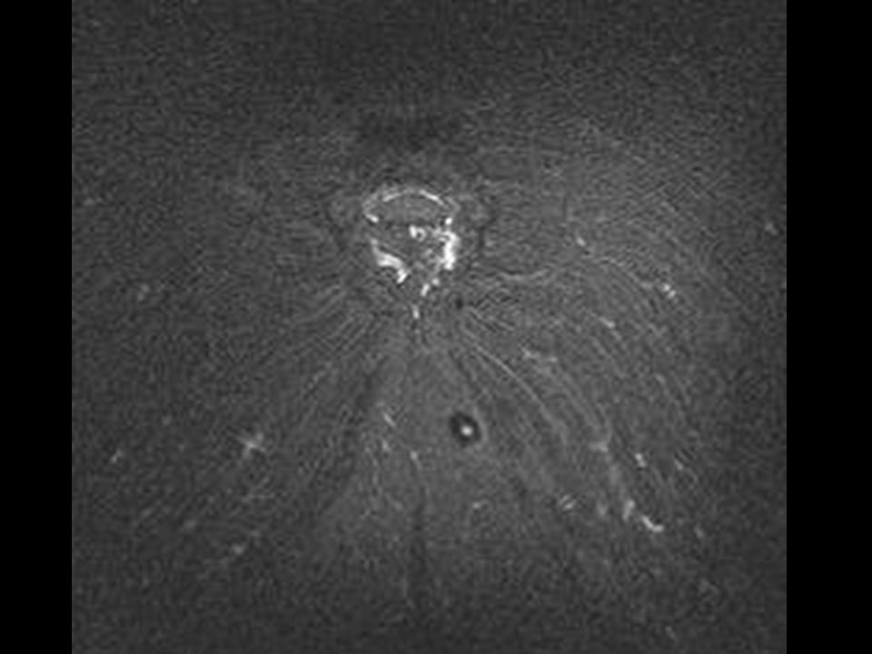
Grade 2- Intersphincteric fistula with collection

40
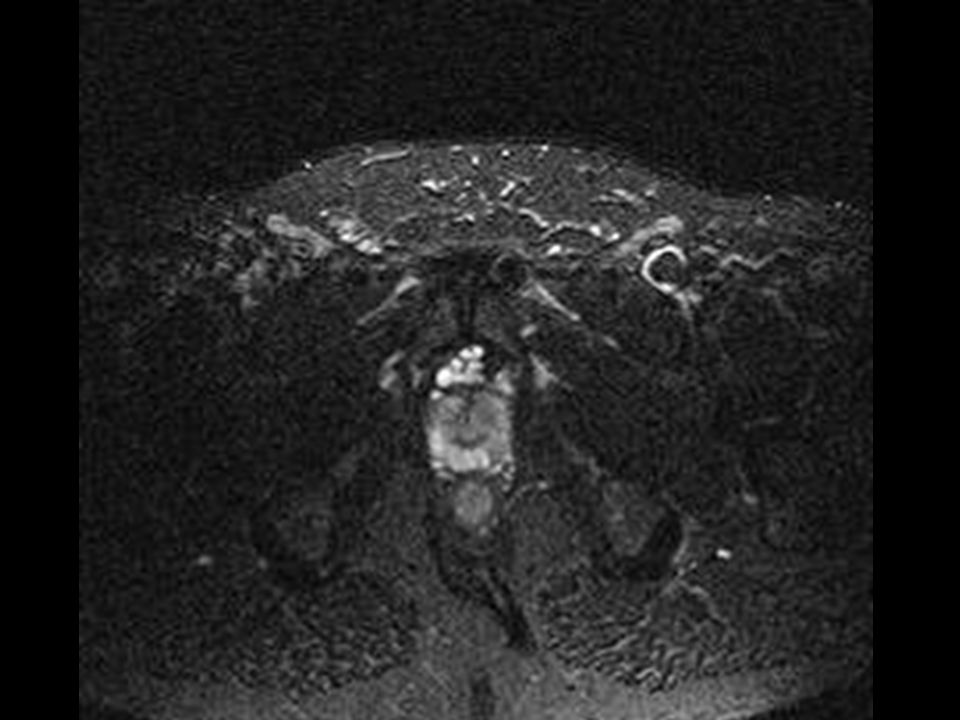
Grade 3- Trans-sphincteric fistula

76
Grade 4- Trans-sphincteric fistula with secondary track

81
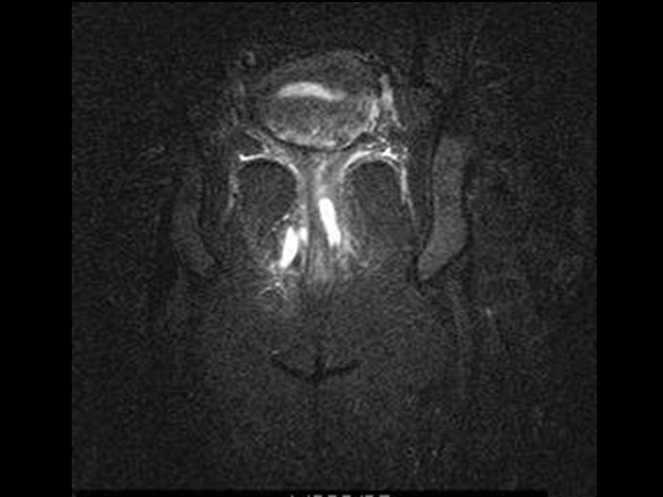
Grade 5- Translevator disease

86
Summary MR is a valuable modality in the assessment of peri-anal fistula Accurately identifies disease complexity

87
References 1: Beets-Tan RGH, Beets GL, Gerritsen van der Hoop A. et al. Preoperative MR Imaging of Anal Fistulas: Does it Really Help the Surgeon? Radiology 2001; 218:75-84 2: Bartram C, Buchanan G. Imaging anal fistula. Radiol Clin N Am 41 (2003) 3: Kuijpers HC, Schulpern T. Fistulography for fistula-in-ano: is it useful? Dis Colon Rectum 1985;28:103-4 4: Buchanan GN, Halligan S, Bartram CI et al. Clinical Examination, Endosonography, and MR Imaging in Preoperative Assessment of Fistula in Ano: Comparison with Outcome-based Reference Standard. Radiology 2004; 233: 5: Spencer JA, Chapple K, Wilson D et al. Outcome After Surgery for Perianal Fistula: Predictive Value of MR Imaging. AJR 1998; 171: 6: Horsthius K, Stoker J. MRI of perianal crohn’s disease. AJR 2004; 183: 7: Morris J, Spencer JA, Ambrose S. MR Imaging Classification of Perianal Fistulas and Its implications for Patient Management. Radiographics 2000; 20:
 : Kuijpers HC, Schulpern T. Fistulography for fistula-in-ano: is it useful Dis Colon Rectum 1985;28: : Buchanan GN, Halligan S, Bartram CI et al. Clinical Examination, Endosonography, and MR Imaging in Preoperative Assessment of Fistula in Ano: Comparison with Outcome-based Reference Standard. Radiology 2004; 233: : Spencer JA, Chapple K, Wilson D et al. Outcome After Surgery for Perianal Fistula: Predictive Value of MR Imaging. AJR 1998; 171: : Horsthius K, Stoker J. MRI of perianal crohn’s disease. AJR 2004; 183: : Morris J, Spencer JA, Ambrose S. MR Imaging Classification of Perianal Fistulas and Its implications for Patient Management. Radiographics 2000; 20:")
Similar presentations