Download presentation
Presentation is loading. Please wait.

1
BIOE 220/RAD 220 REVIEW SESSION January 30, 2011

2
Comments on HW 1 Problem 46-48 (the first set) were actually supposed to refer to a different picture, so don’t think that it’s a representative problem Can tell it’s T1 since CSF is dark You can tell it’s spin echo since edges are well define and there aren’t susceptibility artifacts near the tissue-air boundaries Remember to label left/right whenever it can be identified in the image Other problems should have the correct solution written on your hw if you made a mistake. We can review any problems you’re interested in now The homeworks and exams in this class will generally test whether you properly understand the concepts of the physics, rather than just solving equations

3
How to Identify Modality - Review Are the bones bright? Are we seeing a projection, or a slice? CT Radiograph MRI Are CSF and Gray Matter the Same Brightness? Is gray matter brighter than white matter? Proton Density Projection Slice yes no yes Is CSF very bright? T2 Spin Echo yes no T1 Weighting CSF is dark FLAIR (T2 contrast, dark CSF) no
 no.")
4
X-Ray Overview: Radiography, CT X-ray medical imaging measures x-rays that pass through the body to generate images X-rays are electromagnetic energy in a band of energies much higher than visible light X-rays that pass through the body can do one of several things Pass through – These get detected properly Get absorbed – These aren’t detected Scattered – These can get detected improperly Photoelectric effect – occurs more in high Z (atomic number) materials Compton scattering – dominant effect in soft tissue
 materials Compton scattering – dominant effect in soft tissue")
5
Radiograph schematic

6
Probability of interactions

7
Linear attenuation coefficient

8
Film convention

9
X-ray source properties

10
X-ray absorption

11
Image properties Contrast is determined by the energy of the photons (keV) and the tissue properties SNR is determined by the number of photons that transmit (SNR = sqrt(N) ), which is a function of the transmission properties and the number of photons transmitted (mAs) Scatter degrades SNR by adding detected events where there shouldn’t be any
 and the tissue properties SNR is determined by the number of photons that transmit (SNR = sqrt(N) ), which is a function of the transmission properties and the number of photons transmitted (mAs) Scatter degrades SNR by adding detected events where there shouldn’t be any")
12
Magnification in radiography Objects closer to source get magnified relative to farther objects

14
CT takes a series of radiographs to reconstruct 3 dimensional object

15
CT units are normalized to water attenuation (Hounsfield units) Contrast is the difference in Hounsfield units: C = A - B
 Contrast is the difference in Hounsfield units: C = A - B")
17
CT colormaps are windowed The dynamic range in CT is too large to capture everything with a single colormap The center (“level”) and extent (“window”) are varied to be able to see the type of tissue being imaged Bone window has a high level and wide window Soft tissue or brain window is close to 0 (water) and much smaller in extent
 and extent ( window ) are varied to be able to see the type of tissue being imaged Bone window has a high level and wide window Soft tissue or brain window is close to 0 (water) and much smaller in extent")
21
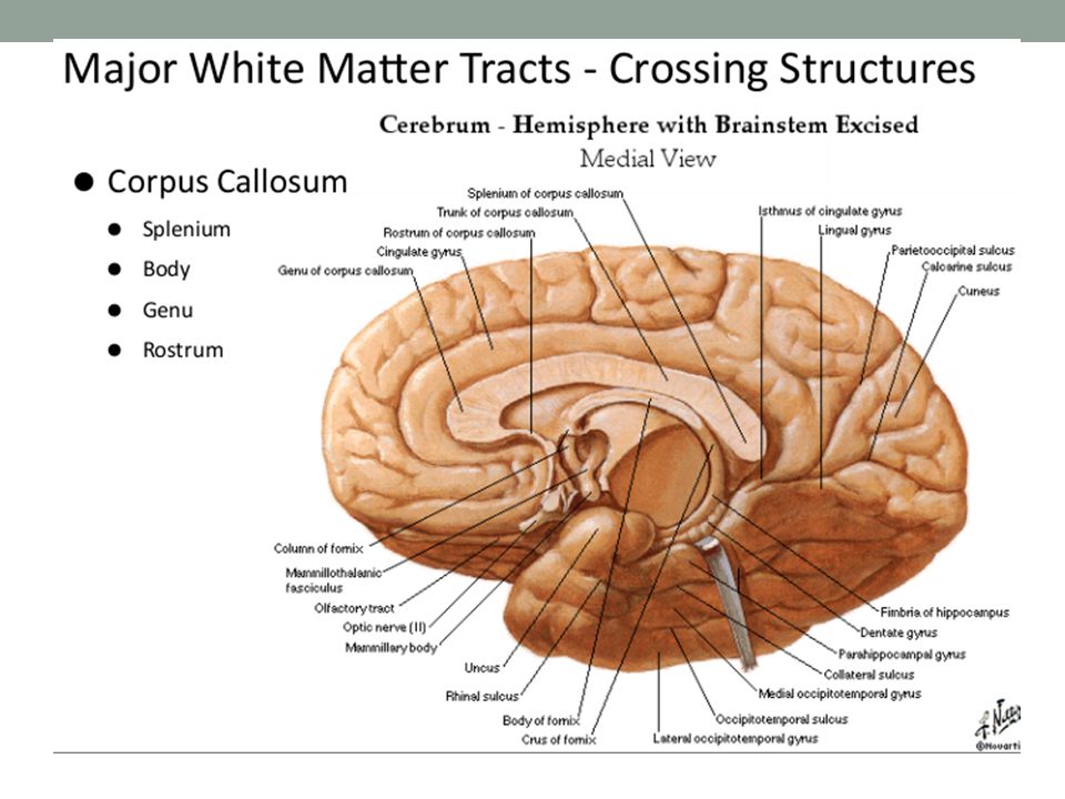
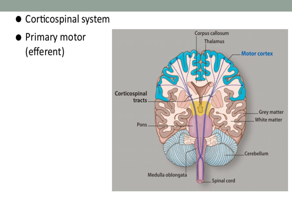
White Matter Tracts Crossing Structures Corpus Callosum Anterior commissure Posterior commissure Connections within brain Optical radiation Connections between brain and the rest of the body Corticospinal tracts

Similar presentations


>")


