Download presentation
Presentation is loading. Please wait.

1
CHLA Case Presentation

2
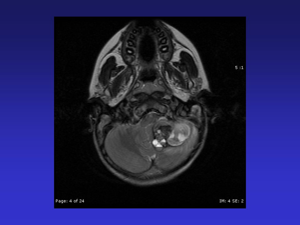
History HPI: 10 year old male with Down syndrome and a 1 week history of headache, nausea, vomiting, dizziness and unsteady gait. PMH: Down syndrome, ASD PSH: PE tubes, orchiopexy Meds: None NKDA

3
Physical Exam Awake and alert CN II-XII intact Motor: 5/5 bilaterally Sensation: Intact bilaterally Reflexes: Symetric, no Babinski CBLM: FTN and RAM intact, gait ataxic

10
Differential Diagnosis Cavernous malformation Teratoma

11
Procedure Posterior fossa craniotomy with gross total resection of mass

13
Diagnosis Cavernous Malformation

14
Also known as cavernous angiomas, cavernomas and hemangiomas Gross appearance is red and lobulated, similar to “mulberries” Size is usually 0.5 to 3 cm Adjacent brain is often hemosiderin stained

15
Cavernous Malformation No large supplying artery or draining vein Low flow No intervening brain Adjacent brain not ischemic Microscopically have blood containing sinusoidal chambers lined by simple epithelium The vascular spaces are separated by fibrous or collagenous tissue rather than brain Often have a gliotic margin

16
Cavernous Malformation 9% of all types of brain vascular malformations Prevalence is 0.4-0.8% M:F ratio is1:1 Age at presentation 20-40 Present with headache, focal neurological deficit, seizures, hemorrhage

17
Cavernous Malformation CM can repetitively hemorrhage resulting in –Gliosis –Tissue discoloration –Hemosiderin-laden macrophages –Microcalcification –Hyalinization –Cysts with blood breakdown products

18
Cavernous Malformation Can be familial –Hispanic families CCM1, 7q11-21 –Non-Hispanic families CCM2, 7p13-15 CCM3, 3q25.2-27

19
Risk of hemorrhage Cantu, C., L. Murillo-Bonilla, et al. (2005). "Predictive factors for intracerebral hemorrhage in patients with cavernous angiomas." Neurol Res 27(3): 314-8. 133 Hispanic patients with 5 year follow-up ICH rate 1.71% per patient per year –Lobar 1.22% –Brainstem 2.33% –Cerebellum 2.39% –Deep hemispheric 2.82% Decreased rate of hemorrhage if family history of epilepsy or lobar location of CM
. Predictive factors for intracerebral hemorrhage in patients with cavernous angiomas. Neurol Res 27(3): Hispanic patients with 5 year follow-up ICH rate 1.71% per patient per year –Lobar 1.22% –Brainstem 2.33% –Cerebellum 2.39% –Deep hemispheric 2.82% Decreased rate of hemorrhage if family history of epilepsy or lobar location of CM.")
20
Association with Venous Malformations Abdulrauf, S. I., M. Y. Kaynar, et al. (1999). "A comparison of the clinical profile of cavernous malformations with and without associated venous malformations." Neurosurgery 44(1): 41-6; discussion 46-7. 55 patients 24% had CM’s associated with VM’s –F>M –Greater risk of symptomatic hemorrhage (62% vs. 38%) –More likely to have lesions in the posterior fossa (P=0.001) –Less likely to present with seizures –Less likely to have family history
. A comparison of the clinical profile of cavernous malformations with and without associated venous malformations. Neurosurgery 44(1): 41-6; discussion patients 24% had CM’s associated with VM’s –F>M –Greater risk of symptomatic hemorrhage (62% vs. 38%) –More likely to have lesions in the posterior fossa (P=0.001) –Less likely to present with seizures –Less likely to have family history.")
21
Association with Down Syndrome There is no known association between Down syndrome and the development of CM Singh et al. (1993) reported on a 30 year-old male with Down sydrome and a cervical intramedullary CM (“chance association”)
 reported on a 30 year-old male with Down sydrome and a cervical intramedullary CM ( chance association ).")
22
Familial Cavernomas Gunel, M., I. A. Awad, et al. (1996). "A founder mutation as a cause of cerebral cavernous malformation in Hispanic Americans." N Engl J Med 334(15): 946-51. Studied 57 Hispanic patients –47 were from 14 different kindreds with familial CMs –10 were sporadic cases Found that all cases could be attributed to inheritance of the same mutation on 7q from a common ancestor with incomplete penetrance
. A founder mutation as a cause of cerebral cavernous malformation in Hispanic Americans. N Engl J Med 334(15): Studied 57 Hispanic patients –47 were from 14 different kindreds with familial CMs –10 were sporadic cases Found that all cases could be attributed to inheritance of the same mutation on 7q from a common ancestor with incomplete penetrance.")
23
Familial Cavernomas Labauge, P., L. Brunereau, et al. (2000). "The natural history of familial cerebral cavernomas: a retrospective MRI study of 40 patients." Neuroradiology 42(5): 327-32. 40 patients with 3.2 year follow-up 232 CMs, 5.9 per patient Hemorrhagic risk 2.5% per lesion per year 27.5% developed new CMs Incidence of new lesions 0.2% per patient year 3.9% of lesions in 22.5% of patients changed significantly in size
. The natural history of familial cerebral cavernomas: a retrospective MRI study of 40 patients. Neuroradiology 42(5): patients with 3.2 year follow-up 232 CMs, 5.9 per patient Hemorrhagic risk 2.5% per lesion per year 27.5% developed new CMs Incidence of new lesions 0.2% per patient year 3.9% of lesions in 22.5% of patients changed significantly in size.")
24
Familial Cavernomas Labauge, P., L. Brunereau, et al. (2001). "Prospective follow-up of 33 asymptomatic patients with familial cerebral cavernous malformations." Neurology 57(10): 1825-8. Prospectively followed 33 asymptomaitic non-Hispanic patients with familial CMs for 2.1 years Total of 234 CMs, mean 7.1 per subject, range 1-85 CMs per subject 2 subjects became symptomatic (hemorrhage, seizure) 30 new lesions appeared in 10 subjects (46%) –0.4 lesions per year Four lesions (1.7%) increased in size in 3 subjects (9.1%)
. Prospective follow-up of 33 asymptomatic patients with familial cerebral cavernous malformations. Neurology 57(10): Prospectively followed 33 asymptomaitic non-Hispanic patients with familial CMs for 2.1 years Total of 234 CMs, mean 7.1 per subject, range 1-85 CMs per subject 2 subjects became symptomatic (hemorrhage, seizure) 30 new lesions appeared in 10 subjects (46%) –0.4 lesions per year Four lesions (1.7%) increased in size in 3 subjects (9.1%).")
Similar presentations


 Changes in cerebral hemodynamics after carotid stenting of symptomatic carotid artery.>")


