Download presentation
Presentation is loading. Please wait.

1
Mitral Valve Prosthesis

2
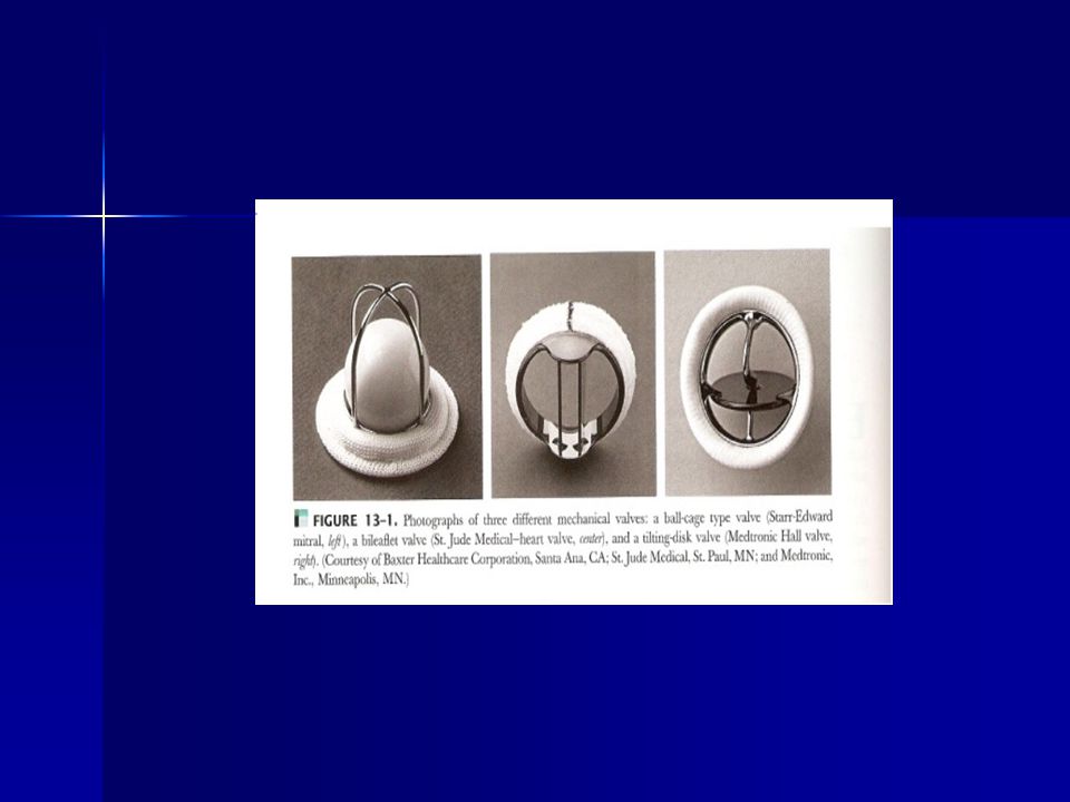
Types of mechanical valves: Ball-cage valve (Starr-Edwards is the most common). - a spherical occluder is contained by metal “ cage ” when the - a spherical occluder is contained by metal “ cage ” when the valve is open & fills the orifice in the closed position. valve is open & fills the orifice in the closed position. Tilting disc valve (Medtronic-Hall is the most common). - Where a single circular disk opens at an angle to the annulus - Where a single circular disk opens at an angle to the annulus plane. Being constrained in its motion by a smaller “ cage ”, a plane. Being constrained in its motion by a smaller “ cage ”, a central strut or a slanted slot in the valve ring. central strut or a slanted slot in the valve ring. Bileaflet valve - Where two semicircular disks hinge open to form 2 large - Where two semicircular disks hinge open to form 2 large lateral orifices. lateral orifices.
. - Where a single circular disk opens at an angle to the annulus - Where a single circular disk opens at an angle to the annulus plane. Being constrained in its motion by a smaller cage , a plane. Being constrained in its motion by a smaller cage , a central strut or a slanted slot in the valve ring. central strut or a slanted slot in the valve ring. Bileaflet valve - Where two semicircular disks hinge open to form 2 large - Where two semicircular disks hinge open to form 2 large lateral orifices. lateral orifices..")
5
2D Echo 2D Echo - helpful in evaluating for the complications of mechanical valve although reverberations make the diagnosis difficult. - can identify gross structural abnormalities of a prosthesis such as dehiscence, vegetations, thrombus or degeneration.

6
Difficulties in 2D: - echo reflectance of the prosthetic material attenuation of the ultrasound beam and multiple ultrasound reverberations from the prosthesis result in difficulties in interpretation. - TEE may be able to visualize normal and abnormal prosthetic valve motion.

7
Doppler: all prosthesis valves are inherently stenotic. The degree of obstruction is a function of prosthesis type, size and site of the prosthetic valve. all prosthesis valves are inherently stenotic. The degree of obstruction is a function of prosthesis type, size and site of the prosthetic valve. evaluate patients soon after surgery (within 30 days) to obtain baseline doppler values. evaluate patients soon after surgery (within 30 days) to obtain baseline doppler values. Heart rate, blood pressure and LV function should be noted as the examinations begins. Heart rate, blood pressure and LV function should be noted as the examinations begins. An average of 5 to 10 beats should be utilized to obtain hemodynamic measurements mainly in AF.. An average of 5 to 10 beats should be utilized to obtain hemodynamic measurements mainly in AF..
 to obtain baseline doppler values. evaluate patients soon after surgery (within 30 days) to obtain baseline doppler values. Heart rate, blood pressure and LV function should be noted as the examinations begins. Heart rate, blood pressure and LV function should be noted as the examinations begins. An average of 5 to 10 beats should be utilized to obtain hemodynamic measurements mainly in AF.. An average of 5 to 10 beats should be utilized to obtain hemodynamic measurements mainly in AF...")
8
Caution: When attempting to rule out significant mitral regurgitation in a prosthetic mitral valve, you must remember that the prosthetic valve will “ mask ” the MR from the doppler ultrasound due to the high acoustic impedance diffirence between the valve and blood. Multiple views and TEE will overcome the problem of masking. When attempting to rule out significant mitral regurgitation in a prosthetic mitral valve, you must remember that the prosthetic valve will “ mask ” the MR from the doppler ultrasound due to the high acoustic impedance diffirence between the valve and blood. Multiple views and TEE will overcome the problem of masking.

9
Bioprosthetic/Mechanical Mitral Valve : Transducer position where the optimal doppler signal was obtained so it may be used for f/u studies (apical views). Transducer position where the optimal doppler signal was obtained so it may be used for f/u studies (apical views). determine the peak velocity; > 2.5m/sec may indicate stenosis or significant regurgitation. determine the peak velocity; > 2.5m/sec may indicate stenosis or significant regurgitation. determine the pressure half-time, > 180msec may be abnormal. determine the pressure half-time, > 180msec may be abnormal. Determine mitral valve area by the pressure half-time method; MVA < 1.8cm2 may be abnormal. Determine mitral valve area by the pressure half-time method; MVA < 1.8cm2 may be abnormal. Determine the the mean pressure gradient; >10mmHg may be abnormal. Determine the the mean pressure gradient; >10mmHg may be abnormal. Determine the mitral valve area by CE. Determine the mitral valve area by CE. Determine the presence and severity of MR. Determine the presence and severity of MR. A perivalvular leak is abnormal. A perivalvular leak is abnormal.
. determine the peak velocity; > 2.5m/sec may indicate stenosis or significant regurgitation. determine the peak velocity; > 2.5m/sec may indicate stenosis or significant regurgitation. determine the pressure half-time, > 180msec may be abnormal. determine the pressure half-time, > 180msec may be abnormal. Determine mitral valve area by the pressure half-time method; MVA < 1.8cm2 may be abnormal. Determine mitral valve area by the pressure half-time method; MVA < 1.8cm2 may be abnormal. Determine the the mean pressure gradient; >10mmHg may be abnormal. Determine the the mean pressure gradient; >10mmHg may be abnormal. Determine the mitral valve area by CE. Determine the mitral valve area by CE. Determine the presence and severity of MR. Determine the presence and severity of MR. A perivalvular leak is abnormal. A perivalvular leak is abnormal..")
12
apical 4 ch - cw

16
Complications: - Thrombus - Pannus (fibrous ingrowth tissue which may lead to regurgitation or stenosis). - Paravalvular leak. - Dehiscence - Infective endocarditis - Hemolysis

20
TEE long axis view of the dehisced sewing ring of a St. Jude MV prosthesis.Color flow imaging shows severe MR.

22
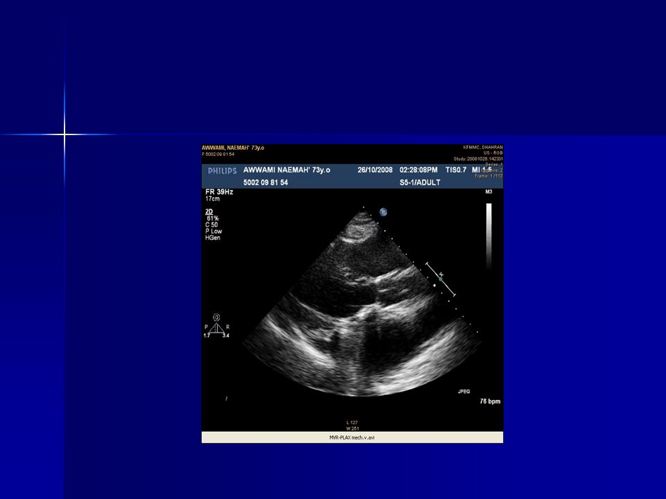
Obstructed mechanical mitral valve prosthesis. At the time of operation thrombus as well as pannus formation was identified along the sewing ring. The pt. underwent a second MVR and post op CW doppler exam showed a peak velocity of 1.6 m/sec, MG of 4 mm Hg and a PHT of 80 ms.

25
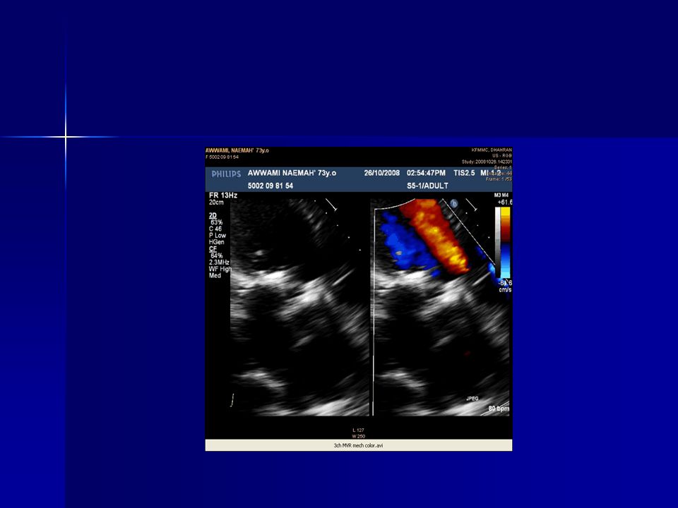
PLAX - color

26
SAX view MVR-metallic

27
4ch 2d

28
4ch-color

33
Obstruction: - when prosthetic valve becomes obstructed the motion of the disk, ball, or leaflets decreases. - most accurate method for detecting and quantitating the degree of prosthetic obstruction is doppler echo. - increased flow velocity not always indicate prosthetic obstruction. - velocity can be increased without stenosis in a high output state and in the presence of the severe MR.

34
Mechanism of Prosthetic Valve dysfunction: - structural failure - thromboembolic complications - endocarditis

35
Structural failure: - Failure of a bioprosthesis valve to open or close properly usually is the result of slowly progressive tissue degeneration & fibrocalcific changes of the leaflets resulting in increase resistance to open (stenosis) or failure to coapt during valve closure. - Typically failure of tissue valve occurs 10 or more years after valve implantation.

36
- Regurgitation occur with a leaflet tear usually adjacent to a region of calcification. - Failure of mechanical valve can occur due to faulty design or wear and tear of prosthetic material resulting in disk escape or incomplete valve closure. This complication seen only with older generation valve. - Current generation mechanical valve are reliable and very durable.

37
- Mechanical valve stenosis or regurgitation is due to thrombus formation or pannus in growth around the valve, impairing disk excursion or closure. - With both bioprosthetic and mechanical valve paravalvular regurgitation can occur around the sewing ring due to loss of suture material post operatively.

38
Thromboembolic complications: - Prosthetic valve particularly mechanical valve are prone to thrombus formation. - Echo evaluation for prosthetic valve thrombus is limited except with very large masses, due to shadowing and reverberations.

39
Endocarditis: - Endocarditis on a bioprosthesis may result in vegetations similar to those seen on native valve, in mechanical valve the infection often is paravalvular and no discrete vegetation may be present.

40
Thank you! Next week topic Aortic Stenosis By: Ayed Onazi

Similar presentations


 Continuous Wave 2) Pulse Wave 3) Color Flow DOPPLER ULTRASOUND.>")


