Download presentation
Presentation is loading. Please wait.

1
CONDUCTIVE TISSUE OF HEART
Lecture – 2 Dr. Zahoor Ali Shaikh

3
CONDUCTIVE TISSUE OF HEART
Action Potential that originates in SA node spread to both Atria through intercalated disc and gap junction. From atria action potential can not pass to ventricle due to fibrous Skelton of heart which separates atria and ventricles. Therefore specialized conducting tissue is required (it is composed of modified Myocardial cells) to conduct the impulse
 to conduct the impulse.")
4
CONDUCTION TISSUE OF THE HEART
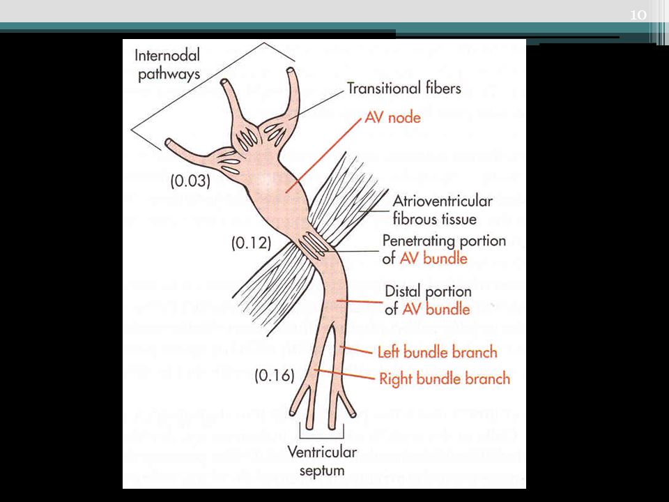
SA Node AV Node Bundle of His or AV Bundle Rt & Lt Bundle Branches Purkinje fiber Ventricle Internodal Fibers

5
CONDUCTION TISSUE OF THE HEART

6
CONDUCTIVE TISSUE OF HEART We will see Anatomical Locations
SA NODE It is 15mm long, 3mm wide, 1mm thick. SA Node – It is pace-maker because it has pace- maker potential or pre-potential – automatic depolarization. SA Node is situated in the right atrium near the opening of superior venaceva. It is supplied by right vagus.

7
CONDUCTIVE TISSUE OF HEART Anatomical Locations
INTERNODAL FIBERS Internodal Fibers – Anterior, Middle and Posterior [Bachman, Wenchkeback, Thorel]. AV NODE It is situated at right posterior part of inter atrial septum. It is supplied by left vagus.

8
CONDUCTIVE TISSUE OF HEART Anatomical Locations
BUNDLE OF HIS [Atrioventricular Bundle] It is a tract of specialized cardiac cells that originate at AV Node and passes through the fibrous ring.

9
CONDUCTIVE TISSUE OF HEART Anatomical Locations
Bundle of His ,after passing through fibrous ring, at the top of interventricular septum, it divides to form right bundle branch and left bundle branch that travel down along the sides of interventricular septum and lye subendocardially. NOTE – Lt Bundle Branch has 2 fascicles Left Anterior Fascicle and Left Posterior Fascicle.

12
CONDUCTIVE TISSUE OF HEART Anatomical Locations
PURKINJE FIBERS From bundle branches, we get Purkinje Fibers, they spread throughout the ventricular myocardium .

14
RATE OF DISCHARGE IN AUTO-RHYTHMIC TISSUE OF HEART

15
CONDUCTIVE TISSUE Why SA-Node is a Pace-maker?
Because its discharge rate is high 70-80/min. This action potential/min drive rest of the heart, therefore, it is known as pace-maker of the heart. It has pre-potential.

16
CONDUCTIVE TISSUE Other auto - rhythmic tissue are firing at slow rate. They can work as pace-maker, if SA-Node is not functioning e.g. if AV Node takes over as pace- maker, heart rate will be about 50/min. Any pace-maker other than SA-Node is called ‘Ectopic Pace-maker’.

17
SPREAD OF CARDIAC EXCITATION
Impulse arise at SA-Node and spread to the atria [via gap junction] – Atrial Syncytium, therefore, both atria depolarize same time. Impulse [AP] goes to AV-Node by Internodal pathway. AV-Node is the only point of electrical contact between atria and ventricle [as atria and ventricle are separated by fibrous ring which is non-conductive].

18
SPREAD OF CARDIAC EXCITATION
AV – Node At AV-Node, there is delay of 0.1 sec [100 milli- sec]. This delay is important because it allows the atria to contract and empty their blood into the ventricle, before impulse reaches the ventricle and causes ventricular depolarization and contraction.

19
SPREAD OF CARDIAC EXCITATION
Ventricular Excitation After AV delay of 0.1sec, impulse [AP] travels quickly via Right Bundle Branch and Left Bundle Branch [branches of Bundle of His] to Purkinje Fibers to the ventricles. Both ventricle depolarize, than contract at same time. Conduction in Purkinje Fiber is fastest 3-5 meter/sec, therefore, both ventricle depolarize quickly and at the same time.

21
CONDUCTION OF IMPULSE There is delay of 0.1 sec in AV node
Important points In Atria – 1 Meter/ sec AV Node slow conduction – 0.03 to 0.05 Meter / sec There is delay of 0.1 sec in AV node Purkinje fiber - 3 to 5 Meter / sec Slowest Conduction at AV – Node Fastest Conduction - Purkinje Fibers

22
SPREAD OF CARDIAC EXCITATION
Why Conduction is slow at AV-Node? Because there are less gap junctions. Diameter of the fiber is small.

23
APPLIED – HEART BLOCKS There are three types of heart blocks:
FIRST DEGREE HEART BLOCK – Every impulse is conducted but very slowly, therefore, there is increase in conduction time [we can see on ECG]. SECOND DEGREE HEART BLOCK – Some impulses are conducted and other are not conducted.

24
APPLIED – HEART BLOCKS THIRD DEGREE HEART BLOCK – Complete heart block, no conduction occurs from SA Node to the ventricle through AV node, therefore, atrial rate is separate [75/min] from the ventricular rate which follows the Purkinje fibers and is about 30/min. IMPORTANT If ventricular rate is very slow e.g. complete heart block, we need artificial pace-maker [implanted device which generates impulse].

25
ECTOPIC PACE-MAKER Any pace-maker other than SA Node is called ECTOPIC Pace-maker. [Ectopic means out of place]. It may be in atria, ventricle. If Ectopic Pace-maker is faster than SA node, it will take over and heart rate will be high. Ectopic pace-maker can occur in heart disease or some factors can precipitate e.g. anxiety, lack of sleep, excess caffeine, alcohol.
![ECTOPIC PACE-MAKER Any pace-maker other than SA Node is called ECTOPIC Pace-maker. [Ectopic means out of place].](http://slideplayer.com/slide/3879072/13/images/25/ECTOPIC+PACE-MAKER+Any+pace-maker+other+than+SA+Node+is+called+ECTOPIC+Pace-maker.+%5BEctopic+means+out+of+place%5D..jpg "It may be in atria, ventricle. If Ectopic Pace-maker is faster than SA node, it will take over and heart rate will be high. Ectopic pace-maker can occur in heart disease or some factors can precipitate e.g. anxiety, lack of sleep, excess caffeine, alcohol.")
26
EFFECT OF SYMPATHETIC & PARASYMPATHETIC ANS
Sympathetic ANS-- Effect on conduction – It increases conduction as it decreases delay at AV node. Parasympathetic – slows the conduction as it increases delay at AV node.

28
EFFECT OF SYMPATHETIC & PARASYMPATHETIC ANS ON HEAR

29
FACTORS AFFECTING CONDUCTION VELOCITY

30
POINT TO PONDER In Transplanted Heart, where there is no sympathetic and parasympathetic nerve supply, what will be the rate of SA Node discharge [Heart Rate] ?

31
What You Should Know From This Lecture?
Conductive Tissue of Heart Its Anatomical Location Rate of Conduction- where is Fastest and slowest Importance of AV Node Delay Effect of Sympathetic & Parasympathetic ANS Factors Affecting Conduction Normal Rate of SA Node, AV Node & Purkinje Fiber Action Potential Discharge Ectopic Pace-maker Heart Blocks

32
Thank you

Similar presentations


 in thorax, in inferior mediastinum>")
>")


>")


>")
