Download presentation
Presentation is loading. Please wait.

2
The Mammary Gland The milk secreting organ Modified sweat gland
Exocrine gland

3
MAMMARY GLANDS thoracic inguinal abdominal

4
MAMMARY GLANDS 4 abdominal; 15 openings / teat

5
MAMMARY GLANDS 2 thoracic, 6 abdominal, ; 3-7 openings / teat

6
MAMMARY GLANDS 2 thor., 6 abdom. 2 ing., ; 8-10 openings/ teat

7
MAMMARY GLANDS 4 thor., 2 abdom. 4 ing., ; 1 opening/ teat

8
MAMMARY GLANDS 2 inguinal ; 1 opening/ teat

9
MAMMARY GLANDS 2 inguinal ; 2 openings/ teat

10
MAMMARY GLANDS 4 thor., 6 abdom. 2 ing., ; 2 openings/ teat

11
MAMMARY GLANDS 4 inguinal ; 1 opening/ teat

12
Anatomy of the Mammary Gland
Mammary gland - milk secreting structure including teats, duct system, lobes, lobules, and secretory tissue Modified sweat gland Exocrine gland Cow Large & in inguinal region 4 teats/quarters = 4 separate glands No mixing of ducts across quarters

13
Anatomy of the Mammary Gland
Rear quartes produce approx. 60% of the milk and the fore quarters produce the remaining 40% The size and shape of udders vary with the 1) producing ability, 2) age, and 3) genetic of the cow
 producing ability, 2) age, and 3) genetic of the cow.")
14
Anatomy of the Mammary Gland
Cow Front & rear quarters separated by fine membrane Left and right separated by median suspensory ligament Supernumerary teats (some with duct and secretory system)
")
15
How much support is enough?
High producing Holstein cow Empty Udder = 25 kg. Milk = 30 kg = 55 kg !!!

16
Udder Support in Cow Skin Fine connective tissue below skin
Connective tissue attaches front quarters to abdominal wall Lateral suspensory ligaments (LSL) Median suspensory ligament (MSL) The subpelvic tendon
 Median suspensory ligament (MSL) The subpelvic tendon.")
18
Udder Support in Cow LSL Sling around udder 2 layers
Inelastic, more fibrous than MSL

19
Lateral Suspensory Ligaments
Like a “hammock” around the udder From the pelvis to the median suspensory ligament Mostly fibrous tissue Collagen Attaches to the alveolar tissue Provides internal framework

20
Udder Support in Cow MSL Primary support Relatively elastic 2 layers
Broken MSL – pendulous udder

21
Median Suspensory Ligament
Primary support of the udder Two adjacent heavy sheets of tissue Mostly elastic, some fibrous tissue Attaches to the abdominal wall Divides the udder into halves Glands on each half are divided by sheets of tissue © Biology of Lactation, Schmidt

23
Udder Support in Cow Lamella septa Connective tissue
Runs between LSL & MSL Divides parenchyma into lobes and lobules

25
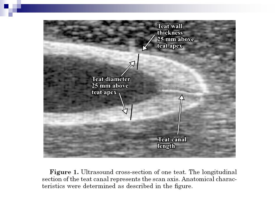
Duct System Teat meatus, the small canal located in the end of each teat is .5 to 1 cm long and is the only sphincter in each gland Seven or eight loose folds of membrane known as furstenburg rosette are located above the teat meatus The teat cistern, the cavity within the teat hold 30 to 90 ml of milk.

28
The Secretory Tissue A Lobe: group of lobules
A Lobule: group of alveoli Alveoli: cluster of alveolus Alveolus: a single layer of epithelial cells surrounding a central lumen

31
Blood Supply to Mammary Gland
400 kg blood to produce 1 kg of milk 2 major arteries Front ½ of udder Rear ½ of udder 4 major veins 2 follow same path as arteries 2 mammary veins

34
© Biology of Lactation, Schmidt
Nervous System Sensory (afferent) nerves in skin and teats Positive stimulation of teats and surrounding area initiates milk let-down reflex via oxytocin © Biology of Lactation, Schmidt
 nerves in skin and teats. Positive stimulation of teats and surrounding area initiates milk let-down reflex via oxytocin. © Biology of Lactation, Schmidt.")
35
Nervous System Sympathetic (efferent) (involuntary) nerves associated with arteries in the gland Control blood flow to the gland Innervation of sphincters muscles in teats Stress causes vasoconstriction decreasing milk secretion and let-down No parasympathetic innervation No nerves to myoepithelial cells or alveolar cells

37
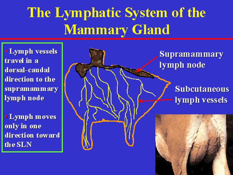
Lymph System of Mammary Gland
What is lymph & what does it do? Supramammary lymph nodes Lymph vessels Factors that influence edema Age Diet (especially NaCl) Exercise Genetics
 Exercise. Genetics.")
40
Mammary Gland Development
Five phases of mammary development Prenatal (teats & cisterns dev.) Prepubertal (limited growth) Postpubertal Pregnancy (most growth) Early lactation
 Prepubertal (limited growth) Postpubertal. Pregnancy (most growth) Early lactation.")
41
Mammary Gland Development
Major development occurs at puberty and during gestation Hormones Estrogen (growth of duct system) Progesterone (development of alveolar tissue in combination with other hormones) GH (growth of duct system) Prolactin (initiation and continuity of lactation)
 Progesterone (development of alveolar tissue in combination with other hormones) GH (growth of duct system) Prolactin (initiation and continuity of lactation)")
50
Mammary Gland Development

54
Mammary Gland Development

58
Anatomy of the Mammary Gland
Goats and sheep 2 teats/ 2 halves (glands) Pig 12-14 teats – 2 glands and duct systems per teat Mare 4 quarters/duct systems but 2 teats
 Pig teats – 2 glands and duct systems per teat. Mare. 4 quarters/duct systems but 2 teats.")
60
Mammary Duct System

63
Epithelial Cell Lumen of alveolus Fat Droplet Fat Droplet migrating
Golgi Body Lysosome Nucleus Mitochondria Mitochondria E.R. Blood Vessel

Similar presentations


>")


