Download presentation
Presentation is loading. Please wait.

1
• Consists of: – Lymph Lymphatic vessels Lymphatic tissue
Lymphatic System • Consists of: – Lymph Lymphatic vessels Lymphatic tissue Lymphatic nodules Lymph nodes Tonsils Spleen thymus

2
Lymphatic System • Lymphatic Vessels • Lymphoid Organs and Tissues
– Functioning in returning interstitial fluid to the vascular system. • Lymphoid Organs and Tissues – House phagocytes and lymphocytes – Activate the immune response

4
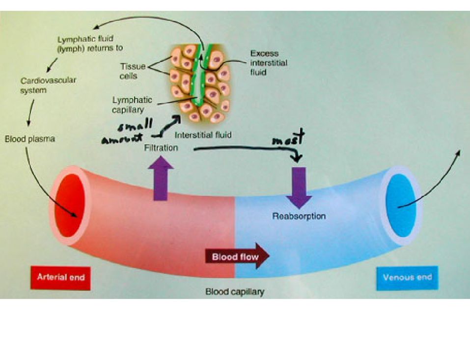
Lymphatic Vessels • Return ISF to the bloodstream
Return leaked plasma proteins to the blood Fluid within them is called lymph 1-way system flowing toward the heart

6
1 way system back towards the heart

7
How does lymph differ from plasma?
Extracellular space Blood Lymphatic vessel PLASMA INTERSTITIAL FLUID or TISSUE FLUID vessel LYMPH How does lymph differ from plasma?

8
• 4 types: – Lymphatic capillaries Lymphatic collecting vessels
Lymphatic Vessels • 4 types: – Lymphatic capillaries Lymphatic collecting vessels Lymphatic trunks Lymphatic ducts

9
Lymphatic Capillaries
• What do they do?

10
Lymphatic Capillaries
• Blind • Lined by endothelium – Loose – Overlapping • Permeability • Flow

11
Lymphatic Capillaries
• Found just about everywhere that blood capillaries are found. – Exceptions

12
Lymphatic Capillaries
• Lacteals – Specialized lymphatic capillaries found in the villi of the small intestine. – Absorb fats from the digestive tract.

14
Lymphatic Capillaries
Collecting Vessels -Similar to veins (in terms of the 3 tunics) -Lots of valves -Both superficial and deep -Pass thru lymph nodes * What happens to the lymph there?
 -Lots of valves. -Both superficial and deep. -Pass thru lymph nodes. * What happens to the lymph there")
16
What feature of lymphatic vessels is visible here? What do they
prevent? Why are they necessary?

17
• Receive lymph from lymphatic collecting • Types:
Lymphatic Trunks • Receive lymph from lymphatic collecting vessels. • Types: – Jugular Subclavian Bronchomediastinal Intestinal Lumbar

18
Lymphatic Ducts • Receive lymph from lymphatic trunks.
• Right lymphatic duct – Receives lymph from the right jugular, right subclavian, and right bronchomediastinal trunks – Empties into the right internal jugular vein • Thoracic duct – Receives lymph from the left jugular, left subclavian, left bronchomediastinal, intestinal, and lumbar trunks – Empties into the left internal jugular vein

21
Lymphatic Vessels Lymphatic Trunks Lymphatic Ducts
Where’s pressure high and where’s pressure low? Lymphatic Trunks What makes lymph flow? Lymphatic Ducts Bloodstream

22
Lymph Flow • Lymph flow will be similar to…
• 3 main factors promote lymph flow: – Skeletal muscle pump – Respiratory pump – Lymphatic smooth muscle

23
1. How does elevating an injured limb affect lymph flow?
2. How would exercise affect lymph flow? 3. How could massage affect lymph flow? 4. In some surgeries for breast cancer, the lymph nodes along thearm are removed in order to assess the spread of the disease. What would be a result of this?

24
What happens when lymph cannot flow?
What could prevent lymph from flowing? Filaria

25
Lymphoid Cells • Lymphocytes • Phagocytes • Dendritic cells
– T lymphocytes • Kill virus-infected and cancerous cells • Coordinate/control immune response – B lymphocytes • Become plasma cells which secrete antibodies • Phagocytes – Eat and kill and activate the rest of the immune system • Dendritic cells – Activate the immune system • Reticular cells – Make reticular fibers that support lymphatic tissues and organs

26
Reticular connective tissue forms the framework of most lymphoid tissues and organs.

27
Lymphoid Tissue • Aggregations of lymphoid cells
Storage/proliferation site for lymphoid cells Surveillance site 2 main types – Diffuse lymphatic tissue – Lymphoid follicles

28
Diffuse Lymphatic Tissue
• What does “diffuse” mean? • Found in lymph nodes and spleen. • Especially prominent in the mucous membranes lining the digestive, respiratory, urinary, and reproductive tracts. – Hence the term MALT.

29
Mucosa Associated Lymphatic Tissue
• Where are the mucosae? • Why is lymphoid tissue needed there? • 2 main types: – GALT – BALT

30
Lymphoid Follicles • A.k.a. lymphoid nodules
• Solid, packed spherical clusters of lymphoid cells and reticular tissues • Often found as parts of larger lymphoid organs (e.g., lymph nodes) • Found in the mucosae • Large number in the distal ileum and appendix
 • Found in the mucosae. • Large number in the distal ileum and. appendix.")
31
Lymphoid follicle in the
stomach

32
• Peyer’s Patches – Aggregates of lymphoid follicles found in the
distal small intestine. • Why there?

33
• Appendix – Blind outpocketing of the cecum.
– Contains aggregates of follicles.

34
Lymphoid Organs • Surrounded by a capsule of dense connective tissue.
• Lymph nodes • Tonsils, thymus, spleen

35
Lymph Nodes • Filter lymph • Clustered along lymphatic vessels
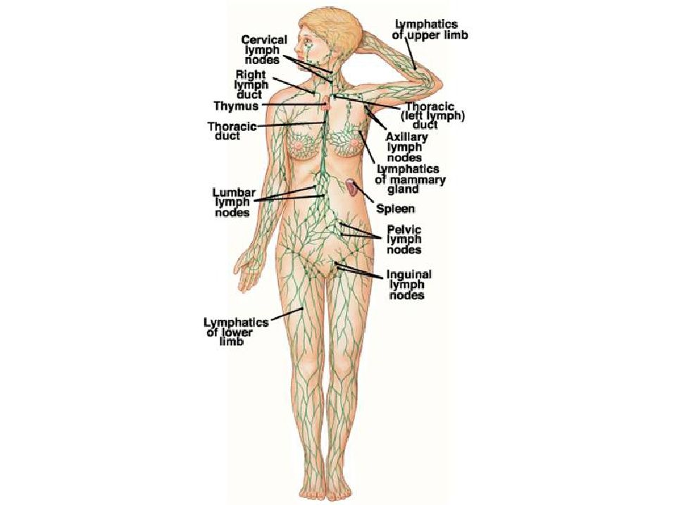
• Large superficial clusters in inguinal, axillary, and cervical regions.

39
Lymph Nodes • Receive lymph from an afferent lymphatic vessel
• Drain lymph into an efferent lymphatic vessel • Surrounded by dense CT capsule – Inward extensions (trabeculae) divide it into compartments • Reticular fibers support the resident macrophages and lymphocytes • Divided into a cortex and a medulla
 divide it into. compartments. • Reticular fibers support the resident macrophages. and lymphocytes. • Divided into a cortex and a medulla.")
41
• Cortex contains: – Subcapsular sinus Cortical sinuses Trabeculae
Lymph Nodes • Cortex contains: – Subcapsular sinus Cortical sinuses Trabeculae Diffuse lymphatic tissue Lymphatic follicles

42
Lymph Nodes • Medulla contains:
– Medullary cords (diffuse lymphatic tissue) – Medullary sinus
 – Medullary sinus.")
44
Flow Through the Node Afferent lymphatic vessel Subcapsular sinus
What happens to the lymph during this journey? What does the lymph flow Cortical sinus past? Medullary sinus Efferent lymphatic vessel

46
Spleen • Largest lymphoid organ • Sits just below the diaphragm,
kind of behind the stomach and above the kidney and colon. • What protects it?

48
Spleen • Major function is blood cleansing
– Removal of aged, broken, or malformed RBCs. • Who does this? – Removal and destruction of pathogens and foreign matter.

49
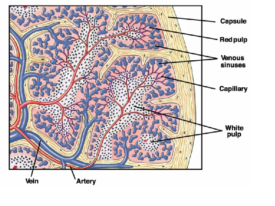
Spleen • Surrounded by a CT capsule w/ inward extending
trabeculae • Internal framework is provide by reticular fibers.

50
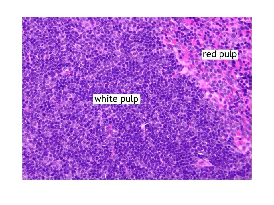
Spleen • Splenic arterioles are surrounded by sheathes
of lymphocytes. – This is known as the white pulp of the spleen. – What will happen here? • The arterioles terminate in splenic capillaries – which are twisty, sinusoidal, and incomplete. – The capillaries and the surrounding splenic tissue is referred to as the red pulp of the spleen. – Macrophages line the capillary surface. – Why?

53
Other Spleen Functions
• Storage of RBC breakdown products • Platelet storage • Fetal RBC production

54
Which would contain more damaged RBCs and more pathogens – the splenic artery or the splenic vein?

55
Thymus • Largest and most active • Involved in T
in fetus and infancy • Involved in T lymphocyte maturation and selection – Also, the removal of those T cells that attack self tissue • Growth ceases during adolescence • No direct fighting.

59
Tonsils • Form a ring of lymphatic tissue at the
entrance to the pharynx. • 3 main types: – Palatine – Pharyngeal – Lingual

61
Palatine – located laterally in the posterior oral cavity.
Largest and most often infected.

62
Pharyngeal – located in the
posterior nasopharynx. A.k.a adenoids.

63
Lingual – located at the
base of the tongue.

65
Not fully encapsulated. Why not?
Contain crypts. What’s their advantage? What’s their disadvantage?

66
Palatine Tonsil

Similar presentations