Download presentation
Presentation is loading. Please wait.

1
Skin and Body Membranes

2
Skin and Body Membranes
Basic Structure Thin sheetlike organs Composed of Epithelial Tissue and Connective Tissue

3
Skin and Body Membranes
Function of body membranes Line or cover body surfaces Protect body surfaces Lubricate body surfaces

4
Classification of Body Membranes
Epithelial membranes Cutaneous membrane Mucous membrane Serous membrane Connective tissue membranes Synovial membrane

5
Cutaneous Membrane Cutaneous membrane = skin A dry membrane
Outermost protective boundary Superficial epidermis Keratinized stratified squamous epithelium Underlying dermis Mostly dense connective tissue Figure 4.1a

6
Cutaneous Membrane Figure 4.1a

7
Mucous Membranes Surface epithelium Moist membranes
Type depends on site Underlying loose connective tissue (lamina propria) Lines all body cavities that open to the exterior body surface Often adapted for absorption or secretion Figure 4.1b
 Lines all body cavities that open to the exterior body surface. Often adapted for absorption or secretion. Figure 4.1b.")
8
Mucous Membranes Figure 4.1b

9
Serous Membranes Surface simple squamous epithelium
Underlying areolar connective tissue Lines body cavities that are closed to the exterior of the body Serous layers separated by serous fluid Figure 4.1c

10
Serous Membranes Figure 4.1c

11
Serous Membranes Specific serous membranes Peritoneum Pleura
Abdominal cavity Pleura Around the lungs Pericardium Around the heart Figure 4.1d

12
Body Cavities

13
Serous Membranes

14
Figure 01.11

15
Serous Membranes

16
Figure 01.12

17
Connective Tissue Membrane
Synovial membrane Connective tissue only Lines fibrous capsules surrounding joints Figure 4.2

18
Integumentary System Skin (cutaneous membrane)
Skin derivatives (accessory organs) Sweat glands Oil glands Hairs Nails
 Sweat glands. Oil glands. Hairs. Nails.")
19
Skin Functions Protects deeper tissues from: Mechanical damage
Bumps, cuts Chemical damage Acids, bases Bacterial damage Infections, disease Thermal damage Heat, cold Ultraviolet radiation Harmful sunlight

20
Skin Functions Desiccation Water loss Aids in heat regulation
Capillaries open to release, or close to hold in heat carried by blood Sweat glands activate to release heat Aids in excretion of urea and uric acid Perspiration Synthesizes vitamin D Sunlight converts cholesterol to vitamin D

21
Skin Structure Epidermis – outer layer Stratified squamous epithelium
Avascular Keratinized (“Cornified”-hardened by keratin) Dermis Dense connective tissue Figure 4.3
 Dermis. Dense connective tissue. Figure 4.3.")
22
Skin Structure Deep to dermis is the hypodermis (subcutaneous)
Not part of the skin Anchors skin to underlying organs Composed mostly of adipose tissue Shock absorber Insulator Loose Connective Tissue Major blood vessels

23
Layer of Epidermis Stratum basale Cells undergoing mitosis
Deepest cell layer Lies next to dermis Stratum spinosum Stratum granulosum

24
Layer of Epidermis Stratum lucidum
Occurs only in thick skin (hairless) Palms, soles Stratum corneum Shingle-like dead cells 20 – 30 cell layers thick Shed constantly New epidermis every days
 Palms, soles. Stratum corneum. Shingle-like dead cells. 20 – 30 cell layers thick. Shed constantly. New epidermis every days.")
25
Melanin Pigment (melanin) produced by melanocytes
Color is yellow to (reddish) brown to black Melanocytes are mostly in the stratum basale Amount of melanin produced depends upon genetics and exposure to sunlight Absorb UV radiation to protect DNA
 brown to black. Melanocytes are mostly in the stratum basale. Amount of melanin produced depends upon genetics and exposure to sunlight. Absorb UV radiation to protect DNA.")
26
Dermis Two layers Papillary layer (upper dermal region)
Projections called dermal papillae (fingerprints) Pain receptors Touch receptors (Meissner’s corpuscles) Capillary loops Reticular layer (deeper dermal region) Blood vessels Glands (sweat & oil) Pressure receptors (Pacinian corpuscles)
 Pain receptors. Touch receptors (Meissner’s corpuscles) Capillary loops. Reticular layer (deeper dermal region) Blood vessels. Glands (sweat & oil) Pressure receptors (Pacinian corpuscles)")
27
Skin Tone

28
Skin Structure Figure 4.4

29
Skin Structure Figure 4.4

30
Normal Skin Color Determinants
Melanin Yellow, brown or black pigments Carotene Orange-yellow pigment from some vegetables Hemoglobin Red coloring from blood cells in dermis capillaries Oxygen content determines the extent of red coloring Cyanosis – skin appears bluish due to low blood oxygen

31
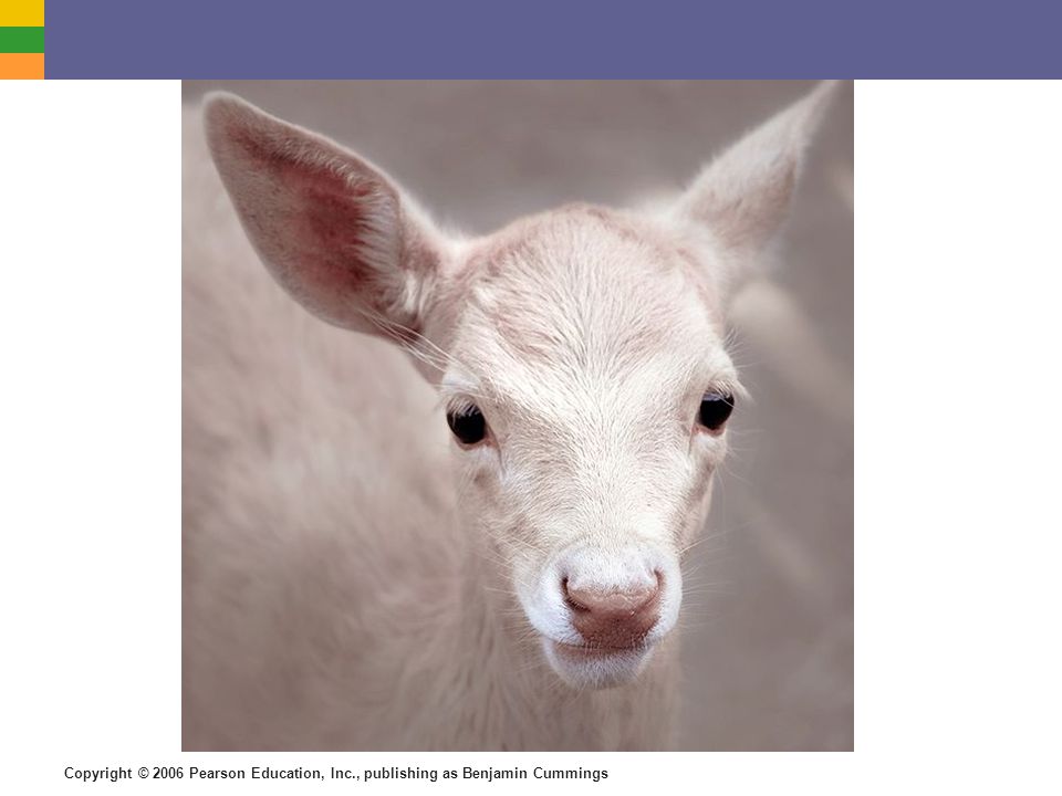
Albinism

32
The eyes of an albino animal appear red because the colour of the red blood cells in the underlying retinal blood vessels shows through where there is no pigment to obscure it

36
Appendages of the Skin Sebaceous glands Produce oil ( sebum )
Lubricant for skin Kills bacteria Most with ducts that empty into hair follicles Glands are activated at puberty Acne – infection of sebaceous gland

37
Figure 06.d

38
Appendages of the Skin Sweat glands Widely distributed in skin
Two types Eccrine Open via duct to pore on skin surface Respond to elevated body temperature Apocrine Ducts empty into hair follicles Respond to emotional stress

39
Sweat and its Function Eccrine
Water, salt, vitamin C, wastes, lactic acid Acidic – inhibits baterial growth Aids in homeostasis of body temperature May loose up to 7 Liters of water in sweat Common on forehead, neck, and back

40
Sweat and its Function Apocrine Fatty acids & proteins
Used as food by bacteria which then cause an odor Axillary and genital areas Function at puberty

41
Appendages of the Skin Hair Produced by hair follicle
Consists of hard keratinized epithelial cells Melanocytes provide pigment for hair color Root – in follicle Shaft – projects from surface Figure 4.7c

42
Hair Anatomy Central medulla Cortex surrounds medulla
Cuticle on outside of cortex Most heavily keratinized Figure 4.7b

43
Associated Hair Structures
Hair follicle Dermal and epidermal sheath surround hair root Arrector pilli Smooth muscle Sebaceous gland Sweat gland Dermal blood vessels nourish hair root Figure 4.7a

44
Associated Hair Structures
Figure 4.7a

45
Appendages of the Skin Nails Scale-like modifications of the epidermis
Heavily keratinized Stratum basale extends beneath the nail bed Responsible for growth Lack of pigment makes them colorless

46
Nail Structures Free edge Body
Lunula – white, half moon; growth occurs Root of nail Eponychium – proximal nail fold that projects onto the nail body Figure 4.9

47
Nail Structures Figure 4.9

48
Skin Homeostatic Imbalances
Infections Athletes foot Caused by fungal infection Boils and carbuncles Caused by bacterial infection Cold sores Caused by virus

49
Skin Homeostatic Imbalances
Infections and allergies Contact dermatitis Exposures cause allergic reaction Impetigo Caused by bacterial infection Psoriasis Cause is unknown Triggered by trauma, infection, stress

50
Skin Homeostatic Imbalances
Burns Tissue damage and cell death caused by heat, electricity, UV radiation, or chemicals Associated dangers Dehydration Electrolyte imbalance Circulatory shock

51
Severity of Burns First-degree burns Only epidermis is damaged
Skin is red and swollen Second degree burns Epidermis and upper dermis are damaged Skin is red with blisters Third-degree burns Destroys entire skin layer Burn is gray-white or black

52
Critical Burns Burns are considered critical if:
Over 25% of body has second degree burns Over 10% of the body has third degree burns There are third degree burns of the face, hands, or feet

53
Skin Cancer Cancer – abnormal cell mass Two types Benign Malignant
Does not spread (encapsulated) Malignant Metastasized (moves) to other parts of the body Skin cancer is the most common type of cancer
 Malignant. Metastasized (moves) to other parts of the body. Skin cancer is the most common type of cancer.")
54
Skin Cancer Types Basal cell carcinoma Least malignant
Most common type Arises from statum basale Squamous cell carcinoma Arises from stratum spinosum Metastasizes to lymph nodes Early removal allows a good chance of cure

55
Skin Cancer Types Malignant melanoma Most deadly of skin cancers
Cancer of melanocytes Metastasizes rapidly to lymph and blood vessels Detection uses ABCD rule

56
Figure 06.b

57
ABCD Rule A = Asymmetry Two sides of pigmented mole do not match
B = Border irregularity Borders of mole are not smooth C = Color Different colors in pigmented area D = Diameter Spot is larger than 6 mm in diameter

58
Figure 06.ba

59
Figure 06.bb

60
Figure 06.bc

Similar presentations


. + Skin Functions Protects deeper tissues from: Mechanical damage (bumps) Chemical damage (acids and bases) Bacterial damage.>")


