Download presentation
Presentation is loading. Please wait.

1
The female genitalia and reproductive system

2
Female reproductive anatomy

3
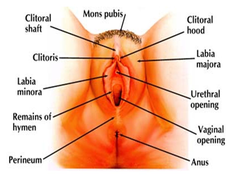
External Genitalia External organs include the sensitive and erotic sex organs: mons pubis, labia唇 and clitoris阴蒂 (also known collectively as the vulva外阴), and the breasts.
, and the breasts.")
5
The Mons Pubis The mons pubis (sometimes called the mons veneris after Venus, the Roman goddess of love) is the region of fatty tissue above the pubic bone. At puberty, the mons pubis enlarges and becomes covered by pubic hair.

6
The Labia Often referred to as "lips," the labia are the fold of tissue surrounding the vaginal and urethral openings, protecting the sensitive orifices from damage. The outer, fleshy hair-covered folds form the labia majora and the inner hairless folds form the labia minora. The labia are rich with nerve endings and generally respond well to stimulation.

7
The Clitoris 阴蒂 The female glans (the male glans is the penis), the clitoris is extremely sensitive and engorges with blood during sexual arousal. Protected by the clitoral hood, the clitoris is above the urethral opening and below the clitoral shaft, which has two arms (called crura) that stretch backwards into the woman's body, under the skin on either side above the vaginal opening. Nerves controlling clitoral muscle contractions travel alongside the walls of the vagina, the bladder and urethra, passing along the sensations produced in any part of the region.
 that stretch backwards into the woman s body, under the skin on either side above the vaginal opening. Nerves controlling clitoral muscle contractions travel alongside the walls of the vagina, the bladder and urethra, passing along the sensations produced in any part of the region..")
8
Internal Structures Internal organs include the uterus子宫, Fallopian tubes输卵管 and ovaries卵巢, which contain the female’s sex cells: the ova, or eggs.

10
The Ovaries The ovaries are paired structures, approximately the size of a walnut, that are anchored to the body cavity and to the uterus by strong cord-like structures known as ligaments. The ovaries contain the ova and produce the female sex hormones estrogen and progesterone孕酮.

11
The Fallopian Tubes The Fallopian tubes, also called the uterine tubes or oviducts, extend from the uterus up around the ovaries. The Fallopian tubes display muscular contractions that help draw the ovum into the uterus. The upper portion of the tubes is where fertilization of an ovum by a sperm commonly occurs; if the zygote does not move down into the uterus, an ectopic pregnancy occurs, which can be fatal to the mother and must be terminated. The Fallopian tubes are surgically altered when a woman undergoes the sterilization procedure known as a tubal ligation.

12
The Uterus The uterus (or womb) is a pear-shaped multi-layered organ located at the base of the pelvic cavity. The opening of the uterus, known as the cervix子宫颈, contains a narrow opening that serves as a passageway for menstrual flow or for a fetus to exit the vagina. The innermost layer of the uterus is the endometrium, which is partially shed with each the menstrual flow. The uterus can be host to a number of sexual health problems, including endometriosis子宫内膜异位, cervical cancer and uterine cancer.

13
The Vagina The vagina is a three to four-inch long, thin-walled mucous-lined tube. In addition to providing an exit for menstrual blood or a fetus, the vagina is the reception site for the penis during intercourse.

14
Important Areas of Examination
External Examination Internal Examination ● Mons pubis ● Vagina, vaginal walls ● Labia majora and minora ● Cervix ● Urethral meatus, clitoris ● Uterus, ovaries ● Vaginal introitus ● Pelvic muscles ● Perineum ● Rectovaginal wall

15
Approach to the Pelvic Examination.
Many students feel anxious or uncomfortable when they begin doing pelvic examinations. At the same time, female patients have their own concerns. Some women have had painful, embarrassing, or even demeaning experiences during previous examinations, whereas others may be facing a pelvic examination for the first time. Some are fearful about what the clinician may find and how findings may affect their lives. Asking the patient’s permission to perform the examination shows courtesy and respect. If a Pap smear is to be collected using the glass-slide technique, time the examination so that it does not occur during menses, because blood can interfere with interpretation.

16
A woman having her first pelvic examination may not know what to expect.
Using three-dimensional models, showing her the equipment and letting her handle the speculum, and explaining each step in advance can help her learn about her body and be more comfortable. Careful and gentle technique is especially important in minimizing any pain or discomfort during the first pelvic examination.

17
The woman’s response to the pelvic examination may reveal clues about her feelings about the examination and her sexuality. If she pulls away, adducts her thighs, or reacts negatively to the examination, you can gently comment “I notice you are having some trouble relaxing. Is it just being here, or are you troubled by the examination? Is anything worrying you?” Behaviors that seem to present an obstacle may lead you to a better understanding of your patient’s concerns. Adverse reactions may signal prior abuse and should be explored.

18
TIPS FOR THE SUCCESSFUL PELVIC EXAMINATION

19
Helping the patient to relax is essential for an adequate examination
Helping the patient to relax is essential for an adequate examination. Adopting the tips above will help ensure the patient’s comfort. Be sure always to wear gloves, both during the examination and when handling equipment and specimens. Plan ahead, so that any needed equipment and culture media are readily at hand.

20
Note that male examiners should be accompanied by female chaperones.
Female examiners should also be assisted if the patient is physically or emotionally disturbed, and to facilitate the examination.

21
Rape Victims. Regardless of age, rape merits special evaluation, usually requiring gynecologic consultation and documentation. Often there is a special rape kit, provided in many emergency departments, that must be used to ensure a chain of custody for evidence. Specimens must be labeled carefully with name, date, and time. Additional information may be needed for further legal investigation.

22
Choosing Equipment. You should have within reach a good light, a vaginal speculum of appropriate size, water-soluble lubricant, and equipment for taking Papanicolaou smears, bacteriologic cultures and DNA probes, or other diagnostic tests, such as potassium hydroxide or normal saline. Review the supplies and procedures of your own facility before taking cultures and other samples.

24
Specula are made of metal or plastic and come in two basic shapes, named for Pedersen and Graves. Both are available in small, medium, and large sizes. The medium Pedersen speculum is usually most comfortable for sexually active women. The narrow-bladed Pedersen speculum is best for the patient with a relatively small introitus,such as a virgin or an elderly woman. The Graves specula are best suited for parous women with vaginal prolapse.

25
Before using a speculum, become thoroughly familiar with how to open and close its blades, lock the blades in an open position, and release them again. Although the instructions in this chapter refer to a metal speculum, you can easily adapt them to a plastic one by handling the speculum before using it.

26
Positioning the Patient
Positioning the Patient. Drape the patient appropriately and then assist her into the lithotomy position. Help her to place first one heel and then the other into the stirrups. She may be more comfortable with shoes on than with bare feet. Then ask her to slide all the way down the examining table until her buttocks extend slightly beyond the edge. Her thighs should be flexed, abducted, and externally rotated at the hips. A pillow should support her head.

27
External Examination Assess the Sexual Maturity of an Adolescent Patient. You can assess pubic hair during either the abdominal or the pelvic examination. Note its character and distribution.

28
Examine the External Genitalia.
Seat yourself comfortably and warn the patient that you will be touching her genital area. Inspect the mons pubis, labia, and perineum. Separate the labia and inspect: ● The labia minora ● The clitoris ● The urethral meatus ● The vaginal opening, or introitus

29
Note any inflammation, ulceration,
discharge, swelling, or nodules. If there are any lesions, palpate them.

30
If any is present, culture it.
If there is a history or an appearance of labial swelling, check Bartholin’s glands. Insert your index finger into the vagina near the posterior end of the introitus. Place your thumb outside the posterior part of the labium majus. On each side in turn, palpate between your finger and thumb for swelling or tenderness. Note any discharge exuding from the duct opening of the gland. If any is present, culture it. PALPATING BARTHOLIN’S GLAND

31
Internal Examination Assess the Support of the Vaginal Walls. With the labia separated by your middle and index fingers, ask the patient to bear down. Note any bulging of the vaginal walls.

32
Insert the Speculum. Select a speculum of appropriate size and shape, and
moisten it with warm, but not hot, water.

33
You can enlarge the vaginal introitus by lubricating one finger with water and applying downward pressure at its lower margin. Check the location of the cervix to help angle the speculum more accurately. Enlarging the introitus greatly eases insertion of the speculum and the patient’s comfort. With your other hand (usually the left),introduce the closed speculum past your fingers at a somewhat downward slope. Be careful not to pull on the pubic hair or pinch the labia with the speculum. Separating the labia majora with your other hand can help to avoid this.

34
Two methods help you to avoid placing pressure on the sensitive urethra.
(1) When inserting the speculum, hold it at an angle (shown below on the left), and then (2) slide the speculum inward along the posterior wall of the vagina, applying downward pressure to keep the vaginal introitus relaxed.
 When inserting the speculum, hold it at an angle (shown below on the left), and then (2) slide the speculum inward along the posterior wall of the vagina, applying downward pressure to keep the vaginal introitus relaxed.")
35
After the speculum has entered the vagina, remove your fingers from the introitus. You may wish to switch the speculum to the right hand to enhance maneuverability of the speculum and subsequent collection of specimens. Rotate the speculum into a horizontal position, maintaining the pressure posteriorly, and insert it to its full length. Be careful not to open the blades of the speculum prematurely.

36
Inspect the Cervix. Open the speculum carefully
Inspect the Cervix. Open the speculum carefully. Rotate and adjust the speculum until it cups the cervix and brings it into full view. Position the light until you can visualize the cervix well. When the uterus is retroverted, the cervix points more anteriorly than illustrated. If you have difficulty finding the cervix, withdraw the speculum slightly and reposition it on a different slope. If discharge obscures your view, wipe it away gently with a large cotton swab.

37
Note the color of the cervix, its position, the characteristics of its surface,
and any ulcerations, nodules, masses, bleeding, or discharge. Inspect the cervical os for discharge.

38
Maintain the open position of the speculum by tightening the thumb screw.

39
Obtain Specimens for Cervical Cytology (Papanicolaou Smears).
Obtain one specimen from the endocervix and another from the ectocervix,or a combination specimen using the cervical brush (“broom”). For best results the patient should not be menstruating. She should avoid intercourse and use of douches, tampons, contraceptive foams or creams,or vaginal suppositories for 48 hours before the examination. In addition to obtaining the Pap smear, for sexually active women age 25 or younger, and for other asymptomatic women at increased risk for infection, plan to culture the cervix routinely for Chlamydia trachomatis.
. For best results the patient should not be menstruating. She should avoid intercourse and use of douches, tampons, contraceptive foams or creams,or vaginal suppositories for 48 hours before the examination. In addition to obtaining the Pap smear, for sexually active women age 25 or younger, and for other asymptomatic women at increased risk for infection, plan to culture the cervix routinely for Chlamydia trachomatis.")
40
OBTAINING THE PAP SMEAR: OPTIONS FOR SPECIMEN COLLECTION
Cervical Scrape and Endocervical Brush Cervical Scrape. Place the longer end of the scraper in the cervical os. Press, turn, and scrape in a full circle, making sure to include the transformation zone and the squamocolumnar junction. Smear the specimen on a glass slide. Set the slide in a safe spot that is easy to reach. Note that doing the cervical scrape first reduces obscuring cells with blood, which sometimes appears after use of the endocervical brush.

41
Endocervical Brush. Take the endocervical
brush and place it in the cervical os. Roll it between your thumb and index finger, clockwise and counterclockwise. Remove the brush and pick up the slide you have set aside. Smear the slide with the brush, using a gentle painting motion to avoid destroying any cells. Place the slide into an ether–alcohol solution at once, or spray it promptly with a special fixative.

42
Cervical Broom Many clinicians use a plastic brush tipped with a broomlike fringe for collection of a single specimen containing both squamous and columnar epithelial cells. Rotate the tip of the brush in the cervical os, in a full clockwise direction, then stroke each side of the brush on the glass slide. Promptly place the slide in solution or spray with a fixative as described above.

43
Perform a Bimanual Examination.
Lubricate the index and middle fingers of one of your gloved hands, and from a standing position, insert them into the vagina, again exerting pressure primarily posteriorly. Your thumb should be abducted, your ring and little fingers flexed into your palm. Pressing inward on the perineum with your flexed fingers causes little if any discomfort and allows you to position your palpating fingers correctly. Note any nodularity or tenderness in the vaginal wall, including the region of the urethra and the bladder anteriorly.

44
Palpate the cervix, noting its position, shape, consistency, regularity, mobility,and tenderness. Normally the cervix can be moved somewhat without pain. Feel the fornices around the cervix. Palpate the uterus. Place your other hand on the abdomen about midway between the umbilicus and the symphysis pubis. While you elevate the cervix and uterus with your pelvic hand, press your abdominal hand in and down, trying to grasp the uterus between your two hands. Note its size, shape, consistency, and mobility, and identify any tenderness or masses.

45
Now slide the fingers of your pelvic hand into the anterior fornix and palpate the body of the uterus between your hands. In this position your pelvic fingers can feel the anterior surface of the uterus, and your abdominal hand can feel part of the posterior surface.If you cannot feel the uterus with either of these maneuvers, it may be tipped posteriorly (retrodisplaced). Slide your pelvic fingers into the posterior fornix and feel for the uterus butting against your fingertips. An obese or poorly relaxed abdominal wall may also prevent you from feeling the uterus even when it is located anteriorly.
. Slide your pelvic fingers into the posterior fornix and feel for the uterus butting against your fingertips. An obese or poorly relaxed abdominal wall may also prevent you from feeling the uterus. even when it is located anteriorly.")
46
Palpate each ovary. Place your abdominal hand on the right lower quadrant,your pelvic hand in the right lateral fornix. Press your abdominal hand in and down, trying to push the adnexal structures toward your pelvic hand.Try to identify the right ovary or any adjacent adnexal masses. By moving your hands slightly, slide the adnexal structures between your fingers, if possible,and note their size, shape, consistency, mobility, and tenderness. Repeat the procedure on the left side.Normal ovaries are somewhat tender. They are usually palpable in slender,relaxed women but are difficult or impossible to feel in others who are obese or poorly relaxed.

47
Do a Rectovaginal Examination.
The rectovaginal examination has three primary purposes: to palpate a retroverted uterus, the uterosacral ligaments, cul-de-sac, and adnexa; to screen for colorectal cancer in women 50 years or older; and to assess pelvic pathology.

48
Do a Rectovaginal Examination.
After withdrawing your fingers from the bimanual examination, change your gloves and lubricate your fingers as needed (see note below on lubricants). Slowly reintroduce your index finger into the vagina and your middle finger into the rectum. Ask the patient to strain down as you do this to relax her anal sphincter. Mention that this may stimulate an urge to move her bowels, but this will not occur. Apply pressure against the anterior and lateral walls with the examining fingers, and downward pressure with the hand on the abdomen.
. Slowly reintroduce your. index finger into the vagina and your. middle finger into the rectum. Ask. the patient to strain down as you. do this to relax her anal sphincter. Mention that this may stimulate an. urge to move her bowels, but this. will not occur. Apply pressure against. the anterior and lateral walls with. the examining fingers, and downward. pressure with the hand on the abdomen.")
49
Note that initially you may use sentences to describe your findings; later you will use phrases. The style below contains phrases appropriate for most write-ups.

54
Thanks

Similar presentations