Download presentation
Presentation is loading. Please wait.

1
Psychoacoustics Riana Walsh riana.walsh@ul.ie

2
Relevant texts Acoustics and Psychoacoustics, D. M. Howard and J. Angus, 2 nd edition, Focal Press 2001 An Introduction to the Psychology of Hearing, B.C.J. Moore, 5 th edition, Academic Press, Elsevier 2004 Fundamentals of Hearing, An Introduction, W. A. Yost, 4 th edition, Academic Press 2000 Listening An introduction to the Perception of auditory events, S. Handel, MIT Press 1989

3
Course outline Structure and function of the auditory system; frequency selectivity of the auditory system; the perception of pitch, loudness and timbre; temporal perception; sound localisation; identification of auditory objects; streaming; organisation of auditory memory; pitch organisation; memory, attention, melody and rhythm.

4
Hearing Psychoacoustics – the study of hearing - relationship between the physical properties of sound and the sensations they produce. Hearing – the process that transforms sound waves into neural signals that can be interpreted by our brain Sound waves – fluctuations in air pressure across time, created by the motion or vibration of an object (e.g. the vibration of vocal chords, oscillating violin string) - physical properties: frequency and amplitude.
 - physical properties: frequency and amplitude..")
5
The peripheral auditory system The peripheral auditory system consists of the outer, middle and inner ear. In brief: The ear drum moves in and out in response to the pressure changes in sound waves – transmitted through the middle to the inner ear – transduced into neural sinals that are interpreted by the brain

7
The path of sound waves through the outer, middle and inner ear Sound waves travel down the auditory canal and cause the ear drum to vibrate. The main function of the ossicles is the efficient transfer of sound waves from air to the fluids of the cochlea. The ossicles of the middle ear vibrate in response to tympanic membrane vibration. They amplify and transmit these vibrations to the oval window. Amplification is necessary as more energy is required to move the fluids (of the inner ear) than air (in middle ear).
 than air (in middle ear)..")
8
The middle ear Achieved: difference in the effective areas of the ear drum and oval window; lever action of the ossicular chain Difference in the area of the eardrum and oval window [pressure = force/area] Middle ear (also acoustic) reflex – muscles attached to the ossicles contract upon exposure to intense sounds (>~80dB SPL) Contraction of these muscles reduces the transmission of pressure through the ossicular chain – may prevent inner ear damage
![The middle ear Achieved: difference in the effective areas of the ear drum and oval window; lever action of the ossicular chain Difference in the area of the eardrum and oval window [pressure = force/area] Middle ear (also acoustic) reflex – muscles attached to the ossicles contract upon exposure to intense sounds (>~80dB SPL) Contraction of these muscles reduces the transmission of pressure through the ossicular chain – may prevent inner ear damage](http://images.slideplayer.com/13/3808216/slides/slide_8.jpg "The middle ear Achieved: difference in the effective areas of the ear drum and oval window; lever action of the ossicular chain Difference in the area of the eardrum and oval window [pressure = force/area] Middle ear (also acoustic) reflex – muscles attached to the ossicles contract upon exposure to intense sounds (>~80dB SPL) Contraction of these muscles reduces the transmission of pressure through the ossicular chain – may prevent inner ear damage")
9
Frequency dependent – most effective < 2 kHz Minimum time for reflex 10-150ms (depends on intensity) – so reflex not effective for sounds with a sudden onset e.g. gunshots This reflex may also function is the reduction of the audibility of self-generated sounds, such as speech. It has been shown to be activated just before vocalisation.

10
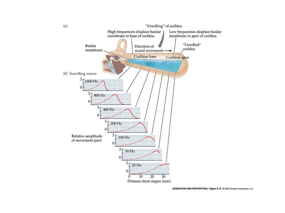
The structure of the inner ear The part of inner ear concerned with hearing is the fluid filled cochlea. Reissner’s membrane and the basilar membrane (BM) divide the cochlea along its length. The start of the cochlea (near oval window) is the base (basal end), the other end of the cochlea is the apex (apical end of the cochlea) Motion of the basilar membrane in response to sound
 divide the cochlea along its length. The start of the cochlea (near oval window) is the base (basal end), the other end of the cochlea is the apex (apical end of the cochlea) Motion of the basilar membrane in response to sound.")
13
The basilar membrane response to sound Movement of the stapes sets the oval window in motion – causes the BM to move. Response of BM to sinusoidal stimulation – travelling wave, which moves from base to apex. The position of the peak in the vibration pattern on the BM depends on the frequency of the sound – this is due to the mechanical properties of the BM High (low) frequencies produce max. BM displacement near the base (apex) – frequency analysis – each point on the BM is sharply tuned
 frequencies produce max. BM displacement near the base (apex) – frequency analysis – each point on the BM is sharply tuned.")
15
BM response to sound Each point on the BM is sharply tuned, responding with high sensitivity to a limited range of frequencies. BM vibration is nonlinear – the magnitude of its response does not grow directly in proportion with the magnitude of the input Linear for low input sound levels ( 90dB SPL)
.")
16
BM response to sound Compressive nonlinearity at midrange levels – a large range of input sound levels is compressed into a smaller range of BM responses Nonlinearity occurs at the base of the BM when the stimulating frequency is close to the BM point being monitored – compression only around the peak of the BM response pattern The nonlinearity and sharp tuning of BM are physiologically vulnerable

17
BM response to sound Compression at the apical end is less than at the basal end – at the apical end compression does not seem to depend on the frequency of the input relative to that of the place (CF) being monitored Frequency-to-place conversion – the distance from the apex to the point of displacement is proportional to the logarithm of the input frequency. For input sounds with more than one frequency the BM vibration pattern depends on the frequency separation of the components

18
Aside Our central nervous system consists of the brain and spinal cord Neurons are the building blocks of our central nervous system Many different types of neuron (e.g. sensory neuron, interneuron, motor neuron) Components of a typical biological neuron
 Components of a typical biological neuron.")
20
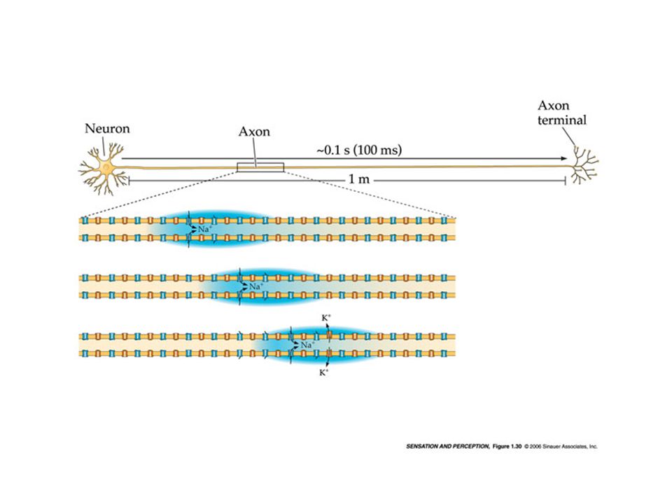
Structure of the neuron Three main sections: dendrites, cell body, and the axon. The function of the dendrites is to receive signals from other neurons at connection points called synapses. The function of the axon is to transmit signals out of the cell body The dendrite is separated from the transmitting axon by a narrow gap called a synapse

21
Structure of the neuron Most neurons have several dendrites to receive stimulation and only one axon to transmit nerve impulses The axon releases chemicals, called neurotransmitters, into the synapse, and these diffuse across to the receiving dendrite and enter the cell body The neurotransmitter may be excitatory or inhibitory - it may excite or inhibit the receiving neuron from firing.

23
The signals received are combined by the cell body If the signal is above a certain threshold, the cell ‘fires’ producing a pulse that propagates down the axon and is passed on to other neurons Towards the end of the axon are multiple branches (axon terminals) each terminating in a synapse In this way a single neuron can excite or inhibit many other neurons
 each terminating in a synapse In this way a single neuron can excite or inhibit many other neurons")
Similar presentations


 Transduction of physical sound waves into brain activity via the ear. Sound is perceptual and subjective. >")


