Download presentation
Presentation is loading. Please wait.

1
Radiographic Critique off the Shoulder
Chapter 4

2
Shoulder AP Contrast Density Controlled by kVp (optimal 70-80)
Adequate to demonstrate bony trabecular patterns and cortical outlines Density Overall not too dark or too light Use grid to absorb scatter Could use compensating filter to better visualize the clavicles and glenoid humarl articulation

3
Shoulder AP Bony trabecular patterns and cortical outlines are sharply defined kVp is correct Respiration and motion are halted Using a small focal spot Short OID

4
Shoulder AP with No Rotation
The glenoid fossa is visualized The superiolateral border of the scapula is not over the thorax The clavicle is horizontal The medial end of clavicle articulates with the vertebral column The glenoid fossa and the medial margin of the humeral head are slightly superimposed

5
Shoulder AP Detecting Rotation Away from the affected shoulder
Clavicles will superimpose the vertebral bodies Increased visualization of glenoid fossa Toward affected shoulder Clavicles will not superimpose the vertebra Scapula will superimpose the thorax

6
Shoulder AP Determining rotation from dislocation
If all factors remain unrotated with the exception of the humeral head Posterior dislocation Humeral head is demonstrated beneath the acromion or spine of the scapula Anterior dislocation Humeral head is demonstrated anteriorly, b beneath the coracoid

7
Shoulder AP Is shoulder in center of the film?
The glenohumeral joint, and coracoid are at the center of the collimated field Should also include the lateral 2/3 of the clavicle, the proximal 1/3 of the humerus and the superior scapula

8
Shoulder AP Determining rotation of the humeral head Neutral rotation
Greater tubercle is partially in profile laterally and humeral head is partially profile medially External rotation Greater tubercle is profile laterally, the humeral head is in profile medially Internal rotation The lesser tubercle is demonstrated in profile medially and the humeral head is superimposed by the greater tubercle

13
Shoulder Y view No Rotation
The medial and lateral borders are superimposed The scapular body does not superimpose the thoracic cavity The scapular body, acromion, and coracoid form a Y

14
Shoulder Y view To determine Rotational Direction
If the lateral border (thick border) is demonstrated next to the ribs the patient was rotated too far toward the film (increase obliquity) pg 183 If the vertebral border is demonstrated over the thorax then the patient is rotated to far away from the film (decrease obliquity) pg 183
 is demonstrated next to the ribs the patient was rotated too far toward the film (increase obliquity) pg 183. If the vertebral border is demonstrated over the thorax then the patient is rotated to far away from the film (decrease obliquity) pg 183.")
15
Shoulder Y view Detecting shoulder dislocation
When the humeral head is not located over the glenoid fossa Posterior dislocation The humeral head is located beneath the acromion (pg 182) Anterior dislocation Shoulder head will be beneath the coracoid (pg 184)
 Anterior dislocation. Shoulder head will be beneath the coracoid (pg 184)")
16
Shoulder Y view Anatomical parts to be included on film
Midscapular body in the center of the film Entire scapula Coracoid Acromion Proximal humerus

18
Clavicle AP True AP projection
The medial end of the clavicle lies next to the lateral edge of the vertebral column, and the thoracic cavity superimposes the vertebral scapular border

19
Clavicle AP Positioning for Kyphosis
Easier to position the patient in the erect position If erect position is not attainable, place shoulders and thorax in the same plane

20
Clavicle AP Detecting rotation
Medial end of the clavicle superimposes the vertebral column the patient is rotated away from affected side Medial end of the clavicle does not superimpose the vertebral column, the patient is rotated toward the affected side. (Pg 186 RAD 24)
")
21
Clavicle AP Anatomy to be included on the film Entire clavicle
Acromion

24
Clavicle AP Axial Bony trabecular patterns and cortical outlines are sharply defined kVp is correct Respiration and motion are halted Using a small focal spot Short OID

25
Clavicle AP Axial Detecting Rotation Determined identical to AP view

26
Clavicle AP Axial Central Ray angulation 15 – 30 degrees cephalically
Clavicle should appear slightly superior to the acromion The medial end should superimpose the 1st and 2nd rib Compare RAD 26 and 27 on page 188 and 189

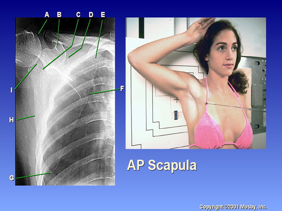
31
Scapula AP Bony trabecular patterns and cortical outlines are sharply defined kVp is correct Respiration and motion are halted Using a small focal spot Short OID

36
THE END

Similar presentations


 Joint>")
 JOINT>")

