Download presentation
Presentation is loading. Please wait.

1
The Biological Crime Scene "Every Contact Leaves a Trace". Dr. Edmond Locard Blood at the Scene is the most visible example of the Locard Exchange Principle

2
Solving Forensic Problems Steps to problem solving Understand the Problem The parameters The issues Knowledge Key to any successful analysis Understand the science Know the technology

3
The Mission: Never Miss Anything The Tools The Evidence Cascade Your Brain Experience The Underlying Science Understanding the Technology Logical & Critical Thinking

4
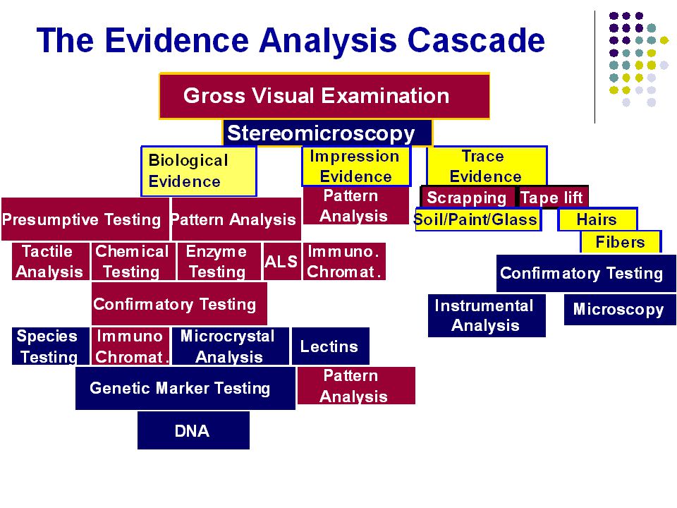
Pattern Analysis The Evidence Analysis Cascade Trace Evidence Presumptive Testing ScrappingTape lift Gross Visual Examination Tactile Analysis Stereomicroscopy Genetic Marker Testing Immuno Chromat. Hairs Fibers Soil/Paint/Glass Pattern Analysis Impression Evidence Biological Evidence Chemical Testing Enzyme Testing Confirmatory Testing Species Testing Microcrystal Analysis ALS Blood, Semen, Saliva Lectins DNA Confirmatory Testing Microscopy Instrumental Analysis Immuno. Chromat. Pattern Analysis

6
Forensically Important Biological Substances What are the they? Blood Semen Saliva Urine Feces Vomit Fingerprint residue Sloughed off cells What else? Bacteria Plant material Pollen Viruses

7
Locating Biological Evidence Your Eye Touch Hi-Intensity & Alternate Light Sources Chemical Tests Enzymatic Tests Immunological Tests

8
Blood

9
CellsLiquid Red Blood Cells White Blood Cells Plasma Serum

10
Plasma Cells Blood Complex Connective Tissue Salts Blood Group Antigens Drugs Hormones Antibodies DNA HLA Antigens Blood Group Substances White CellsRed Cells Enzymes Isoenzymes Genetic Markers Individual Specific Antibodies Forensically Speaking: What is Blood?

11
Genetic Markers Drugs of Abuse Prescriptions Identification Psychological Behavior Disease Susceptibility Individual Identity AncestrySexing Racial Identity Forensically Critical Information from Blood DNA Profiling Antibody Profiling

12
The Scientific Faces of Forensic Biology Chemistry Immunology/Chemistry Serology/Biochemistry Molecular Biology Population Genetics

13
The Investigators Job Find Those Stains! Presumptive Testing Tools Tactile/Visual Analysis Biological Evidence Chemical Testing Enzyme Testing ALS Blood, Semen, Saliva

14
Using Alternate Light Sources To Find Biological Evidence

15
The Electromagnetic Spectrum Using Light to Find Biological Evidence 190-290290-400 Ultraviolet Region Short wave Long wave Visible Region 400-455455-492 >700 492-577577-597597-622622-700 Infrared Region VioletBlueGreenYellowOrangeRed IR

16
ALS Wavelengths Applications to Finding Biological Evidence Bone 455/CSS/515OrangeOrange Teeth Fingernails Body FluidsCSSOrange 1-2 Orange Dk Surfaces UV Clear/Yellow None “ w/crust White/obliqueClear None Evidence Type MiniScope 400 settingsGoggle Camera Filter Hair untreated Blk White/oblique ClearNone treated-red/bld 415/CSS Yellow/Orange Yellow/Orange Blood 415, 455 Clear/Yellow None

17
Choosing a Goggle Color Color RangeALS Setting (nm)Goggle Long wave UV 300-400Clear Violet515-445Yellow Blue/green455-515Orange Green-red536Red - - - - - -CSSOrange
Goggle Long wave UV Clear Violet Yellow Blue/green Orange Green-red536Red CSSOrange")
18
Detecting Blood @ the Scene

19
Historical Overview Classification 1862 Chemistry

20
Chemical Testing Catalytic Tests

21
Blood – Presumptive tests Based on the peroxidase properties of hemoglobin globin heme

22
Blood – Presumptive tests heme porphyrin iron

23
Catalytic Tests: Presumptive Testing for Blood Van Deen’s or Day’s Test 1862 Kastle Meyer 1901 Benzidine 1904 Tetramethylbenzidine 1976

24
Common Presumptive Tests phenolphthalin (Kastle-Meyer) leucomalachite green (LMG) Luminol (BlueStar) 3,3’,5,5’-Tetramethylbenzidine (TMB) leucocrystal violet (LCV) o-tolidine Benzidine: Carcinogenic o-toluidine hydrogen peroxide: Bubbles
 leucomalachite green (LMG) Luminol (BlueStar) 3,3’,5,5’-Tetramethylbenzidine (TMB) leucocrystal violet (LCV) o-tolidine Benzidine: Carcinogenic o-toluidine hydrogen peroxide: Bubbles")
25
Presumptive Testing Practical Variations One step All reagents added together Considered to be most sensitive Doesn’t allow for identifying false positives Two Step Reagent added to the stain Peroxide added last Three Step Alcohol added first Reagent second Peroxide last

26
Kastle-Meyer reagent phenolphthalin Organic molecule that becomes an indicator potassium hydroxide makes solution basic Ethanol/water used to dissolve stain zinc dust Used to reduce Phenolphthalein to phenolphthalin Continued presence in final solution slows spontaneous oxidation of phenolphthalein

27
Kastle-Meyer Test How run the Test Dissolve the stain in water/ethanol KM reagent added to stain color change at this point: false positive Add 3% H 2 O 2 Pink: KM positive

28
Blood – Presumptive tests General Considerations heme has peroxidase activity 2H 2 O 2 2O· + 2H 2 O Heme Fe+++ Oxygen free radicals cleaved from peroxide group Heme Fe++

29
Blood – Presumptive tests General Considerations Free radicals interact with organic chemicals (dyes) O· chemical oxidized Presumptive test detects oxidized organic dyes + Chemical reduced
 O· chemical oxidized Presumptive test detects oxidized organic dyes + Chemical reduced")
30
Kastle-Meyer Test Rare Some substances inhibit reaction Blood can mask the color change Acidic solutions can mask the reaction false negatives

31
BlueStar TM Luminol 3-aminophthalate +N 2 +light

32
Using IR to Search for Blood Camera Viewfinder

33
Semen

34
Plasma Cells Semen Complex Connective Tissue Salts Blood Group Antigens Drugs Hormones Antibodies DNA HLA Antigens Blood Group Substances White Cells Sperm Cells Enzymes Isoenzymes Genetic Markers Individual Specific Antibodies Pre-Sperm Cells Genetic Markers Isoenzymes Forensically Speaking: What is Semen?

35
Contributing glandPercentage of ejaculate Testicles and epididymus5 per cent Seminal vesicles46 to 80 per cent Prostate gland13 to 33 per cent Bulbourethral and urethral glands2 to 5 per cent Semen Origins

36
Identifying Semen Test Type Specificity Microscopic Tests Spermatozoa ………………19 th Century…….............. Specific Visual Tests Crusty stains …………………………………………….Nonspecific Color of stain …………………………………………….Nonspecific UV Light Phosphorescence ……1950’s, 1970’s……..Nonspecific Alternate Light Sources …………1980’s……………...Nonspecific Chemical / Crystals Tests Florence Test …………19 th Century..…………………Nonspecific Barberio Test …………19 th Century..…………………Nonspecific Fructose/Zn …………..1950’s………………………..Nonspecific Enzyme/Protein Tests Acid Phosphatase ………………1950’s……………….Nonspecific Lactic Dehydrogenase-X ……… 1980’s……………….Specific for sperm Sperm/seminal Esterases ………1980’s………………Nonspecific Gamma seminoprotein ……………1980’s…………….Specific for seminal plasma Immunological Tests Prostate Specific Antigen (p30)......1980’s..............Specific for seminal plasma ABACard p30 Test ………………….1990’s………….Specific for seminal plasma
 ’s Specific for seminal plasma ABACard p30 Test ………………….1990’s………….Specific for seminal plasma.")
37
Identifying Semen Historical Perspectives Medico legal identification of semen in sexual assault cases Early part of 19 th century First systematic efforts used chemical tests 1826 Ollivier, D’angers & Barruel Stain ext’d w/water & ETOH Stain extract in ETOH had “Spermatic” odor Suspect had claimed stain was fat from uncooked animal meat.

38
Identifying Semen Historical Perspectives 1827 Orphilia – Case of 13 year old girl Testified 1. as defense expert after a physician said that he’d isolated semen from vagina after 9 days 2. No chemical tests available to ID semen 3. Girl had mucous discharge Designed tests Based on appearance of the stains Changes in color & consistency on heating, immersion in water, odor emitted by moistened stain & behavior of aqueous extract toward Compared with vaginal discharges, nasal mucus & saliva stains Found sperm in fresh seminal samples & in 18 year old dried semen (van Leeuwenhoek who credited medical student Ham with the discovery in 1677)
.")
39
Identifying Semen Historical Perspectives 1837 Rattier Published paper on ID of sperm in semen stains Suggested the procedure for medico-legal proceedings 1839 Devergie Identified sperm in 10 month old semen stains Suggested that sperm ID was a more certain criteria for identifying semen than chemical tests Orphilia disagreed Bayard Published extensive paper on the use of the microscope in examining semen stains for sperm Procedures became widely accepted & used from this point. Early chemical methods slowly abandoned.

40
Identifying Semen Historical Perspectives 1858 Lassaigne Series of reagents to use with semen stains 1896 Crystal tests First non-morphological test for semen that persisted Still used in very few laboratories

41
Identifying Semen Historical Perspectives Crystal tests Florence Test 1896 1.65g KI, 2.54g I 2 in 30mL water Reagent allowed to diffuse under cover slip on microscope slide Florence considered it a useful presumptive test Characteristic crystals not found with: nasal, vaginal mucus, urine, sweat, saliva, tears, milk, cerebral fluid or leucorrheal discharge Seminal component called virispermine 1897 Richter Positive crystals from vaginal & uterine mucus from dead bodies Found that lecithin gave the test & postulated that it was choline in semen that gave the crystals Whitney 2 ½ year old semen stains Positive test with: morphnine, strychnine & other alkaloids Thought it was the choline 1902 Bocarius established that it is choline in semen that gives the test

42
Identifying Semen Historical Perspectives Crystal tests Florence Test 1937 Kahane & Levy Conducted biochemical studies & tissue distribution of choline 11.2-14.4 mg choline/100 mL semen Any tissue with this concentration will give the Florence test

43
Identifying Semen Historical Perspectives Crystal tests Barberio Test 1905 Microscope slide test Saturated picric acid (water or abs ETOH): refractive yellow crystals Positive reaction with semen, semen stains, putrefied seminal material & semen heated to 150 deg C 1906 Cevidalli Proposed test carried out in glycerin w/picric in ETOH Neg rxn with dog, horse, pig semen 1907 Lecha-Marzo Regarded the test more specific than the Florence Test 1913 Baecchi Suggested the crystals were spermine picrate 1924 Rosenheim Confirmed crystals as spermine picrate Noticed that spermine phosphate crystals form spontaneously if semen left to stand – first described by van Leeuwenhoek in 1678
: refractive yellow crystals Positive reaction with semen, semen stains, putrefied seminal material & semen heated to 150 deg C 1906 Cevidalli Proposed test carried out in glycerin w/picric in ETOH Neg rxn with dog, horse, pig semen 1907 Lecha-Marzo Regarded the test more specific than the Florence Test 1913 Baecchi Suggested the crystals were spermine picrate 1924 Rosenheim Confirmed crystals as spermine picrate Noticed that spermine phosphate crystals form spontaneously if semen left to stand – first described by van Leeuwenhoek in 1678")
44
Identifying Semen Historical Perspectives Crystal tests Barberio Test 1932 Harrison – confirmed that crystals were spermine picrate Proposed modified test using 2.5% TCA to extract stain & centrifuge protein N C OH H 2 C H 2 CH 3 OH- + Spermine: 20-250mg/100 mL semen

45
Locating Dried Semen Visual Examination First Stain Appearance Crusty stains Yellow stains on aging Mixed with blood Light red (diluted appearing) or streaks with blood Menstrual blood Blood from trauma Blood of assailant Tactile Feel the “crust” of the stain Stereomicroscopic Examination Characteristic “look” of dried biological material Extremely small stains
 or streaks with blood Menstrual blood Blood from trauma Blood of assailant Tactile Feel the crust of the stain Stereomicroscopic Examination Characteristic look of dried biological material Extremely small stains")
46
Semen – Presumptive tests visual exam touch UV light (λ ~ 495nm or CSS) Long wave UV w clear goggles enzymatic test acid phosphatase (AP) reagent crystal test Florence test - Choline Barberio test - Spermine
 Long wave UV w clear goggles enzymatic test acid phosphatase (AP) reagent crystal test Florence test - Choline Barberio test - Spermine")
47
high concentration in semen not unique to semen vaginal secretions blood of males with prostate cancer Enzymatic activity drops after three months keep in mind during exam! Negative results must be questioned Acid phosphatase test Semen – Presumptive tests

48
Sodium a-naphthyl phosphate broken down by AP frees naphthyl group Fast Blue o-dianisidine combines with naphthyl group produces scarlet red color Acid phosphatase test Semen – Presumptive tests a-naphthyl phosphate Acid Phosphatase o-dianisidine Scarlet Color

49
Choline (Florence) test detects choline iodine solution microscopic choline-iodine crystals Barberio test detects spermine picric acid microscopic spermine picrate crystals Crystal tests Semen – Presumptive tests
 test detects choline iodine solution microscopic choline-iodine crystals Barberio test detects spermine picric acid microscopic spermine picrate crystals Crystal tests Semen – Presumptive tests")
50
ArticleColorFiberW LightUVLaserOld ALS pantsblueCot/poly--------- Bed shtwhiteCotton1-4 1-161-8 pantieswhiteNylon1-41-21-41-2 shirtCream/rdacetate1-21-16 shirtY/brownpolyester--------- 1-2--------- sweaterGray/blkPoly/ct/ray--------- 1-4--------- sweaternavyNylon/acryneat1-2 sockgrayPoly/ct/ray---------1-4 sockwhitePoly/ct/ray1-21-41-8 Detection of Semen Using Light Sources Auvdel, M: Comparison of Laser & UV Techniques Used in Detection of Body Secretions JFS: 32(2) 1987, 326-345.
 1987,")
51
Using the ALS To Locate Semen Stains 1968 Case Analyzed in 2005 CSS setting – Orange Goggles

52
Vaginal Secretions

53
Electrophoretic Separation of Vaginal & Seminal Acid Phosphatases

54
SAP/VAP Electrophoresis SAP VAP Bacterial Laboratory Technique for Separating Seminal from Vaginal Acid Phosphatase

55
Immunological Tests for Semen Prostate Specific Antigen (p30) Cross-over electrophoresis Rocket electrophoresis Elisa Immunological Chromatography ABAcard p30 Test Card RSID (semenogelin )
 Cross-over electrophoresis Rocket electrophoresis Elisa Immunological Chromatography ABAcard p30 Test Card RSID (semenogelin )")
56
Saliva

57
Oral Fluid Cells Saliva Complex Connective Tissue Salts Blood Group Antigens Drugs Hormones Antibodies DNA Blood Group Substances Epithelial Cells Enzymes Isoenzymes Genetic Markers Individual Specific Antibodies Genetic Markers Forensically Speaking: What is Saliva?

58
Using the ALS To Locate Saliva Stains 1968 Case Analyzed in 2005 CSS setting – Orange Goggles

59
An Historical Overview

60
Total AMY Levels in Semen & Saliva Saliva ………………………………………………… 94x10 Semen (vasectomized) …………………………….. 10 Semen (aspermatic) ………………………………... 18 3 Body Fluid Avg. [AMY]
 ……………………………… Body Fluid Avg. [AMY].")
61
Two Amylases in the Human Body AMY 1 Blood Saliva AMY 2 Blood Pancreas Vag. Sec.

62
Identifying Saliva Presumptive Tests for Amylase Identification of High Levels of Amylase Phaedebas – Scene versions of the test Diffusion into Starch Agarose – Scene adaptable Differentiation of AMY1 v AMY2 Plant Extracts - Lectins Monoclonal Antibodies agains AMY 1 & 2 mmunologyical Chromatograplhy ABA Card Saliva Test – Amy 1 RSID – Amy A

63
ArticleColorFiberW LightUVLaserOld ALS pantsblueCot/poly--------- Bed shtwhiteCotton1-21-81-16 pantieswhiteNylon--------- neat--------- shirtCream/r d acetate--------- 1-2--------- shirtY/brownpolyester--------- sweaterGray/blkPoly/ct/ray--------- sweaternavyNylon/acryneat sockgrayPoly/ct/ray--------- sockwhitePoly/ct/ray--------- Detection of Saliva Using Light Sources

64
Urine

65
Liquid Cells Urine SaltsDrugs Hormones DNA Epithelial Cells Genetic Markers Forensically Speaking: What is Urine?

66
Locating Urine Stains Based on the detection of inorganic anions & organic compounds typically found in urine Inorganic anions Phosphate Sulfate Organic compounds Creatine Creatinine Steroid derivatives Urinary indican Urochrome Free purine & pyrimidine Urea

67
SubstanceUrineSerumSalivaSemenSweat phosphate70-1052.4-3.767.4-21.111.009-.043 sulfate14.5-122.5.45--0.7-7.4 creatinine105-2100.60.275-0.455-0.1-1.3 creatine0-142.7-20- urea 1400- 3500 16-350-18.17212-57 uric acid5.6-211.6-3.90.5-8.760.07-0.25 Concentrations of Components of Urine -v- Other Fluids

68
Locating Urine Stains Microscopic, UV & Odor Fluorescence UV Light: can help locate color varies ALS: fluoresces weakly under different wavelengths Odor Gentle heating Kirk (1953) said most specific test for urine
 said most specific test for urine")
69
Identifying Urine Stains Urea – Xanthydrol Crystal Test 1914 Policard:Suggested using xanthydrol for urea crystals Test carried out on few threads of stained material 1915 MaiocchiFound false positives with serum, saliva, tears 1922 Balthazard Negative with blood, egg white, semen, milk, feces 1947 IshlerFalse positives with xanthydrol crystal test Alcoholic xanthydrol & Acetic Acid + fibers Crystals form w/in 30 minutes Kirk (1953): Didn’t put much stock in identifying urea to ID Urine
: Didn’t put much stock in identifying urea to ID Urine")
70
Identifying Urine Stains Urea – Enzymatic Tests: Urease Urease catalyzes decomposition of Urea urea + water CO 2 + 2 NH 3

71
Feces Odor Color Visual appearance Dissolution in water followed by heating Cellular Material (vegetable – cells w/DNA)
")
72
Confirming Human Origin Immunology in a Card Format Immunological Chromatography Blood Semen Saliva - ID

73
Human Blood Cross-reaction with Ferret Blood Anti-human Hemoglobin

74
Human Blood Cross-reaction with Ferret Blood Anti-human Hemoglobin

75
Human Blood – RSID Glycophorin-A

76
Saliva – RSID Card Amy A

77
Human Blood – OBTI Human-Ferret Cross-reaction

78
Old Stains Cold Cases Never Trust a Negative Presumptive Test Typical Problems 1.Degradation 2.Oxidation 3.Contamination 4.Solubility 5.Stupidity

79
Quality Assurance Reagent:Test Date1:10,0001:100,0001:1,000,000Comments Kastle Meyer:4/15/09++WkCut off @ 100,000 Kastle Meyer:9/15/09++WkCut off @ 1,000,000 Kastle Meyer: 1/15/10+wk-Does not meet standard Kastle Meyer: 1/15/10++= Fresh reagent – Meets Standard

Similar presentations