Download presentation
Presentation is loading. Please wait.

1
Pediatrics Review Gina Neto, MD FRCPC Pediatric Emergency Medicine

2
Objectives To review Pediatric Emergency in 45 min! Review significant pediatric presentations from newborn to older children Present evidence based management of key pediatric conditions

3
Neonatal Jaundice Most common medical presentation in 1 st week of life Unconjugated vs Conjugated Physiologic jaundice 3 rd day of life RBC mass, Immature liver conjugation, Increased enterohepatic circulation Poor feeding and dehydration “Not Enough Breastfeeding Jaundice” Increased enterohepatic circulation, Decreased bilirubin clearance

4
Neonatal Jaundice Breast milk jaundice Starts Day 5-7, Well baby May last for several weeks ? Component in milk that inhibits conjugation Blood destruction Immune (Hemolysis) ABO incompatibility, Rh Disease Non-Immune Blood Disorders (G6PD, Spherocytosis) Hematoma, Polycythemia Sepsis Other (Gilbert, Crigler-Najjar, Hypothyroidism)
 ABO incompatibility, Rh Disease Non-Immune Blood Disorders (G6PD, Spherocytosis) Hematoma, Polycythemia Sepsis Other (Gilbert, Crigler-Najjar, Hypothyroidism).")
5
Neonatal Jaundice Conjugated hyperbilirubinemia is always pathologic Liver Disease Biliary obstruction (atresia, choledochal cyst) Hepatitis Sepsis STORCH infection Syphilis, Toxoplasmosis, Rubella, CMV, HSV, Hep B Metabolic disorders Galactosemia, Tyrosinemia
 Hepatitis Sepsis STORCH infection Syphilis, Toxoplasmosis, Rubella, CMV, HSV, Hep B Metabolic disorders Galactosemia, Tyrosinemia")
6
Neonatal Jaundice Bilirubin Induced Encephalopathy Basal ganglia involvement Early High-pitched cry, lethargy, hypotonia Late Hypertonia, extensor rigidity, seizures, coma, death Long term Athetoid cerebral palsy, deafness

7
Neonatal Jaundice Labs Bilirubin – total and conjugated STAT ! CBC, Blood group, Direct antibody test (Coombs) Consider Septic Workup, Lytes, BUN, Cr, Glu, VBG Start phototherapy immediately Converts unconjugated bilirubin into water soluble isomers If treatment needed: Consider IV hydration, Keep baby warm Catheter Urine ~8% will have a UTI
 Consider Septic Workup, Lytes, BUN, Cr, Glu, VBG Start phototherapy immediately Converts unconjugated bilirubin into water soluble isomers If treatment needed: Consider IV hydration, Keep baby warm Catheter Urine ~8% will have a UTI.")
8
Neonatal Jaundice Remember to also plot on Exchange Transfusion Graph if in treatment range

9
Septic Appearing Newborn Signs and symptoms Fever, hypothermia Lethargy, irritability Seizures Tachypnea, grunting Apnea Poor feeding Vomiting, diarrhea Gastric distension

10
Septic Appearing Newborn “THE MISFITS” (Causes of Shock in the Newborn) Trauma Non-accidental Heart Duct dependent lesions Coarctation of aorta, Hypoplastic left heart, Aortic stenosis Arrythmias – SVT Endocrine Congenital Adrenal Hyperplasia Thyrotoxicosis
 Trauma Non-accidental Heart Duct dependent lesions Coarctation of aorta, Hypoplastic left heart, Aortic stenosis Arrythmias – SVT Endocrine Congenital Adrenal Hyperplasia Thyrotoxicosis")
11
Septic Appearing Newborn “THE MISFITS” (Causes of Shock in the Newborn) Metabolic - Hypoglycemia, Hyponatremia Inborn errors of metabolism Sepsis Formula errors Intestinal catastrophes - Volvulus, Necrotizing Enterocolitis Toxins Seizures
 Metabolic - Hypoglycemia, Hyponatremia Inborn errors of metabolism Sepsis Formula errors Intestinal catastrophes - Volvulus, Necrotizing Enterocolitis Toxins Seizures")
12
Sepsis Risk factors Prematurity PROM >18 hrs fever in mother or infant at delivery multiple births previous sibling with GBS infection Pathogens Group B Strep 30% E. coli 30-40%, Other Gram neg 15-20% Gram pos (including Listeria monocytogenes) 10% Viral (HSV, HZV, RSV, Coxsackie), Chlamydia
 10% Viral (HSV, HZV, RSV, Coxsackie), Chlamydia.")
13
Sepsis ABC’s, Fluid Resuscitation, Glucose “Septic work up” CBC, Blood C&S Cath urine R&M, C&S LP - if stable CXR - if resp sx's Viral serology Treatment Ampicillin & Gentamycin or Cefotaxime If suspect herpes – Add Acyclovir

14
Congenital Adrenal Hyperplasia Most common is 21 hydroxylase deficiency (95%) Aldosterone and cortisol deficiency Excess production of testosterone girls - ambiguous genitalia boys - adrenal crisis/shock at ~1 week Management: Correct fluid and electrolyte imbalances Low Glu, Low Na, High K Hydrocortisone Fludrocortisone
 Aldosterone and cortisol deficiency Excess production of testosterone girls - ambiguous genitalia boys - adrenal crisis/shock at ~1 week Management: Correct fluid and electrolyte imbalances Low Glu, Low Na, High K Hydrocortisone Fludrocortisone")
15
Cardiogenic Shock Acyanotic duct dependent lesions Critical left heart obstruction Poor systemic blood flow Acidosis and shock Hypoplastic left heart syndrome Coarctation of the aorta Aortic stenosis Total anomalous pulmonary venous return

16
PDA Closure Increased O2 sat with first breath contraction of ductus arteriosus physiologic closure at ~12 hrs anatomic closure at 2-3 wks Duct dependent lesions No pulmonary blood flow cyanosis No systemic blood flow shock

17
Cardiogenic Shock Acyanotic duct dependent lesions ABC’s Fluid resuscitation Consider Intubation Maintain relative hypoxia and hypercarbia Start Prostaglandin E 1 Infusion at 0.05-0.1 g/kg/min Complications: Apnea in ~15%, Hypotension, Fever, Seizures Improvement within 15-30 min

18
Cyanotic Newborn Cyanotic heart lesions Decreased pulmonary blood flow Tetralogy of Fallot (TOF) Pulmonary atresia or stenosis Desaturated blood shunted to systemic circulation Transposition of great vessels (TGA) Truncus arteriosus Tricuspid atresia Total anomalous pulmonary venous return (TAPVR)
 Pulmonary atresia or stenosis Desaturated blood shunted to systemic circulation Transposition of great vessels (TGA) Truncus arteriosus Tricuspid atresia Total anomalous pulmonary venous return (TAPVR)")
19
Cyanotic Newborn Upper airway obstruction Choanal atresia, laryngeal web, vascular ring Pulmonary disease Pneumonia, pneumothorax Diaphragmatic hernia Persistent pulmonary hypertension Neurologic hemorrhage, hydrocephalus, infection, seizures neuromuscular disorders Polycythemia Methemoglobinemia

20
Cyanotic Newborn Management ABC’s, Glucose 100% oxygen test Poor response to oxygen suggests cyanotic cardiac lesion Use only diagnostically O2 promotes closure of PDA Maintain relative hypercarbia and hypoxia pCO2 45-50, O2 sat <90% Start Prostaglandin E 1 Infusion at 0.05-0.1 g/kg/min

21
Congestive Heart Failure Left to Right shunts Presentation at 1 month Decreasing pulmonary vascular resistance 1 st month of life Symptoms Irritability, Diaphoresis Poor feeding (early fatigue), Failure to thrive Signs Tachypnea, Tachycardia Respiratory distress, Poor perfusion Enlarged liver
, Failure to thrive Signs Tachypnea, Tachycardia Respiratory distress, Poor perfusion Enlarged liver")
22
Congestive Heart Failure VSD most common 25% Other: ASD 5-10%, PDA 10% Diagnosis Pansystolic Murmur, Hyperactive precordium ECG – LVH CXR – cardiomegaly, vascular redistribution Management ABC’s, Glucose Furosemide CPAP

23
Congenital Heart Disease - Age of presentation

24
Volvulus 40% present in first week, 80% present by 1 month Malrotation Short small bowel mesentery, ligament of Treitz poorly fixed Twisting of the bowel around the superior mesenteric artery Sudden onset of bilious vomiting Acute abdomen with shock Bowel ischemia and necrosis, GI bleeding ABC’s, Fluid resuscitation, Glucose Upper GI series Emergent surgery

25
Pyloric Stenosis 4-6 weeks of age Male to female 4:1, first born males 5% of siblings and 25% if mother was affected Symptoms of gastric outlet obstruction Non-bilious vomiting Emesis increases in frequency and eventually becomes projectile Peristaltic wave, palpable mass in epigastrium “olive” Labs – low K, low Cl, metabolic alkalosis Ultrasound

26
Intussusception Usually invagination of ileum into cecum (75%) 6 months to 3 yrs 75% less than 2 years old 40% present between 3-9 months old Males to female 3:2 90% are idiopathic Post viral illness – hypertrophy of Peyer patches Pathologic causes Meckel diverticulum, polyps, hematoma (Henoch-Schonlein Purpura), lymphoma/leukemia, cystic fibrosis
 6 months to 3 yrs 75% less than 2 years old 40% present between 3-9 months old Males to female 3:2 90% are idiopathic Post viral illness – hypertrophy of Peyer patches Pathologic causes Meckel diverticulum, polyps, hematoma (Henoch-Schonlein Purpura), lymphoma/leukemia, cystic fibrosis")
27
Intussusception Classic triad present in 10-30% Intermittent, crampy abdominal pain Vomiting “Currant jelly" stools Late sign, Intestinal edema and mucosal bleeding Lethargy in 25% Ultrasound (Sens 97-100%, Spec 88-100%) AXR (Sens 45%, Spec 21%) Lack of air in RLQ, obstruction Target sign, Crescent sign
 AXR (Sens 45%, Spec 21%) Lack of air in RLQ, obstruction Target sign, Crescent sign")
28
Target sign Intussusception

29
Crescent Sign Intussusception

30
Air Contrast Enema Success rate 95% Bowel perforation in 1-3% Recurrence rate 10-15% 50% within first 24 hrs Other 50% within 10 mos

31
Henoch-Schonlein Purpura IGA mediated vasculitis 2-11 yrs Rash 100% Palpable petechiae/purpura, can be urticarial Arthritis 70% Ankles > knees >wrists > elbows Abdominal pain 50% Intussusception 2% Nephritis 40% (ESRD in ~1%)
")
32
Henoch-Schonlein Purpura Investigations CBC, PT PTT, Lytes, BUN, CR; Urinalysis Prot, Alb, Immunoglobulins Strep testing – Throat swab, ASOT Weekly U/A and BP checks until symptoms resolved then monthly for 6 months Treatment NSAID’s for pain relief Consider steroids for abdominal, testicular, CNS involvement Not effective for renal complications Nephrology consult if hypertension, nephrotic sx’s

33
Kawasaki Disease Usually < 4 yrs old peak 1-2 yrs Fever for > 5 days and 4 of: Bilateral non-purulent conjunctivitis Rash Changes of peripheral extremities Initial stage: reddened palms and soles Convalescent stage: desquamation of fingertips and toes Changes of lips and oral cavity Cervical lymphadenopathy ( >1.5 cm)
")
34
Kawasaki Disease Subacute phase - Days 11-21 Desquamation of extremities Arthritis Convalescent phase - > Day 21 If untreated ~ 25% coronary artery aneurysms Other manifestations: Uveitis, Pericarditis, Myocarditis Hepatitis, Gallbladder hydrops Aseptic meningitis

35
Kawasaki Disease Incomplete (Atypical) Fever >5 d with 2-3 criteria AAP Kawasaki statement Newburger et al. Pediatrics, 2004

36
Kawasaki Disease Supplemental Lab Criteria ESR >40 CRP >3 WBC > 15 000/mm Anemia Platelets after 7 days > 450 Elevation of ALT Albumin < 3 Urine >10 WBC/hpf

37
Kawasaki Disease Treatment IV Immunoglobulin (2 g/kg) Reduces coronary aneurysms to 3% if given within 10 days of onset of illness Defervescence with 48 hrs ASA During acute phase high dose (80-100 mg/kg/day) then low dose (3-5 mg/kg/day) for 6-8 weeks Stop if normal ECHO
 Reduces coronary aneurysms to 3% if given within 10 days of onset of illness Defervescence with 48 hrs ASA During acute phase high dose ( mg/kg/day) then low dose (3-5 mg/kg/day) for 6-8 weeks Stop if normal ECHO")
38
Fever in most children is from self-limited viral illness ~1% have serious illness < 1 month have highest rate of SBI (serious bacterial illness) SBI in ~ 12% Bacteremia - 2%, Meningitis - 1%, UTI - 10% < 2 weeks - SBI in ~30% Fever
 SBI in ~ 12% Bacteremia - 2%, Meningitis - 1%, UTI - 10% < 2 weeks - SBI in ~30% Fever")
39
1-3 months Overall Rate of Infection is ~8% Bacteremia 2% Meningitis 0.8% UTI 5% Low Risk Infant Rate of Infection is 1% Bacteremia 0.5% Meningitis <0.1%

40
Low Risk Criteria for Febrile Infants 1-3 mos Previously healthy Born at term (> 37 weeks) No hyperbilirubinemia, No hospitalizations No chronic or underlying diseases No focal bacterial infection (except otitis media) Laboratory parameters: WBC count 5-15/mm 3 Urinalysis WBC count < 5/hpf Stool WBC count < 5/hpf (if infant has diarrhea) Fever
 No hyperbilirubinemia, No hospitalizations No chronic or underlying diseases No focal bacterial infection (except otitis media) Laboratory parameters: WBC count 5-15/mm 3 Urinalysis WBC count < 5/hpf Stool WBC count < 5/hpf (if infant has diarrhea) Fever")
41
3-36 months UTI in 5% Highest risk in girls, uncircumcised boys Bacteremia < 0.5% S.pneumoniae, N.meningitidis, Gram negatives Before HIB and Pneumococcal vaccine bacteremia rate 5% S.pneumoniae 75%, H.flu 20%, N.meningitidis 1% Pneumonia Occult pneumonia can occur with no resp sxs CXR if persistent fever

42
Periorbital Cellulitis Focus of infection is the skin Conjunctivitis Dacrocystitis Skin lesion – Bite, Laceration, Chicken Pox Treat as any skin infection – IV or PO depending on severity Does not become an orbital cellulitis Insect bites can be very swollen Itchy, doughy, non-tender

43
Orbital Cellulitis Sinus infection – usually ethmoid Through thin ethmoid bone (lamina papyracea) into orbit Inside orbital septum (limited by periosteum of frontal bone) Unwell, fever, eye pain Limitation of ocular movements, proptosis late Admit IV antibiotics (Ceftriaxone and Clinda) Monitor vision, EOM regularly
 into orbit Inside orbital septum (limited by periosteum of frontal bone) Unwell, fever, eye pain Limitation of ocular movements, proptosis late Admit IV antibiotics (Ceftriaxone and Clinda) Monitor vision, EOM regularly")
44
Diabetic Ketoacidosis pH<7.30 and/or HCO 3 <15 mmol/L Ketonuria Symptoms Polyuria/Polydipsia, Wt loss Abdominal pain, Fatigue Signs Kussmaul respirations Ketotic breath “Look dry”; usually mild-mod dehydration CNS changes – headache, confusion, irritability, lethargy

45
Diabetic Ketoacidosis Cerebral edema in 0.7-3.0% Patient risk factors Age < 5 years New onset DM, Longer duration of Sx High initial urea, Low initial pC0 2, pH < 7.1 Treatment risk factors Rapid administration of hypotonic fluids IV bolus of insulin Early insulin infusion Failure of serum Na to rise during treatment Use of NaHCO 3

46
Diabetic Ketoacidosis Diabetic ketoacidosis in children and adolescents with diabetes. Wolfsdorf J et al. Pediatric Diabetes 2009

47
Diabetic Ketoacidosis Fluid bolus only if hypotensive 10 ml/kg over 30-60 min Calculate fluids based on 10% dehydration replaced over 48 hrs NS + 40 meq KCl/L (if voiding and K<5)
")
48
Diabetic Ketoacidosis Start Insulin infusion 1-2 hrs after IV fluids Insulin 0.1 units/kg/hr No Insulin bolus No Bicarbonate Monitor hourly VS, neurovitals, glucose Gas, lytes, osm, urine ketones q2-4h

49
Diabetic Ketoacidosis Cerebral Edema For headache alone Raise head of the bed to 30 o Decrease fluids to maintenance Consider treatment of sxs don’t improve If GCS < 10 3% Saline 5 cc/kg over 20 min OR Mannitol 0.5 gm/kg iv over 20 min Prepare for intubation STAT CT scan

50
Asthma Decide treatment based on severity Pediatric Respiratory Assessment Measure (PRAM)
")
51
Asthma Mild: PRAM 1-3 Salbutamol MDI x 3 doses prn Moderate Asthma: PRAM 4-7 Salbutamol MDI x 3 doses then prn Dexamethasone 0.15-0.3 mg/kg PO or Prednisone 1-2 mg/kg PO

52
Asthma Severe Asthma: PRAM 8-12 Salbutamol via nebulization with Ipratropium 250 mcg x 3 doses q20 min then prn Dexamethasone 0.15-0.3 mg/kg PO (max 12 mg) or Prednisone 1-2 mg/kg PO (max 60 mg)
 or Prednisone 1-2 mg/kg PO (max 60 mg)")
53
Asthma If not improving within 60 min or signs of impending respiratory failure (PRAM > 12) Magnesium Sulfate 50 mg/kg/dose IV (max 2g) Give over 20-30 min May cause severe hypotension IV NS 20 bolus ml/kg Methylprednisolone 1-2 mg/kg IV
 Magnesium Sulfate 50 mg/kg/dose IV (max 2g) Give over min May cause severe hypotension IV NS 20 bolus ml/kg Methylprednisolone 1-2 mg/kg IV")
54
Asthma

55
Bronchiolitis RSV - Respiratory Syncytial Virus most common Parainfluenza, Influenza, Adenovirus, Human metapneumovirus Peak in winter More serious illness < 2 months Hx of prematurity < 35 weeks Congenital heart disease Bronchiolitis with fever < 3 mos Bacteremia rare, UTI in 5%

56
Bronchiolitis Nebulized Epinephrine Short term relief Dexamethasone 1 mg/kg on Day 1 then 0.6 mg/kg for 5 days Nebulized Hypertonic Saline Consider using Benefit only in inpatient studies No benefit of Salbutamol, CXR not helpful

57
Croup Parainfluenza most common RSV, influenza, adenovirus Hoarse voice, barky cough, stridor Young children, Peak fall and spring Neck soft tissue xray if atypical, severe, not improving Dexamethasone (0.6 mg/kg) for all, effect by 6 hrs Nebulized Epinephrine effect by 30 min Consider Nebulized Budesonide if severe Difficult airway!!!
 for all, effect by 6 hrs Nebulized Epinephrine effect by 30 min Consider Nebulized Budesonide if severe Difficult airway!!!")
58
Mild 0-3, Moderate 4-7, Severe >8 Respiratory failure if >12 Croup

59
Bacterial Tracheitis Toxic, unwell appearing Severe Croup sxs – non responsive to treatment Mortality 4% Streptococcus, Staphylococcus aureus Also H.influenza, M.catarrhalis, C.diphtheriae ICU admission Consult anesthesia if need intubation IV Ceftriaxone and Clindamycin (or Clox, or Vanco)
")
60
Retropharyngeal Abscess Complication of Pharyngitis, Head & Neck infections, Penetrating trauma Grp A strep, oral anaerobes and S. aureus < 6 yrs Retropharnygeal lymph nodes regress Stridor, sore throat, muffled voice Neck pain and stiffness, torticollis Trismus Fever, unwell appearance

61
Retropharyngeal Soft Tissues * Age (yrs)Maximum (mm) 0-11.5 x C2 1-30.5 x C2 3-60.4 x C2 6-140.3 x C2 Age (yrs)Maximum (mm) 0-12.0 x C5 1-21.5 x C5 2-31.2 x C5 3-61.2 x C5 6-141.2 x C5 Retrotracheal Soft Tissues * * *
Maximum (mm) x C x C x C x C2 Age (yrs)Maximum (mm) x C x C x C x C x C5 Retrotracheal Soft Tissues * * *")
62
Retropharyngeal Abscess Complications Airway compromise Erosion into carotid artery Aspiration pneumonia Mediastinitis Lateral pharyngeal space rupture Extension into spine IV Ceftriaxone and Clindamycin Consult ENT Consider CT

63
Epiglottitis Involves all supraglottic tissues GAS, Strep pneumoniae H.influenza rare Heat injury from hot liquids, crack cocaine Rapid onset of severe sore throat, stridor, drooling, tripod position Do not disturb patient Consult Anesthesia, ENT- Intubate in OR IV Ceftriaxone and Clindamycin

64
Foreign Body Aspiration Highest risk between 1 -3 yrs old Immature dentition, poor food control More common with food than toys peanuts, grapes, hard candies, sliced hot dogs Acute respiratory distress (resolved or ongoing) Witnessed choking Cough, S tridor, W heeze, Drooling Uncommonly…. Cyanosis and resp arrest

65
Inspiratory Expiratory

66
Child Abuse Suspect if history vague, changing, inconsistent with degree of injury or child’s development Bruises Can not date bruises by color “If they don’t cruise they don’t bruise” Uncommon for toddlers to bruise buttocks, genitalia, inner arms or legs, neck or trunk Patterned marks – linear, hand prints Bites – adult if > 3 cm

67
Child Abuse Fractures Metaphyseal (corner, bucket handle) shearing force from shaking usually < 1yr Posterior ribs Femoral in non-ambulatory child Scapular, sternal, spinous process Multiple fractures, different ages Low risk – clavicle, tibia in toddler
 shearing force from shaking usually < 1yr Posterior ribs Femoral in non-ambulatory child Scapular, sternal, spinous process Multiple fractures, different ages Low risk – clavicle, tibia in toddler")
68
Child Abuse Head trauma Direct contact injuries scalp hematoma, subgaleal hematoma depressed skull fracture epidural hematoma Rotational acceleration injuries subdural hemorrhages cerebral edema retinal hemorrhages

69
Child Abuse Retinal hemorrhages Multiple layers with peripheral extension most specific for abuse Bilateral, flame shaped uncommon in accidental trauma (<1.5%) Other causes 40% of normal newborns Glutaric aciduria II (retinal and subdural hemorrhages) CPR – prolonged duration, coagulopathy Infectious – CMV, malaria, endocarditis
 Other causes 40% of normal newborns Glutaric aciduria II (retinal and subdural hemorrhages) CPR – prolonged duration, coagulopathy Infectious – CMV, malaria, endocarditis")
70
Child Abuse Admit all children < 2 yrs Skeletal survey for < 2 yrs (consider for 2-5 yrs) CT head if < 1 yr Abdominal CT, LFT’s, lipase - if abdominal injury Opthalmologic exam Any suspect head injury, ALTE Ideally within 24 hours (must be <72 hrs) Consider bone disease or coagulopathy Arrange clinical photography of marks/bruises Mandatory reporting to child welfare agency
 CT head if < 1 yr Abdominal CT, LFT’s, lipase - if abdominal injury Opthalmologic exam Any suspect head injury, ALTE Ideally within 24 hours (must be <72 hrs) Consider bone disease or coagulopathy Arrange clinical photography of marks/bruises Mandatory reporting to child welfare agency")
71
Minor Head Injury CATCH - Canadian Assessment of Tomography for Childhood Head Injury Inclusion criteria Witnessed loss of consciousness or disorientation Definite amnesia Persistent vomiting (two or more distinct episodes of vomiting 15 minutes apart) Persistent irritability in the ED if < 2 yrs GCS > 13 in the ED Injury within the past 24 hours. Osmond et al, CMAJ 2010

72
Minor Head Injury

73
Identification of children at very low risk of clinically-important brain injuries Inclusion criteria Any child with injury < 24 hours Looks at who does not need a CT rather than who needs a CT Kupperman et al, Lancet 2009

74
Minor Head Injury < 2 yrs

75
Minor Head Injury > 2 yrs

76
Febrile Seizure Simple Febrile Seizure T>38.5 6 mo-5 yr Generalized seizure, < 15 min One seizure within 24 hours Neurologically normal before and after Occur in ~ 5% of children Recurrence in 30%

77
Febrile Seizure Risk of epilepsy is 1% Epilepsy in general population 1% Higher risk (2.4%) if: Multiple febrile seizures < 12 mos at the time of first febrile seizure Family history of epilepsy
 if: Multiple febrile seizures < 12 mos at the time of first febrile seizure Family history of epilepsy")
78
Seizure Management Highest in children > 2 yrs 10% of epilepsy will have at least 1 episode of SE Mortality 3-8% Usually related to acute neurological condition (infection, trauma, stroke) Convulsive Status Epilepticus Seizure or recurrent seizures, without return to baseline for 20-30 min Neuronal injury if >30 min “Impending” Status Epilepticus Seizures without return to baseline for > 5 min
 Convulsive Status Epilepticus Seizure or recurrent seizures, without return to baseline for min Neuronal injury if >30 min Impending Status Epilepticus Seizures without return to baseline for > 5 min")
79
Seizure Management Non-structural causes Febrile seizure Infection Epilepsy Metabolic (Hypoglycemia, Hyponatremia, Hypocalcemia) Toxins Hypoxia Structural causes Trauma AVM Stroke Tumor
 Toxins Hypoxia Structural causes Trauma AVM Stroke Tumor")
80
Seizure Management ABC’s Check Glucose Extended lytes Anticonvulsant levels (Phenytoin, Phenobarbitol, Valproic acid, Carbamazepine) ?Septic work up ? Imaging Consider ingestion Anion gap, Osm gap, ASA, Acet, Toxic alcohols

81
Status Epilepticus CPS Guidelines Paediatr Child Health 2011;16(2):91-7
:91-7")
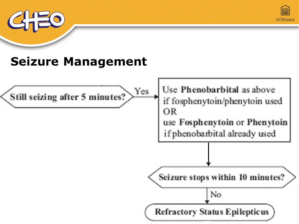
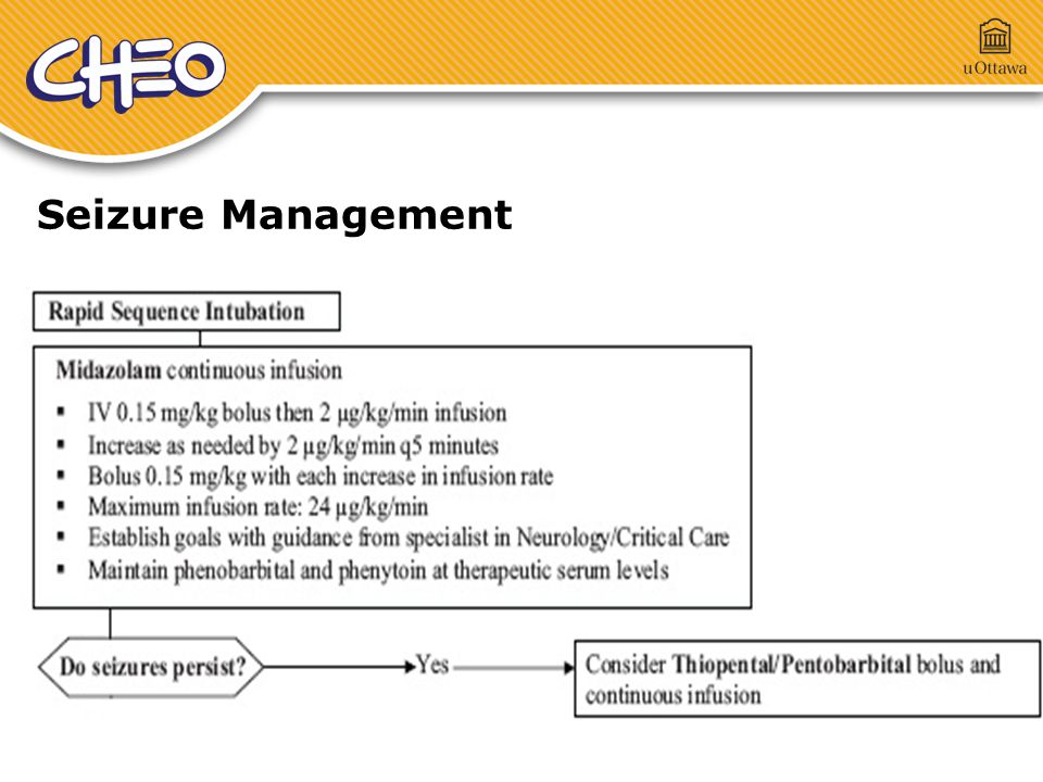
82
Seizure Management

87
Summary Quick tour through PEM Hope this talk triggered some of the areas you need to focus on studying What is completely missing…..lots of things Pediatric Orthopedics Fracture differences Limping child Hip problems Rashes, Viral exanthems Gastroenteritis, Appendicitis

88
Questions ?

Similar presentations