Download presentation
Presentation is loading. Please wait.

1
Body Composition

2
2 component model Fat tissue Fat free tissue

3
Body Composition Why the interest? Excess body fat Hypertension Type 2 diabetes Hyperlipidemia Certain cancers Poor performance/function Poor self-image Incident of overweight is ↑ng Want to “look good”

4
Body Composition Terminology Depot or storage fat – fat stored in adipose cells as nutritional reserve Essential fats – fats necessary for normal physiologic function

5
Body Composition Terminology Lean body mass (wt) – body mass minus depot fat Fat free mass (wt) – body mass minus all fat (depot & essential)
 – body mass minus depot fat Fat free mass (wt) – body mass minus all fat (depot & essential)")
6
Body Composition Relative %age of body weight that is fat & fat-free tissue Lab & field tests vary in complexity, expense

7
Anthropometry Measurement of the human body

8
Anthropometry Ht/wt Circumference/girths Skinfolds Hydrostatic weighing Bioelectrical impedance DXA TOBEC X-ray

9
Body Composition - %Fat Norms Essential Fat 11.0 - 14.03.0 - 5.0 Athletes 12.0 – 22.05.0 – 13.0 Fitness 16.0 - 25.012.0 – 18.0 Potential Risk 26.0 – 31.019.0 – 24.0 Obese ≥ 32.0 ≥ 25.0 Classification Women Men

10
Body Composition - Tests Densitometry –Two component model Fat & fat free mass –Ratio of body mass to body volume (D B =BW/BV) –Body mass determined form body weight –Body volume from under water weighing or plethysmography
 –Body mass determined form body weight –Body volume from under water weighing or plethysmography")
11
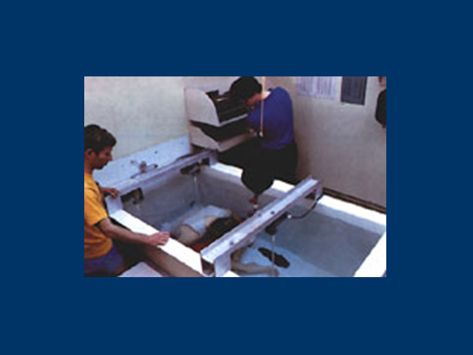
Densitometry Hydrostatic (under water) weighing –Criterion, gold standard
 weighing –Criterion, gold standard")
12
Densitometry Hydrostatic (under water) weighing –Archimedes’ principle – when a body is immersed in water, it is buoyed by a counterforce equal to the weight of the water displaced –The loss of weight in water, corrected for density of the water, allows calculation of body volume
 weighing –Archimedes’ principle – when a body is immersed in water, it is buoyed by a counterforce equal to the weight of the water displaced –The loss of weight in water, corrected for density of the water, allows calculation of body volume")
15
Densitometry RV Density of H 2 O Trapped gas in GI tract (100 ml) Body weight (dry) Body weight (wet)
 Body weight (dry) Body weight (wet)")
16
Body Composition - Densitometry Body density = weight in air (weight in air – weight in water) – Residual Density of the water volume
 – Residual Density of the water volume")
17
Body Composition - Densitometry Body density = bone & muscle more dense than water, fat tissue less dense

18
Body Composition - Densitometry Plethysmography air displacement – Δs in pressure in a closed chamber

19
Bodpod

20
Body Composition - Densitometry %fat = 457 - 414.2 Body Density %fat = 495 - 450 Body Density

21
Body Composition – Anthropometric Methods Height Weight Circumferences Skinfolds

22
Body Composition – Anthropometric Methods Reliability & validity –Skill of the measurer –Type of caliper or tape measure –Subject factors related to skinfold compressibility, edema, & variability in fat pattern & distribution –Prediction equation used to estimate fatness

23
Body Composition – Anthropometric Methods Body Mass Index (BMI) –Assess weight relative to height –[body weight (kg)/height (m 2 )] –BMI > 25 – increased health risk –25.0 to 29.9 = Overweight –> 30 = Obese –See Table 4-1 pg.58 & Table 4-2 pg.59
![Body Composition – Anthropometric Methods Body Mass Index (BMI) –Assess weight relative to height –[body weight (kg)/height (m 2 )] –BMI > 25 – increased health risk –25.0 to 29.9 = Overweight –> 30 = Obese –See Table 4-1 pg.58 & Table 4-2 pg.59](http://images.slideplayer.com/12/3588146/slides/slide_23.jpg "Body Composition – Anthropometric Methods Body Mass Index (BMI) –Assess weight relative to height –[body weight (kg)/height (m 2 )] –BMI > 25 – increased health risk –25.0 to 29.9 = Overweight –> 30 = Obese –See Table 4-1 pg.58 & Table 4-2 pg.59")
24
Body Composition – Anthropometric Methods Waist-to-hip circumference –Body fat distribution –More fat on trunk (abdominal fat) = greater risk of HTN, type 2 diabetes, hyperlipidemia, CAD, premature death –Health risk is high W/H ratio > 0.94 in young men W/H ratio > 0.82 in young women W/H ratio > 1.03 in men 60-69 yrs W/H ratio > 0.90 in women 60-69 yrs See Box 4-1 & Table 4-3 pgs.60-61 ACSM GET&P
 = greater risk of HTN, type 2 diabetes, hyperlipidemia, CAD, premature death –Health risk is high W/H ratio > 0.94 in young men W/H ratio > 0.82 in young women W/H ratio > 1.03 in men yrs W/H ratio > 0.90 in women yrs See Box 4-1 & Table 4-3 pgs ACSM GET&P")
25
Classification of Disease Risk Based on Body mass Index (BMI) and Waist Circumference (Table4-1) Disease Risk Relative to normal Weight and Waist Circumference
 and Waist Circumference (Table4-1) Disease Risk Relative to normal Weight and Waist Circumference")
26
Body Composition – Anthropometric Methods Skinfolds –Amount of subcutaneous fat is proportional to total body fat

27
Box 4-2. Standardized Description of Skinfold Sites & Procedures Continued Procedures –All measurements on the right side of the body –Caliper should be placed 1 cm away from the thumb & finger, perpendicular to the skinfold, & halfway b/n the crest & the base of the fold

28
Box 4-2. Standardized Description of Skinfold Sites and Procedures Continued –Pinch should be maintained while reading the caliper –Wait 1 to 2 s (& not longer) before reading caliper –Take duplicate measures at each site & retest if duplicate measurements are not w/n 1 to 2 mm –Rotate through measurement sites or allow time for skin to regain normal texture & thickness
 before reading caliper –Take duplicate measures at each site & retest if duplicate measurements are not w/n 1 to 2 mm –Rotate through measurement sites or allow time for skin to regain normal texture & thickness.")
29
Box 4-2. Standardized Description of Skinfold Sites & Procedures Skinfold Site –AbdominalVertical fold; 2cm to the right side of the umbilicus –TricepsVertical fold; on the posterior midline of the upper arm, halfway between the acromion & the olecranon processes, w/ the arm held freely to the side of the body –BicepsVertical fold; on the anterior aspect of the arm over the belly of the biceps muscle, 1 cm above the level used to mark the triceps site

30
Box 4-2. Standardized Description of Skinfold Sites & Procedures Continued Skinfold Site Continued –Chest/PectoralDiagonal fold; one-half the distance between the anterior axillary line & the nipple (men) or one-third the distance b/n the anterior axillary line & the nipple (women) –Medial CalfVertical fold; at the maximum circumference of the calf on the midline of its medial border
 or one-third the distance b/n the anterior axillary line & the nipple (women) –Medial CalfVertical fold; at the maximum circumference of the calf on the midline of its medial border.")
31
Box 4-2. Standardized Description of Skinfold Sites & Procedures Continued Skinfold Site Continued –MidaxillaryVertical fold; on the midaxillary line at the level of the xiphoid process of the sternum (An alternate method is a horizontal fold taken at the level of the xiphoid/sternal in the midaxillary line –SubscapularDiagonal fold (at a 45 angle); 1 to 2 cm below the inferior angle of the scapula
; 1 to 2 cm below the inferior angle of the scapula.")
32
Box 4-2. Standardized Description of Skinfold Sites & Procedures Continued SuprailiacDiagonal fold; in line w/ the natural angle of the iliac crest taken in the anterior axillary line immediately superior to the iliac crest ThighVertical fold; on the anterior midline of the thigh, midway b/ the proximal border of the patella & the inguinal crease (hip)
.")
33
Table 4-5. Body Composition (%Body Fat) For Men*
 For Men*")
34
Table 4-6. Body Composition (%Body Fat) For Women*
 For Women*")
35
Body Composition – Other Techniques Bioelectrical Impedance Analysis (BIA) –Volume of FFM is proportional to the electrical conductivity of the body –Small electrical current is passed through body –Measures resistance to current Fat is poor conductor – 14 to 22% water Lean tissue is good conductor - >90% water –Assumes normal hydration
 –Volume of FFM is proportional to the electrical conductivity of the body –Small electrical current is passed through body –Measures resistance to current Fat is poor conductor – 14 to 22% water Lean tissue is good conductor - >90% water –Assumes normal hydration")
36
Body Composition – Other Techniques Bioelectrical Impedance Analysis (BIA) –Does not require a high degree of technical skill –More comfortable –Requires minimal cooperation –Intrudes less on privacy
 –Does not require a high degree of technical skill –More comfortable –Requires minimal cooperation –Intrudes less on privacy")
37
Body Composition – Other Techniques Dual Energy X-ray Absorptiometry (DEXA) –Assess total bone mineral, bone, fat, & lean tissues –Uses three component model (fat, solids, water) Near-infrared interactance (NIR) –Light absorption & reflection –Chemical composition of the body –Accuracy 4 to 11%
 –Assess total bone mineral, bone, fat, & lean tissues –Uses three component model (fat, solids, water) Near-infrared interactance (NIR) –Light absorption & reflection –Chemical composition of the body –Accuracy 4 to 11%")
38
Dual X-Ray Absorptiometry

39
Prediction Equations Population specific –Derived on homogeneous population General equations –Diverse, heterogeneous samples that account for differences in age, sex, race, ethnicity, etc.

40
Prediction Equations Equation Selection –To whom is the equation applicable –Was equation developed on an appropriate reference model? –Was a representative sample studied? –How were predictor variables measured? –Was the equation cross-validated? –Does equation give accurate estimates? (2.5 to 3.5% - %fat) (2.5 to 3.5 kg – FFM)
 (2.5 to 3.5 kg – FFM).")
Similar presentations


 Body volume(l) %body fat.>")


