Download presentation
Presentation is loading. Please wait.

1
Lecture 4 Skull

2
Objectives: learn the bones of the braincase
learn the bones of the face and palate learn the cavities of the skull and associated structures

3
Overview: Skull The bones of the skull protect the brain and the special sense organs (sight, smell, hearing, equilibrium and taste) They form the boundaries to the entrance of the digestive and respiratory systems They also provide attachment to the facial muscles and the powerful muscles of mastication

4
Overview: Different breeds of dogs have different shapes and lengths of the skull. Dogs with long skulls are called dolichocephalic (e.g., Greyhound) Those with short skulls are called brachycephalic (e.g., Bulldog). The skulls of the intermediate breeds are called mesaticephalic (e.g., Dachshund).
 Those with short skulls are called brachycephalic (e.g., Bulldog). The skulls of the intermediate breeds are called mesaticephalic (e.g., Dachshund).")
6
Skull The facial bones: The braincase bones:
The facial bones form the boundaries of the nasal cavity, bony orbit, and the roof and lateral walls of the oral cavity. The braincase bones: The bones of the braincase (neurocranium) form the boundaries of the cranial cavity that encloses the brain and the meninges
 form the boundaries of the cranial cavity that encloses the brain and the meninges.")
7
The facial bones The facial bones can be classified into two groups:
A. Paired bones of the facial bones: 1. Lacrimal 2. Nasal 3. Maxilla 4. Zygomatic 5. Incisive 6. Palatine 7. Pterygoid 8. Dorsal nasal concha 9. Ventral nasal concha 10. Mandible bone Unpaired bones of the facial bones: 1. Vomer 2. Hyoid

10
A. Paired bones of the facial bones
1. Lacrimal located in the rostromedial aspect of the orbit. At its center there is the fossa for the lacrimal sac, where the osseous lacrimal canal begins.

11
The lacrimal bone articulates
With: frontal bone, maxilla, palatine bone, zygomatic bone and ethmoid bone.

12
A. Paired bones of the facial bones
2. Nasal: The nasal bone is very short in brachycephalic skull. Its internal surface is covered by mucous membrane in live animal.

13
The nasal bone articulates with:
the frontal , maxilla and incisive bone.

14
A. Paired bones of the facial bones
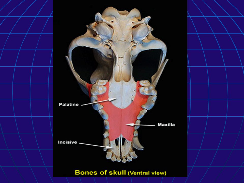
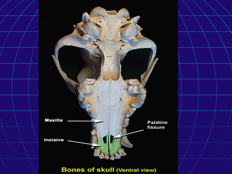
3. Maxilla: The maxilla is the largest bone of the face. Together with the incisive bone, the maxilla forms the upper jaw. On its external surface there is the infraorbital foramen for the passage of infraorbital nerve, vein and artery.

15
A. Paired bones of the facial bones
infraorbital canal: The canal begins at the maxillary foramen and ends at the infraorbital foramen. The short infraorbital canal lies dorsal to the upper fourth premolar.

19
A. Paired bones of the facial bones
4. Zygomatic: The zygomatic bone forms the zygomatic arch (rostral part) together with the zygomatic process of the temporal bone and Maxilla. It articulates with the maxilla, lacrimal and temporal bones.
 together with the zygomatic process of the temporal bone and Maxilla. It articulates with the maxilla, lacrimal and temporal bones.")
21
A. Paired bones of the facial bones
5. Incisive (Premaxilla): The incisive bone contains three alveoli for the upper incisor teeth. It articulates with the maxilla, vomer and nasal bone.
: The incisive bone contains three alveoli for the upper incisor teeth. It articulates with the maxilla, vomer and nasal bone.")
25
A. Paired bones of the facial bones
6. Palatine: The palatine bone forms the caudal part of the hard palate. It is divided into horizontal and perpendicular laminae. Each horizontal lamina has two surfaces, palatine and nasal.

26
A. Paired bones of the facial bones
6. Palatine: palatine canal: Running through the palatine bone is the palatine canal, which provides passage for the major palatine artery, vein and nerve. The palatine canal begins at the caudal palatine foramen in the pterygopalatine fossa and terminates in the hard palate through the major and minor palatine foramina.

30
A. Paired bones of the facial bones
7. Pterygoid: The pterygoid is small four-sided bone that articulates with the medial surface of the pterygoid process of the sphenoid bone.

32
A. Paired bones of the facial bones
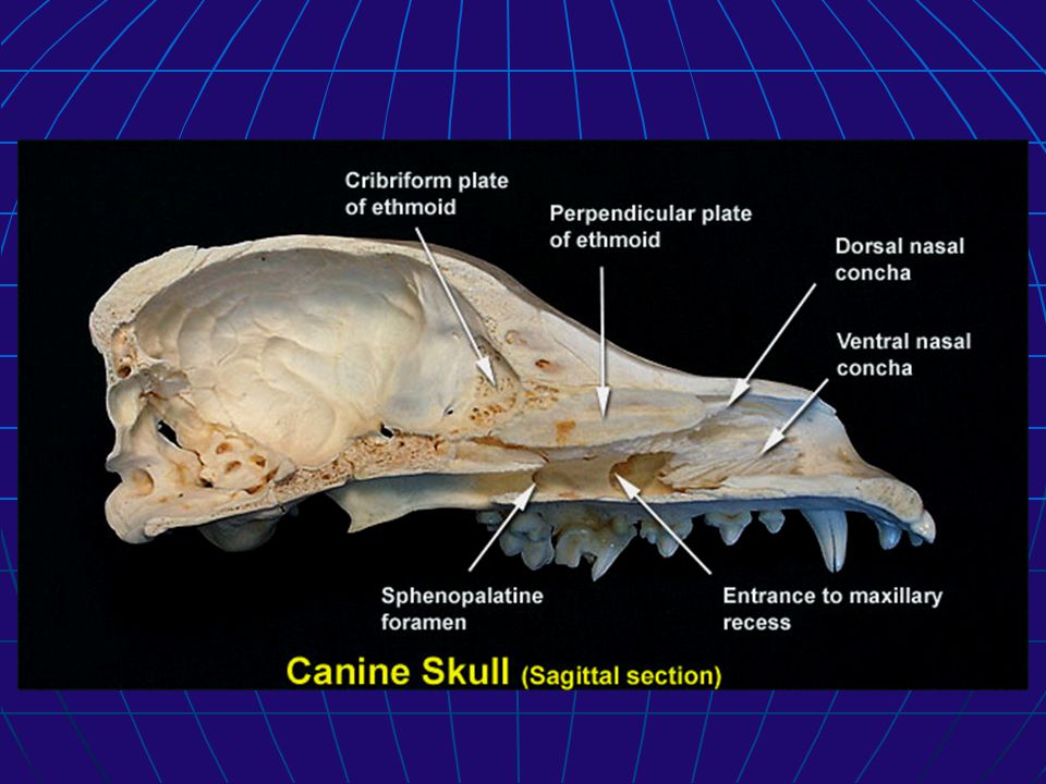
8. Dorsal nasal concha: The dorsal nasal concha is attached to the ethmoidal crest on the inner wall of the nasal bone. The dorsal nasal concha is a simple curved shelf of bone. The space ventral to the dorsal nasal concha is the middle meatus and the space dorsal to it is the dorsal meatus.

35
A. Paired bones of the facial bones
9. Ventral nasal concha: The ventral nasal concha is attached to the conchal crest on the medial wall of the maxilla. It is formed of primary and secondary bony scrolls. The space between the conchae and the nasal septum is the common meatus, whereas the space dorsal to the conchae is the middle meatus and the space ventral to it is the ventral meatus.

39
A. Paired bones of the facial bones
10. Mandible: The mandible consists of two parts that are united rostrally at the symphysis. Each part is divided into a horizontal body, and a vertical ramus. The body carries the lower teeth, and the ramus articulates with the temporal bone.

40
A. Paired bones of the facial bones
10. Mandible: The dorsal (alveolar) border of the mandible bears alveoli for the lower incisors, canine, premolars and molar teeth. The lateral surface of the ramus presents a triangular depression, the masseteric fossa, for the attachment of the masseter muscle.
 border of the mandible bears alveoli for the lower incisors, canine, premolars and molar teeth. The lateral surface of the ramus presents a triangular depression, the masseteric fossa, for the attachment of the masseter muscle.")
42
A. Paired bones of the facial bones
10. Mandible: The dorsal end of the ramus is represented by the coronoid process. The condylar process of the ramus articulates with the temporal bone to form the temporomandibular joint. The coronoid process and the condylar process are separated by the mandibular notch.

44
A. Paired bones of the facial bones
Mandibular canal : The mandibular canal begins at the mandibular foramen on the medial side of the ramus. It perforates the mandible rostrally and ends at the three mental foramina (caudal, middle, rostral) on the rostrolateral part of the body. The mandibular canal provides passage way for the inferior alveolar artery, vein and nerve.
 on the rostrolateral part of the body. The mandibular canal provides passage way for the inferior alveolar artery, vein and nerve.")
47
B. Unpaired bones of the facial bones
1. Vomer: The vomer is a single bone that extends obliquely from the base of the cranial cavity to the upper surface of the hard palate. It forms the caudoventral part of the nasal septum. The vomer articulates with the sphenoid bone, ethmoid bone, palatine bones, maxilla and incisive bones.

49
B. Unpaired bones of the facial bones
2. Hyoid bones: hyoid apparatus extend from the mastoid process of the skull to the thyroid cartilage of the larynx. They support and stabilize the tongue and the larynx.

50
B. Unpaired bones of the facial bones
The hyoid apparatus consists of : stylohyoid Epihyoid Ceratohyoid basihyoid thyrohyoid The basihyoid is the only single bone that connects the paired bones from each side at the root of the tongue. Attaching to the free end of the stylohyoid is the tympanohyoid cartilage, which articulates with the mastoid process.

Similar presentations