Download presentation
Presentation is loading. Please wait.

1
Edema, Hyperemia and Congestion

2
Cardiovascular disease
Most important cause of morbidity and mortality in developed nations In US, 81 million affected in 2005, causing 35 to 40% of deaths Components of cardiovascular system include Heart Vessels Blood Water, salts, proteins, clotting factors, cells

3
Blood Hemodynamics Edema Hyperemia Congestion Shock
Water build-up in interstitial spaces and cavities Hydrodynamic transudate is dilute, protein-poor Inflammatory exudate is concentrated, protein rich Hyperemia Acute, actively increased blood flow—arteriole dilation, increased heart rate Tissues look red (erythema) Congestion Chronic, passively reduced outflow—venule dilation Tissues look pale or blue (cyanosis) Shock Circulatory failure
 Congestion. Chronic, passively reduced outflow—venule dilation. Tissues look pale or blue (cyanosis) Shock. Circulatory failure.")
4
Fluids—water This unit addresses factors affecting the distribution between intravascular and interstitial spaces

5
Factors affecting intravascular and interstitial water movement
Concentration of solutes Albumin and other proteins (huge difference) Sodium and other ions (small difference) Hydrostatic pressure Higher on arteriolar side Lower on venular side Lowest in interstitium Blood volume decreased b.p. Water intake/deprivation Water loss from skin or gut Perspiration, vomiting, diarrhea Blood loss; acute hemorrhage
 Sodium and other ions (small difference) Hydrostatic pressure. Higher on arteriolar side. Lower on venular side. Lowest in interstitium. Blood volume decreased b.p. Water intake/deprivation. Water loss from skin or gut. Perspiration, vomiting, diarrhea. Blood loss; acute hemorrhage.")
6
Fluid transit FIGURE 4-1 Factors influencing fluid transit across capillary walls. Capillary hydrostatic and osmotic forces are normally balanced so that there is no net loss or gain of fluid across the capillary bed. However, increased hydrostatic pressure or diminished plasma osmotic pressure will cause extravascular fluid to accumulate. Tissue lymphatics remove much of the excess volume, eventually returning it to the circulation via the thoracic duct; however, if the capacity for lymphatic drainage is exceeded, tissue edema results

7
Edema Localized or generalized accumulation of fluid in interstitial spaces Anasarca: severe, generalized edema ana = throughout, sark = flesh Most commonly used to describe fetal or neonatal whole-body, subcutaneous swelling Effusions into body cavities Hydrothorax: within thorax, around lungs; also pleural effusion Hydropericardium: Fluid in the pericardial sac Hydroperitoneum or ascites: Fluid in the peritoneal cavity Extravasate: (v.) to move out of the vasculature
 to move out of the vasculature.")
8
Responses to edema Skin: swells according to elasticity
dependent edema: distribution affected by gravity (ankles, sacrum) dependent = hanging down in this context Brain: compresses without room to swell Lung: alveoli fill preventing gas exchange
 dependent = hanging down in this context. Brain: compresses without room to swell. Lung: alveoli fill preventing gas exchange.")
9
Appearance of edema Swollen tissues (not cells—fluid is outside the cells) Heavy tissues Wet tissues Widening of fascial planes or interlobular septa Filled cavities

10
Causes of edema Increased hydrostatic pressure
Focal impairment—deep vein thrombosis Generalized impairment—right heart failure Decreased plasma osmotic pressure Hypoproteinemia Decreased protein synthesis—serum albumin Increased protein loss to nephrotic syndrome Sodium (and water) retention Increased capillary permeability inflammation or injury (burns) Lymphatic obstruction Filariasis, breast carcinoma
 retention. Increased capillary permeability. inflammation or injury (burns) Lymphatic obstruction. Filariasis, breast carcinoma.")
11
Appearance and causes of hyperemia
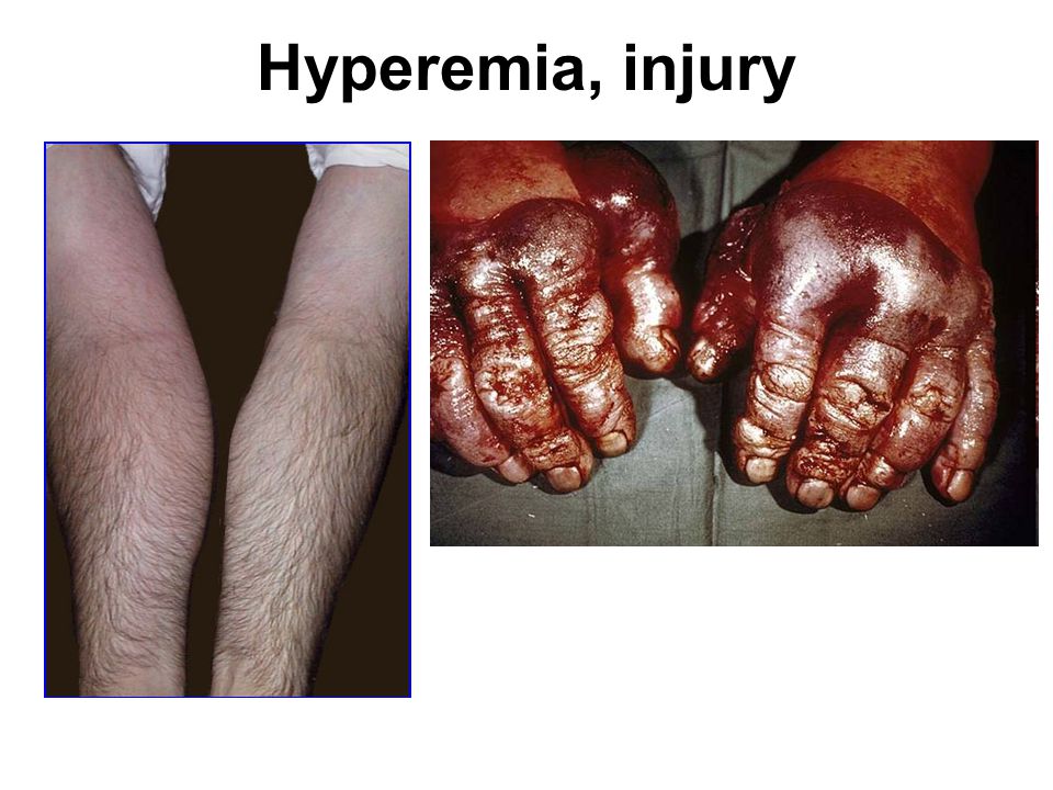
Increased flow of blood into tissue Local process of arteriole dilation greater than venule dilation Appearance of blood flow is RED Normal physiological examples: Exercise Blushing Erection Inflammatory response (rubor) Small contribution to edema
 Small contribution to edema.")
12
Congestion Impaired venous outflow from tissue
Local increases in venous pressure Central congestive heart disease increases diastolic (venous) b.p. Right-side failure congests portal drainage, liver, generalized edema Left-side failure congests pulmonary drainage, lungs, hypoxemia
 b.p. Right-side failure congests portal drainage, liver, generalized edema. Left-side failure congests pulmonary drainage, lungs, hypoxemia.")
13
FIGURE 4-2 Pathways leading to systemic edema from primary heart failure, primary renal failure, or reduced plasma osmotic pressure (e.g., from malnutrition, diminished hepatic synthesis, or protein loss from nephrotic syndrome).
.")
14
Congestive heart failure
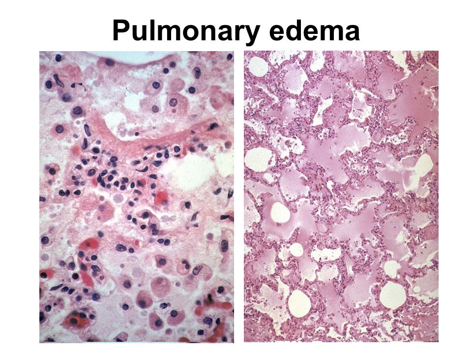
Right side failure—volume backs up behind pulmonary circulation Generalized edema Portal edema—nutmeg liver Left side failure—volume backs up behind systemic circulation Pulmonary edema and pleural effusion Distension of alveolar capillaries leading to capillary rupture and red cells in alveoli Intra-alveolar macrophages phagocytose red cells and accumulate hemosiderin Fibrosis of the interstitium with hemosiderin deposition Serous or serosanguinous effusions surrounding lungs

15
Congestive heart failure
Reduced cardiac output Renal hypoperfusion Activation of renin-angiotensin-aldosterone axis Early response is beneficial sodium and water retention increased vascular toneelevated antidiuretic hormone (ADH) improved cardiac outputrestored renal perfusion Ongoing response increases edema Volume of blood exceeds volume of vasculature Fluids build up in tissues
 improved cardiac outputrestored renal perfusion. Ongoing response increases edema. Volume of blood exceeds volume of vasculature. Fluids build up in tissues.")
16
Anasarca

17
Hyperemia, injury
18
Trivial and life-threatening edema
This example of a fluid collection, a friction blister of the skin, is an almost trivial example of edema. This example of edema with inflammation is not trivial at all: there is marked laryngeal edema such that the airway is narrowed. This is life-threatening. Thus, fluid collections can be serious depending upon their location

19
Pitting edema From GRIPE

20
Elephantiasis--lymphedema
From GRIPE Left: elephantiasis; Right: elephantiasis secondary to carcinoma

21
Peau d’orange and post-mastectomy lymphedema

22
Ascites due to portal congestion
American Gastroenterological Association, AGA Teaching Project, 1975 UI Medical Library call # WI, 720, P8423, 1975 Ascites with "caput madusae" (medusa head), also known as "Cruveilhier-Baumgarten Syndrome". Cirrhosis and portal hypertension sometimes create anastomoses of portal drainage with the umbilical vein. Also note protrusion of the naval from abdominal pressure.
, also known as Cruveilhier-Baumgarten Syndrome . Cirrhosis and portal hypertension sometimes create anastomoses of portal drainage with the umbilical vein. Also note protrusion of the naval from abdominal pressure.")
23
Nutmeg liver Here is an example of a "nutmeg" liver seen with chronic passive congestion of the liver. Note the dark red congested regions that represent accumulation of RBC's in centrilobular regions. Microscopically, the nutmeg pattern results from congestion around the central veins, as seen here. This is usually due to a "right sided" heart failure.

24
Hepatic congestion If chronic hepatic passive congestion continues for a long time, a condition called "cardiac cirrhosis" may develop in which there is fibrosis bridging between central zonal regions, as shown below, so that the portal tracts appear to be in the center of the reorganized lobule. This process is best termed "cardiac sclerosis" because, unlike a true cirrhosis, there is minimal nodular regeneration. If the passive congestion is pronounced, then there can be centrilobular necrosis, because the oxygenation in zone 3 of the hepatic lobule is not great. The light brown pigment seen here in the necrotic hepatocytes around the central vein is lipochrome.

25
Effusions Extravascular fluid collections can be classified as follows: Exudate: extravascular fluid collection that is rich in protein and/or cells. Fluid appears grossly cloudy. Transudate: extravascular fluid collection that is basically an ultrafiltrate of plasma with little protein and few or no cells. Fluid appears grossly clear. Effusions into body cavities can be further described as follows: Serous: a transudate with mainly edema fluid and few cells. Serosanguinous: an effusion with red blood cells. Fibrinous (serofibrinous): fibrin strands are derived from a protein-rich exudate. Purulent: numerous PMN's are present. Also called "empyema" in the pleural space.
: fibrin strands are derived from a protein-rich exudate. Purulent: numerous PMN s are present. Also called empyema in the pleural space.")
26
Pleural effusions and edema
This is a right pleural effusion (in a baby). Note the clear, pale yellow appearance of the fluid. This is a serous effusion. Here is an example of bilateral pleural effusions. Note that the fluid appears reddish, because there has been hemorrhage into the effusion. This is a serosanguinous effusion.
. Note the clear, pale yellow appearance of the fluid. This is a serous effusion. Here is an example of bilateral pleural effusions. Note that the fluid appears reddish, because there has been hemorrhage into the effusion. This is a serosanguinous effusion")
27
Fibrinous exudate Fibrinous exudate of pericardium

28
Pleural effusion

29
Pleural effusion

30
Pulmonary edema http://peir.path.uab.edu:3555/images/med/00011603.jpg
31
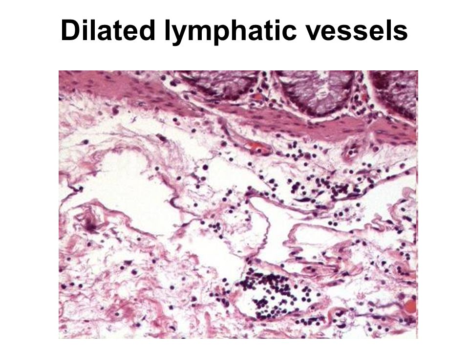
Dilated lymphatic vessels
Similar presentations


 ~5% of total.>")
>")


 Chapter: Hemodynamic disorders, Thrombosis and Shock - Edema - Hemorrhage - Hyperemia.>")

 Congestion Chronic, passively reduced.>")

 intracellular. (1/3)extracellular (interstitial fluid) 5% blood plasma. edema = an accumulation of interstitial.>")

 saying that her legs have been swollen for a month. On examination you find that she has.>")


