Download presentation
Presentation is loading. Please wait.

1
Tuesday Case Conference May 2009

2
Biopsy finding LM –Glomeruli are normal in size to mildly enlarged Mild enlargement of the mesangial areas with occasional nodular appearance –Tubulointerstitial Interstitial fibrosis and tubular atrophy, involving approximately 50% –Artery Severe intimal fibrosis of arcuate artery Sever cirucumferential hyalinosis of arterioles IF –Negative for C4D, BKV, and CMV EM –Podocyte effacement invovling ~40% of the surface area Diagnosis: –Early Diabetic Nephropathy –IFTA related to CNI

3
Objectives What are different causes of allograft failure? –What is Interstitial Fibrosis and Tubular Atrophy - Chronic Allograft Nephropathy? A new biomarker for IFTA?

4
Transplantation The preferred prescription for ESRD patients

5
Mortality on Dialysis Annual Death RateN All dialysis patients16 per 100 patient years~300,000 Patients on list6 per 100 patient years~50,000 Cadaver transplant patients 3 per 100 patient years~25,000

6
Graft survival increases with less time on dialysis Goldfarb-Rumyantzev A, et al. Nephrol Dial Transplant 2005;20:167–175

7
Risk of Death with Transplantation OJO, A. O. et al. J Am Soc Nephrol 2001;12:589-597

8
Renal Transplantation Transplant is preferred prescription of choice for ESRD patients –Sooner the better Dramatic improvement, since 1980s, 1-year allograft survival rates: 90-95% Long-term outcomes have changed little –Death with a functioning graft –Chronic allograft nephropathy

9
Recurrent glomerulonephritis and other causes of graft failure 2002_NEJM_Briganti-Chadban_risk of renal allograft loss from recurrent glomerulonephritis

10
The most common cause of graft failure, after the first year

11
What is CAN – IFTA? Incompletely understood clincopathological entity –chronic rejection, transplant nephropathy, chronic renal allograft dysfunction, trnasplant glomerulopathy, or chronic allograft nephropathy Chronic Allograft Nephropathy –Banff 1991 used interchangeably with ‘chronic rejection’ –Banff 1997 term to be used when it is impossible to precisely define the etiology of chronic allograft damage –Banff 2005 “interstitial fibrosis and tubular atrophy, without evidence of any specific etiology”

12
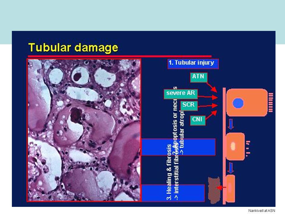
IF/TA (CAN) Interstitial fibrosis Fibrointimal proliferation Nankivell at ASN
 Interstitial fibrosis Fibrointimal proliferation Nankivell at ASN")
13
Electron microscopic appearances in chronic allograft nephropathy

14
Some possible immune and nonimmune mechanisms of injury leading to chronic allograft nephropathy

15
IF/TA score Nankivell at ASN

16
Onset of IFTA Very common Progressive Associated with –Proteinuria –Decreased GFR –hypertension 2003_NEJM_Nankivell-Chapman_antural history of chronic allograft nephropathy

17
Nankivell at ASN

19
Clinical Risk Factors for long-term allograft failure IF/TA is a major cause of late allograft loss Challenge is to dissect the identifiable causes and to develop cause-specific treatment Existing risk factors/markers –Increased serum creatinine –Proteinuria –HTN

20
An innovative biomarker for IFTA?

21
Background In kidney, interstitial fibrosis has been considered a common mechanism of disease progression –No effective treatment to revert established fibrosis and diagnosis is often late in the disease course –Fibroblasts are the pivotal effector cells in fibrogenesis –Studies looking to identify activated fibroblast found abnormal expression of mesenchymal markers (fibroblast) by tubular epithelial cells Epithelial phenotypic changes (EPC)
 by tubular epithelial cells Epithelial phenotypic changes (EPC)")
22
Background Epithelial-mesenchymal transformation (EMT) –where cell shifting between epithelial and mesenchymal phenotype is well recognized process that characterizes the embryonal plasticity –Key element in metastasis of tumors –Observed in the process of wound healing and fibrotic remodeling after inflammatory injury
 –where cell shifting between epithelial and mesenchymal phenotype is well recognized process that characterizes the embryonal plasticity –Key element in metastasis of tumors –Observed in the process of wound healing and fibrotic remodeling after inflammatory injury")
23
Four key events during EMT 2006_ArthritisResTx_Zvaifler_Relevance of the stroma and epithelial mesenchymal transition_REV

24
Examined whether EPC of tubular cells predict the progression of fibrosis in the allograft –Cytoplasmic translocation of β-catenin –Expression of Vimentin –intermediate filament typically expressed by mesenchymal cells 83 kidney tranplant with protocol graft biopsy at both 3 and 12 mo 2008_JASN_Hertig-Dubois_Early epithelial phenotypic changs predict graft fibrosis

25
Role of the Cadherins in Establishing Molecular Links between Adjacent Cells NEJM, 1996

26
Immunohistochemical staining epithelial phenotypic change β-cateninVimentin 2008_JASN_Hertig-Dubois_Early epithelial phenotypic changs predict graft fibrosis

27
IF/TA score and EPC status 2008_JASN_Hertig-Dubois_Early epithelial phenotypic changs predict graft fibrosis

28
Change in Serum Creatinine by EPC status 2008_JASN_Hertig-Dubois_Early epithelial phenotypic changs predict graft fibrosis

29
Conclusion EPC (+) –an early and independent marker to predict the progression of IF/TA lesions between 3 and 12 mo after transplantation –Associated with poorer graft function from the time point of 18 mo and thereafter Risk factors for renal graft fibrosis –3-mo EPC score and 12-mo t scores were significantly higher in progressors as compared with nonprogressors
 –an early and independent marker to predict the progression of IF/TA lesions between 3 and 12 mo after transplantation –Associated with poorer graft function from the time point of 18 mo and thereafter Risk factors for renal graft fibrosis –3-mo EPC score and 12-mo t scores were significantly higher in progressors as compared with nonprogressors")
30
Treatment

31
Strategies to prevent and treat IFTA

32
Reference Goldfarb-Rumyantzev A, et al. Nephrol Dial Transplant 2005;20:167–175 OJO, A. O. et al. J Am Soc Nephrol 2001;12:589- 597 Briganti EM, Russ GR, McNeil J, et al: Risk of renal allograft loss from recurrent glomerulonephritis. N Engl J Med 2002;347:103– 109 Nankivell-Chapman, Natural history of chronic allograft nephropathy, NEJM, 2003 Briganti-Chadban, Risk of renal allograft loss from recurrent glomerulonephritis, NEJM, 2003

Similar presentations


 RENAL DISEASE: OVERVIEW AND ACUTE RENAL FAILURE Pathophysiology of Disease: Chapter 16 (388-394) Jack.>")