Download presentation
Presentation is loading. Please wait.

1
SHEEP BRAIN DISSECTION
parts, parts, parts

4
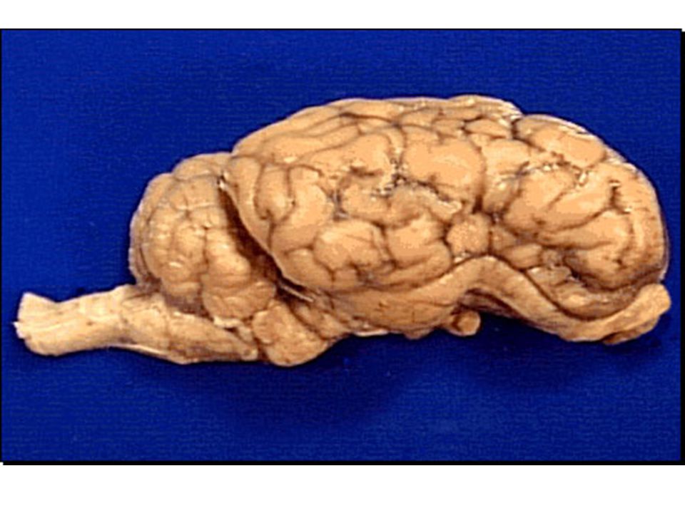
cerebral cortex Sheep brains have parts comparable to human brains. Notice the two hemispheres. Identify the lobes of the cerebral cortex: frontal, temporal, parietal, occipital. What are their functions? Notice the sulci (singular - sulcus) and gyri (singular - gyrus). These increase the surface area of the cortex.
 and gyri (singular - gyrus). These increase the surface area of the cortex.")
5
Frontal lobe

6
Temporal Lobe

7
Parietal Lobe

8
Occipital Lobe

9
Sulcus

10
Gyrus

11
Cerebellum

12
Dorsal view

13
Just take a look!

14
Cerebral cortex

15
Cerebellum

16
Corpus Callosum

17
Thalamus

18
Hypothalamus

19
Midbrain

20
Pons

21
Medulla oblongata

22
Ventricular System

23
Pineal Body

24
Why does the spinal cord of the sheep project horizontally instead of vertically as in a human?

25
1) cerebellum, 2) occipital lobe, 3) cerebral cortex, 4) corpus callosum, 5) pineal gland, 6) thalamus, 7) hypothalamus, 8) optic nerve, 9) pons, 10) spinal cord

26
Name the parts with pins

27
red pin = corpus callosum blue pin = thalamus green pin = cerebellum

28
sheep - ventral view

29
ventral view ~optic nerve

30
ventral view~olfactory bulb and tract

31
ventral view~brain stem

33
Cranial nerves Olfactory bulb nerve I. Optic nerve II.
Oculomotor nerve III. Trochlear nerve IV. Trigeminal nerve V Abducens nerve VI.

34
Oculomotor nerve The oculomotor nerve, cranial nerve III, is found on the the medial surface of each cerebral peduncle. This nerve supplies four muscles of the eye. Movement of the eyes up (elevation), down (depression), up and toward the temple, and inward toward the nose (adduction). Controls constriction of the pupil and accommodation of the lens.
, down (depression), up and toward the temple, and inward toward the nose (adduction). Controls constriction of the pupil and accommodation of the lens.")
35
Trigeminal nerve The trigeminal , cranial nerve V, is a mixed nerve that carries both sensory and motor information. The motor component controls the muscles of mastication ( chewing), and the sensory component carries sensory information from the face and jaw. Tooth pain for example, is carried by the trigeminal nerve.
, and the sensory component carries sensory information from the face and jaw. Tooth pain for example, is carried by the trigeminal nerve.")
36
Trochlear nerve The trochlear, cranial nerve IV, serves the superior oblique muscle of the eye. The superior oblique allows you to move your eyes down and in toward the nose (intorsion). This nerve is often obscured by the trigeminal nerve.
. This nerve is often obscured by the trigeminal nerve.")
37
Abducens The abducens, cranial nerve VI, serves the external rectus muscle of the eye. The external rectus allows you to move your eyes outward toward the temple (abduction). This nerve can be located at the anterior limit of the medulla (that section of the brain located caudal to the pons and anterior to the spinal cord).
. This nerve can be located at the anterior limit of the medulla (that section of the brain located caudal to the pons and anterior to the spinal cord).")
38
ventral view~name the nerves

39
Cerebral cortex, Gray, White, Thalamus

40
Cerebral Cortex The cerebral cortex is a highly ordered gray matter structure composed of six distinct layers. In spite of its complexity, it is composed of only three cell types: pyramidal cells, stellate cells and fusiform cells.

41
White Matter White matter is composed of fiber tracts. The "white" is due to myelin sheaths around the fibers which appear white.

42
Gray Matter

43
Thalamus

44
Do you know . . . Which cranial nerve is the largest?
Which cranial nerve is the only one that exits the "posterior" side of the brainstem? How many cranial nerves are responsible for eye movements? What does "abducens" refer to? Which cranial nerves carry gustatory (taste) information? Which cranial nerve is the longest? What two cranial nerves carry sensory information about blood pressure to the brain? Which cranial nerve is responsible for pupillary constriction?
 information Which cranial nerve is the longest What two cranial nerves carry sensory information about blood pressure to the brain Which cranial nerve is responsible for pupillary constriction")
45
Which cranial nerve is the largest?
CN V (Trigeminal) Which cranial nerve is the only one that exits the "posterior" side of the brainstem? CN IV (Trochlear) How many cranial nerves are responsible for eye movements? Three: CN III (Oculomotor), IV (Trochlear), and VI (Abducens). What does "abducens" refer to? The abducens nerve carries motor impulses to the lateral rectus eye muscle which moves the eye laterally causing abduction of the eye. Which cranial nerves carry gustatory (taste) information? CN VII (Facial), CN IX (Glossopharyngeal) and CN X (Vagus). Which cranial nerve is the longest? CN X (Vagus) which reaches from the medulla to the digestive and urinary organs. What two cranial nerves carry sensory information about blood pressure to the brain? CN IX (Glossopharyngeal) and CN X (Vagus). Which cranial nerve is responsible for pupillary constriction? CN III (Oculomotor).
 Which cranial nerve is the only one that exits the posterior side of the brainstem CN IV (Trochlear) How many cranial nerves are responsible for eye movements Three: CN III (Oculomotor), IV (Trochlear), and VI (Abducens). What does abducens refer to The abducens nerve carries motor impulses to the lateral rectus eye muscle which moves the eye laterally causing abduction of the eye. Which cranial nerves carry gustatory (taste) information CN VII (Facial), CN IX (Glossopharyngeal) and CN X (Vagus). Which cranial nerve is the longest CN X (Vagus) which reaches from the medulla to the digestive and urinary organs. What two cranial nerves carry sensory information about blood pressure to the brain CN IX (Glossopharyngeal) and CN X (Vagus). Which cranial nerve is responsible for pupillary constriction CN III (Oculomotor).")
46
References trc.ucdavis.edu/.../Nervous/ grosscns/brain2/brain4.html

Similar presentations


>")


