Download presentation
Presentation is loading. Please wait.

1
Ankle & Foot Fracture/Dislocations Shawn Dowling

2
ANATOMY 101 ANKLE 3 Primary Joints Medial malleolus w/medial talus Tibial plafond w/talar dome Lat malleolus w/lat talus 3 Bones: Tibia, Fibula and Talus 3 sets of Ligaments: Lateral collaterals (ATFL, CFL, PTFL) Syndesmotic Ligaments Medial collaterals (Deltoid)
 Syndesmotic Ligaments Medial collaterals (Deltoid)")
3
Tibia Fibula Talus BONES

4
Medial Collateral Ligaments Lateral Collateral Ligaments Syndesmotic Ligaments LIGAMENTS

5
Tibia Fibula Talus JOINTS Fibulotalar Tibiotalar (mid) Tibiotalar (lateral)
 Tibiotalar (lateral)")
6
FOOT Complicated (28 bones, 57 articulations) Subdivided in 3 segments & mvts Hindfoot - inv/ever Midfoot - abd/add Forefoot – flex/ext Joints Talo-crural jnt Inversion/eversion Hindfoot – mid foot (Choparts) Inversion/eversion Midfoot – forefoot (Lisfranc’s)* Abd/adduction MTP-IP Flex/extension
 Subdivided in 3 segments & mvts Hindfoot - inv/ever Midfoot - abd/add Forefoot – flex/ext Joints Talo-crural jnt Inversion/eversion Hindfoot – mid foot (Choparts) Inversion/eversion Midfoot – forefoot (Lisfranc’s)* Abd/adduction MTP-IP Flex/extension")
7
HINDFOOT MIDFOOT FOREFOOT calcaneus talus Medial navicular cuneiforms cuboid metatarsals phalanges sesamoids

8
Choparts Lisfrancs MTP IP

9
A B C D F E

11
What are stable fractures? Ankle forms a ring Disruption of only 1 structure is stable Disruption of > 1 is unstable

12
Approach to Ankle/Foot X- rays Go through complete approach (ABC’s) 3 views- AP, lat, Mortise (15-20° int rot) ankle, Direct evidence of injury: assess bones Indirect evidence of injuries: are all ankle measurements normal? Joint effusion? Describe x-ray, rather than simply naming it

13
Management In general Chip/avulsion #’s <3mm = Tx as sprain Non-displaced, non-intra-articular, stable #’s 3 wks NWB cast, 3-5 wks WB cast, f/u with cast clinic Unstable #’s, intra-articular # - speak with Ortho Open – saline soaked dsg, IV ABx, Td, Ortho urgently NV compromise – reduce and call Ortho Urgently

14
Diagnosis?Classification?Treatment? Does it change you mgmt if they have a tender deltoid ligament?

15
Lateral Malleoli #’s MC ankle #, MOI: usu inversion injury Weber classification – used to determine risk of syndesmosis injury and therefore need for operative repair Management NWB x 3wks, WB x 3-5wks* Refer B’s or C’s, Functional bimalleolar’s to ortho

16
Stable ? Is the location significant? Management? What measurements/lines do you look at in the ankle? What do they signify?

18
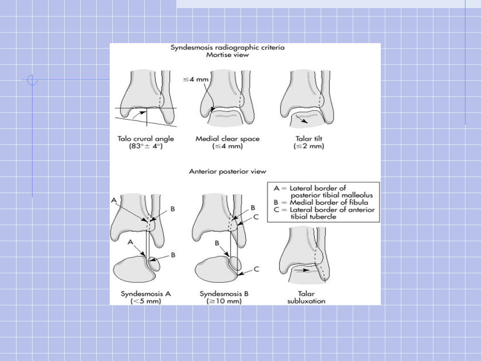
Medial clear Space <5mm A B A-B = talar tilt <3 is normal Syndesmosis injury >10 mm 1 2 3 4

19
Point out 3 abnormalities. Diagnosis? Stable? Treatment?

20
Maisonneuve

21
Diagnosis? Treatment?

22
Bimalleolar/Trimalleolar #’s Involve the medial, lateral and/or posterior malleoli Splint, pain control, NPO Need to speak to ortho as they will likely need OR

23
Mechanism of injury? Associated injuries? Management?

24
PILON # Mechanism of injury- axial load? Associated injuries- calcaneus, C,T & L spine, pelvis, intra-abdominal. Management- OR, approx 50% are open fractures

25
Description? What do you want to know/assess? What do you want to do? How?

26
Ankle Dislocations Relatively common, usually assoc w/# Describe the position of foot/talus to tibia If open, Tx as such X-rays should not delay reduction if NV compromise or skin tenting present Analgesia/PS, Reduce, splint, post-red films

27
Pediatric Ankle Injuries Not just little adult # The ligament attachments are stronger than the physis therefore more #’s, less sprains Overall management is similar to adults Although with fractures you can accept more angulation (little to no displacement) LLC casts are the initial choice for most #’s
 LLC casts are the initial choice for most #’s")
29
Can we apply OAR/OFR in children? Six studies looked at validating OAR in peds Different age groups (2-18, 6-16) Sens 85*-100*% Considered all # Some considered all #, others only “significant #”)
 Sens 85*-100*% Considered all # Some considered all #, others only significant # ).")
30
BMJ 2003. Accuracy of OAR to exclude fractures of the ankle and mid-foot: A systematic review This study references all of the OAR done in children as well as adults

31
Problems with the studies Haven’t come up with a common definition of significant # Unsure of what to do with SH-1, inconsistent Dx Local practice (and Edmonton) – variable some apply it, some use rule + discretion, others use clinical judgement
 – variable some apply it, some use rule + discretion, others use clinical judgement")
32
Conclusion This needs to be further studied Need to determine which #’s are significant But I think they will likely be validated Although I think they’ll have to Tx SH1 as distinct injuries

33
Describe fracture? Classification? Management? SH-2 LLC x 3 wks, then SLC X 1-3 wks

34
Describe fracture? Classification? Management? SH2 Reduction/immbolize (air cast)
")
35
SALTR S traight A bove (metaphysis) L ower (epiphysis) T hru the Physis R am (Crush)
 L ower (epiphysis) T hru the Physis R am (Crush)")
36
Non-operative management for SH 1-2 Attempt closed reduction, can accept more angulation Long leg cast x 3wks, followed by SLC x 3wks SH 3-4 -> OR SH 5 ->poor fx prognosis Complications for SH 3-5 include growth arrest, limb length discrepancy

37
Ankle # Complications Acute Skin necrosis NV injury Compartment syndrome # Blisters Wound infection/osteo Chronic Mal-union Non-union Post-traumatic arthritis AVN Chronic pain Chronic instability

38
Describe # Do you need to speak to Ortho? ?ottawa ankle rules Talar Dome # Yes – Ortho to see in cast clinic

39
Describe # Anything special about this bone? Blood flow distal to proximal like scaphoid and proximal femur, therefore inc AVN risk Is there a classification system for these #’s?

40
Talus Body Neck Head Chopart’s joint

41
Talar fractures Minor talar fractures Chip and avulsion fractures of neck,head, and body. Usually same mechanism as ankle sprains Talar neck fractures 50% of major talar injuries. extreme dorsiflexion force Hawkins classification Talar body fractures 23% of all talar fractures (including minor fractures) Major talar body fractures are uncommon usually axial loading (e.g. falls) Talar head fractures Uncommon (5-10%) compressive force transmitted up through the talonavicular joint applied on a plantarflexed foot
 Major talar body fractures are uncommon usually axial loading (e.g. falls) Talar head fractures Uncommon (5-10%) compressive force transmitted up through the talonavicular joint applied on a plantarflexed foot.")
42
Hawkins Classification of Talar Neck Fractures Type 1: = nondisplaced; Type 2: subtalar subluxation Type 3: dislocation of the talar body (50% open #’s) Type 4: dislocation of the talar body & distraction of the talonavicular joint. Fracture type influences management & prognosis Thanks Moby

43
Describe injury. Name this injury.v Management?

44
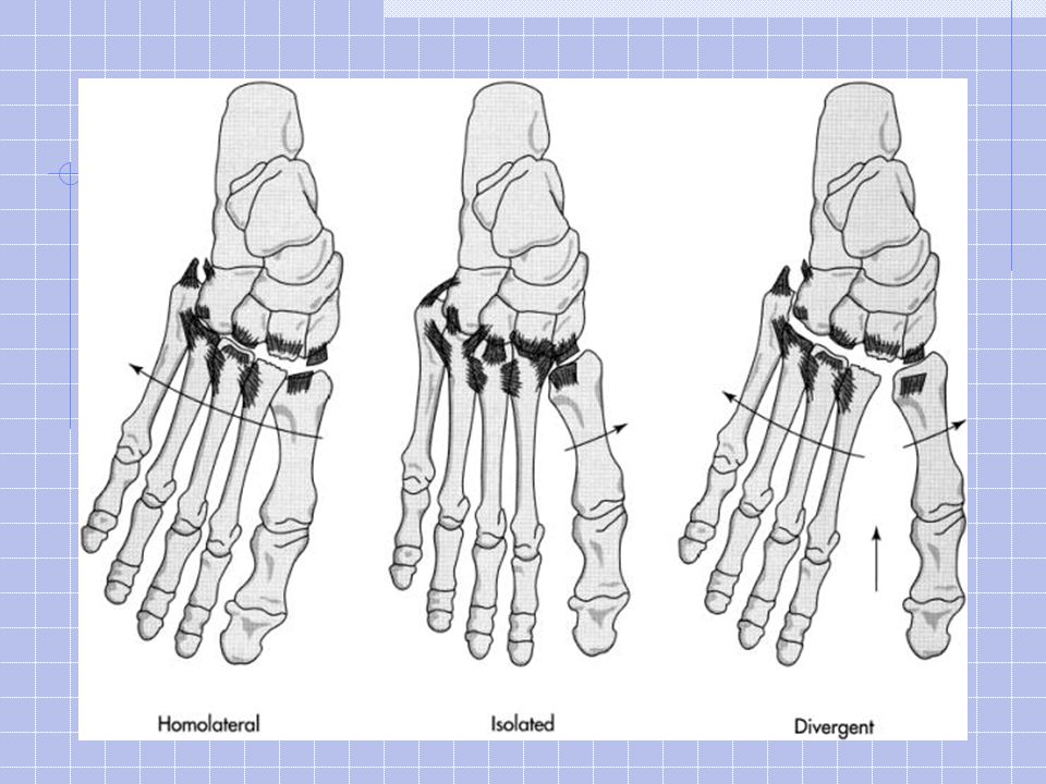
Describe injury. Name this injury Lisfranc Management? OR

46
What to look for on x-ray: Normally, medial aspect of metatarsals 1-3 should align with medial borders of cuneiforms Metatarsals should be aligned dorsally with tarsals on lateral view Medial 4 th metatarsal should align with medial cuboid Any fracture or dislocation of the navicular or cuneiforms or widening between metatarsals 1-3 Proximal 2 nd metatarsal # is pathognomonic Thanks Dave

47
Normal Lisfranc joint alignment Tx: Need to speak to ortho May try closed reduction

48
Describe. Management NWB cast # usu from direct trauma

49
Describe. Management Walking cast x 2-3 weeks Avulsion type #

50
Metatarsal # Treatment: Nondisplaced or min displaced fractures of metatarsal 2-4 stiff shoe, casting, or fracture brace. Non displaced 1 st metatarsal NWB BK walking cast (cuz it’s a major WB surface) Displaced 1 st or 5 th metatarsal ER ortho Attempt closed reduction if >3mm displacement or 10 degrees angulation Thanks Dave
 Displaced 1 st or 5 th metatarsal ER ortho Attempt closed reduction if >3mm displacement or 10 degrees angulation Thanks Dave.")
51
Phalangeal #’s Non-displaced: buddy tape, (air cast if hallux involved as they are painful) Significant displacement/angulation: closed reduction -> speak with ortho if reduction is inadequate (esp w/hallux) If subungal hematoma is present with tuft # - evacuate hematoma and repair nail bed
 Significant displacement/angulation: closed reduction -> speak with ortho if reduction is inadequate (esp w/hallux) If subungal hematoma is present with tuft # - evacuate hematoma and repair nail bed")
53
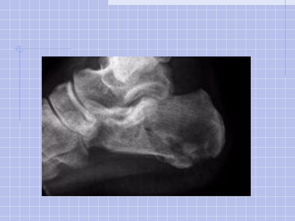
10°

54
apex of anterior process apex of posterior facet Posterior tuberosity

55
Calcaneus # Management Order Harris (axial view), may need CT Probably should speak to Ortho for all since x-rays under-estimate extent of injury But…non-displaced, extra-articular – NWB cast x 6-8 wks Otherwise, Tx varies considerably and is best determined by Ortho
, may need CT Probably should speak to Ortho for all since x-rays under-estimate extent of injury But…non-displaced, extra-articular – NWB cast x 6-8 wks Otherwise, Tx varies considerably and is best determined by Ortho")
56
Summary Ankle #’s If #/injury disturbs>1 structure in ring = unstable or if intra-articular – ortho Otherwise: NWB cast x 3wks Foot Stable, extra-articular, wgt bearing surface NWB cast Unstable, or intra-articular – ORTHO Stable, extra-articular, non-wgt bearing surface: conservative mgmt (rigid shoe, walking cast, buddy tape) If in doubt, Look up management of # - too many particularities to memorize
 If in doubt, Look up management of # - too many particularities to memorize")
57
References Emergency Medicine Reports Management of Acute Foot and Ankle Disorders in the Emergency Department: Part I—The Ankle. Management of Acute Foot and Ankle Disorders in the Emergency Department: Part II—The Foot. Rosens www.wheelessonline.com Moritz and Dave Dyck’s Rounds Google Images

Similar presentations


>")


 Radiographic Evaluation of the Ankle>")


>")


: Brian M. Fuller MD, Maine Medical Center.>")

