Download presentation
Presentation is loading. Please wait.

2
CBC and Peripheral Blood Smears Morey A. Blinder, M.D. Associate Professor of Medicine and Laboratory Medicine Department of Internal Medicine Divisions of Hematology and Laboratory Medicine

3
Objectives Automated cell counting Peripheral blood morphology

4
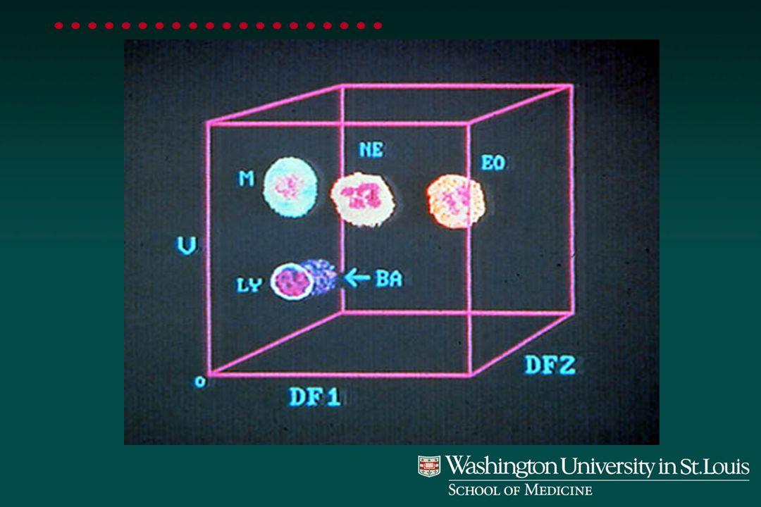
Coulter Principle

5
Red Cell Parameters

7
Red Cell Histogram and Count

8
Calculation of the RDW RDW = Coefficient of variation of red cell volume distribution Normal range = 11.5% - 14.5% RDW = X 100 S.D. Mean

9
Red Cell Distribution Width - RDW

10
Comparison of RDW in Iron Deficiency and Anemia of Chronic Disease

17
CBC Report

19
Automated Cell Counting: Deficiencies Abnormalities and inclusions in WBC RBC shape abnormalities RBC inclusions Platelet abnormalities and clumping

20
Peripheral Blood Morphology

21
Normal Peripheral Smear

22
“More information can be gained from examining the blood smear than from any single hematologic procedure”

23
Reticulocyte: Polychromasia

24
Reticulocyte Manual Count by Supravital Stain: Normal Count

25
Reticulocytes: Elevated Count

26
Erythrocyte Inclusions with Wright’s Stain InclusionCompositionAppearance Condition Basophilic PrecipitatedEvenly dispersedLead poisoning stipplingribosomesfine or coarse granulesthalassemia other anemias Howell-Jolly Nuclear Dense, round Post-splenectomy bodiesfragmentblue granule PappenheimerIron-containingSmall blue granulesAnemias bodiesgranulesin clusters OrganismSmall blue inclusionMalaria Babesiosis

27
Basophilic Stippling

28
Howell-Jolly Body

29
Malaria

30
RBC Inclusions: Composite

31
Erythrocyte Distribution Abnormalities Rouleaux formationStacking of RBCs due to increased plasma proteins coating RBCs AgglutinationAntibody-mediated clumping; temperature dependent

32
Rouleaux Formation

33
Agglutination Reaction

34
Variations in RBC Size and Shape AnisocytosisVariations in size (e.g. microcytes) PoikilocytosisVariations in shape (e.g. target cells) HypochromiaIncreased central pallor due to decrease in hemoglobin
 PoikilocytosisVariations in shape (e.g. target cells) HypochromiaIncreased central pallor due to decrease in hemoglobin.")
35
Hypochromic Microcytic RBC

36
Normal Hypochromic microcytic

37
Hypochromia without Anisocytosis: Thalassemia Trait

38
Severe Hypochromia: Iron Deficiency Anemia

39
Mixed Population: Treated Iron Deficiency Anemia

40
Microcytic Hypochromia: Alpha Thalassemia ( -/--)
")
41
Microcytic Hypochromia: Beta Thalassemia Major

43
Macrocytic Anemia: Macro-Ovalocytes

44
Shape Abnormalities of Erythrocytes TerminologyDescriptionCondition Target cellsCentral hemoglobin; target-shapedLiver disease; thalassemia: Abnormal Hgb; iron deficiency EchinocyteShort spicules, equally-spacedUremia, hypokalemia, artifact AcanthocyteSpiculated, irregularLiver disease (alcohol), Post-splenectomy SpherocyteSpherical, no central pallorHS, Immune hemolytic anemia SchistocyteFragmented RBC, helmet cells MAHA, burns OvalocyteOval/elliptical shapedHereditary elliptocytosis, Megaloblastic anemia Sickle cellbipolar spiculated shapeHgb S-containing “banana” shapedhemoglobinopathy Teardrop cellsingle elongated extremityMyelophthistic changes Bite cellsIrregular gap in membrane G6PD deficiency
, Post-splenectomy SpherocyteSpherical, no central pallorHS, Immune hemolytic anemia SchistocyteFragmented RBC, helmet cells MAHA, burns OvalocyteOval/elliptical shapedHereditary elliptocytosis, Megaloblastic anemia Sickle cellbipolar spiculated shapeHgb S-containing banana shapedhemoglobinopathy Teardrop cellsingle elongated extremityMyelophthistic changes Bite cellsIrregular gap in membrane G6PD deficiency")
45
Target Cells Diagnostic possibilities Liver disease Hemoglobinopathy Thalassemia Iron deficiency Post-splenectomy Lipid disorders

46
Echinocytes (Burr Cells)
")
47
Acanthocytes (Spur Cells)
")
48
Target Cells Spur Cells Morphologic Changes in Liver Disease

49
Hepatorenal Syndrome: Burr + Spur Cells

50
Spherocytes

51
Spherocytes: Autoimmune Hemolytic Anemia

52
Spherocytes: Hereditary Spherocytosis

53
Schistocytes: Microangiopathic Hemolytic Anemia

54
Elliptocytes: Hereditary Elliptocytosis

55
Sickle Cell Anemia: Hgb SS

56
Hemoglobin SC Disease

57
Hemoglobin S-Beta Thalassemia

58
Homozygous Hemoglobin C Disease (Hgb CC)
")
59
Teardrop Cells

60
Bite Cells

61
Heinz Bodies

62
Morphology of Leukocytes Normal WBC populations Neutrophils (Granulocytes) Lymphocytes Monocytes Eosinophils Basophils
 Lymphocytes Monocytes Eosinophils Basophils")
63
Neutrophil

64
Eosinophil

65
Neutrophil Eosinophil

66
Monocytes

68
Small Lymphocyte

69
Small Intermediate Large Lymphocytes

70
Basophils

71
Granulocyte Inclusions or Variants Terminology Description Condition Dohle bodies Pale blue areas in Infections, pregnancy, cancer neutrophil cytoplasm Toxic Large purple granules Infection Granulation in neutrophil cytoplasm Vacuoles Transparent areas Infection, Toxin in neutrophil cytoplasm Hypersegmented ≥ 6 nuclear lobes Megaloblastic anemia Auer rods Reddish long needle-like Acute myeloid leukemia inclusions Ehrlichia Blue inclusions in Ehrlichia sp. monocytes/neutrophils

72
Dohle Bodies

73
Toxic Granulation

74
Toxic Granulation and Vacuole Formation

75
Hypersegmented Neutrophils

76
Auer Rod: Acute Myeloid Leukemia

77
Ehrlichia

78
Myeloid Leukemias and Leukemoid Reaction Bone marrow exam is almost always indicated Cytogenetic analysis Flow cytometry analysis

79
Neutrophilia: Leukemoid Reaction

80
Neutrophilia: CML

81
Pelger-Huet Abnormality

82
Acute Myeloid Leukemia: M1 Myeloblasts without Differentiation

83
Acute Myeloid Leukemia: M2 Myeloblasts with Some Differentiation

84
Acute Myeloid Leukemia: M3 Promyelocytic Leukemia

85
Acute Myeloid Leukemia: M4 Myelomonocytic Leukemia

86
Acute Myeloid Leukemia: M5 Monocytic Leukemia

87
Acute Myeloid Leukemia: M6 Erythroleukemia

88
Acute Myeloid Leukemia: M7 Megakaryocytic Leukemia

89
Abnormalities of Lymphocytes VariantMorphologic categories Atypical lymphsAbundant cytoplasm, RBC “skirting” Abnormal lymphsNuclear abnormalities i.e. clefts, folds, notches Plasmacytoid lymphsAbundant cytoplasm Hairy cellsCytoplasmic projections Sezary cellsDeeply folded nucleus ProlymphocyteLarge lymph with prominent nucleolus

90
Atypical (Reactive) Lymphocytes
 Lymphocytes")
92
Abnormal Lymphocytes

93
Plasmacytoid Lymphocytes

94
Plasma Cell: Plasma Cell Leukemia

95
Hairy Cell: Hairy Cell Leukemia

96
Sezary Cell

97
Prolymphocytes

98
Chronic Lymphocytic Leukemia (CLL)
")
99
CLL: Smudge Cells

100
CLL: Balloon Cells

101
Acute Lymphocytic Leukemia: L1

102
Acute Lymphocytic Leukemia: L2

103
Acute Lymphocytic Leukemia: L3 (Burkitts)
")
Similar presentations


. Complete Blood Count ( CBC)>")


>")


>")