Download presentation
Presentation is loading. Please wait.

1
Case of the Year Lyn Callaghan Advanced Neonatal Nurse Practitioner
Wishaw General Hospital

2
Background Baby boy born by SVD at 40+2 weeks
Spontaneous onset of labour No Infection risks Crash Call at delivery due to Shoulder Dystocia Baby born in good condition Apgars 9/1 9/5 Mum’s second baby Healthy pregnancy Blood Group 0 Negative Anti D during pregnancy

3
Newborn Examination Baby now 15 hours of age Mother – no concerns
Breast feeding well Passed urine and meconium Mother – no concerns Midwife – feels baby intermittently tachypnoeic On examination Pale No respiratory distress Normal tone Normal examination Note baby becomes tachypnoeic on handling Heart rate +/- 150 when settled

4
Do you think anything is wrong with the baby?
What do you think it could be? What do you do next?

5
What happened next? Chased Cord bloods Repeated FBC and bilirubin
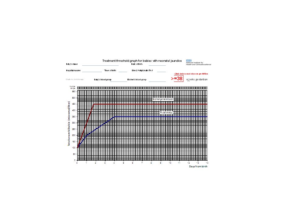
Informed mum of rationale At Birth Hb 15.5 HCT 0.56, WCC 19.0, Plat 204, Retics 5.5% Bili 24. Blood Group O pos DCT Negative At 15 hrs of age Hb 8.2 HCT 0.23, WCC 20.1, Plat 143, Retics 7.1%. Bili 28

7
Repeat FBC, Partial sepsis screen, DCT, Coagulation
Admit to NNU Repeat FBC, Partial sepsis screen, DCT, Coagulation Chest x-ray – normal Echo – normal structure Baby examines well Pale Tachypnoeic Tachycardia Mean BP normal At 17 hrs of age Hb 5.1, HCT 0.19, WCC 22, Plat 98. Retics 8.2% Bili 29 CRP <6 Coag mildly deranged Blood Group O pos DCT Neg

8
By 21hrs of age baby developed abdominal tenderness and guarding
Stabalisation Progress TUT Platlet transfusion FFP CRUSS Abdominal X-ray Abdominal Scan By 21hrs of age baby developed abdominal tenderness and guarding Differential diagnosis Haemorrhage ? Bowel ?Liver Transferred to Surgical Centre MRI Confirmed Diagnosis

9
Haemangioma of Liver Can cause Treatment Heart failure Anaemia
Thrombocytopenia Corticosteroids Embolization Hepatic Artery Ligation Liver Resection

10
Congenital Hepatic Haemangioma
Hepatic tumours account for 1-5% of all Paediatric tumours Hepatic Haemangioma is the third most commonest tumour of childhood Occasionally diagnosed on antenatal scan Often never detected Rarely present as large abdominal mass Cardiac failure due to massive atreriovenous shunting Jaundice from compression of bile ducts GI Bleeding Fever/illness resembling systemic inflammatory process

11
What is a haemangioma? A vascular birthmark caused by abnormal blood vessels in or under the skin Most common benign tumour of the vascular endothelium in infancy Can occur anywhere outside and inside of the body

12
What are the common types found on the newborn examination?
What do we tell the parents?

13
Salmon Patch Stork Bite/Mark Angels Kiss

14
Salmon Patch Pink maculae Dilated superficial capillaries
Commonly seen at nape of the neck, mid forehead and upper eyelids Most common vascular malformation Usually fades within a year

15
Strawberry Haemangioma
Bright red and sharply demarcated

16
Strawberry Haemangioma
Bright red vascular tumour Dilated mass of capillaries Usually protrudes above the skin Can appear anywhere on the body Can start as a flat red superficial lesion Can increase in size over next year

17
Strawberry Haemangioma
Usually, growth is complete and involution has commenced by twelve months. Half of all infantile haemangioma have completed involution by age five. 70% by age seven, and most of the remainder by age twelve In more severe cases haemangioma may leave residual tissue damage.

18
Strawberry Haemangioma Complications
Psychological Haemangioma near the eyes, nose, mouth, or on throat may interfere with vital functions and therefore require removal Some may break down and ulcerate Bleed Rarely can cause heart failure if large and blood being diverted into the excess blood vessels Lesions next to bone can also cause erosion of the bone The vast majority of hemangiomas are not associated with complications. Hemangiomas may break down on the surface, called ulceration. If the ulceration is deep, significant bleeding may occur in rare occasions. Ulceration on the deeper area can be painful and problematic. If a hemangioma develops in thelarynx, breathing can be compromised. A hemangioma can grow and block one of the eyes, causing an occlusionamblyopia. Very rarely, extremely large hemangiomas can cause high-output heart failure due to the amount of blood that must be pumped to excess blood vessels. Lesions adjacent to bone can also cause erosion of the bone

19
Strawberry Haemangioma Treatment
Oral corticosteriods Smaller lesions sometimes injected Propranalol Timolol gel Interferon Vincristine Surgical removal Pulsed dye laser For very early flat lesions Hogeling M et al. (August 2011). "A randomized controlled trial of propranolol for infantile hemangiomas".Pediatrics 128 (2): e259–e266. doi: /peds PMID
. A randomized controlled trial of propranolol for infantile hemangiomas .Pediatrics 128 (2): e259–e266. doi: /peds PMID")
20
Port Wine Stain Nevus Flammeus
Usually present at birth and on face, but can occur anywhere Permanent capillary angioma Does not blanch with pressure A vascular lesion – capillary malformation in the skin Does not disappear over time Also associated more rarely with Klipper Trenaunay Syndrome

21
Port Wine Stain Grows in proportion to general growth
Early stains are usually flat and pink – as child matures colour can deepen to a dark red or purple colour In adulthood can become bumpy and raised

22
Port Wine Stain Complications
Psychological Eye problems is on eyelid or next to eye If over forehead and upper lip Sturge Weber Syndrome must be excluded Spine abnormalities Varicose veins If the port-wine stain is around the eye or on the eyelid, a referral may be made to an optometrist or ophthalmologist for a test of the ocular pressures in that eye. If swelling occurs in the port-wine stain, it may cause vision problems,glaucoma, or blindness. 1 in 10 babies born with a port wine stain on the face will have problems with the eye or the brain The majority of children have no complications Brain abnormalities: are an uncommon association with port-wine stains of the face. This is due to extensive blood vessel abnormalities in the brain (the Sturge-Weber syndrome). Epilepsy and other problems may then develop.
. Epilepsy and other problems may then develop.")
23
Port Wine Stain Treatment
Some may improve over time Laser treatment variable results depending on skin colour Works best in young children Works best on smaller stains Skin camouflage still common treatment Some provided free on NHS Support Groups Birthmark Support Group British Association of Skin Camouflage Changing Faces Generally, the paler the port-wine stain, the greater the chance of excellent results. The laser can cause an unpleasant stinging. Therefore, younger children usually have laser treatment under a general anaesthetic or with sedation. Local anaesthetic is normally sufficient for older children and adults. Up to ten treatments are needed depending on the size. Treatments are given about eight weeks apart. It is best to have completed all sessions before a child reaches the age of 5 years. There may be pain, bruising and swelling over the treated area for a while after each session. If lasers don't work, surgery is sometimes required

24
Mongolian Blue Spot Congenital Dermal Melanocytosis
Benign , flat birthmark Irregular borders and shape Commonest colour is blue, but can also be blue-grey, blue-black or deep brown Normally disappears by yrs of age Almost certainly by puberty Important to document on record Prevalent among Mongolians Asians Malay Polynesians East African Latin Americans Turkish The Mongolian spot is a congenital developmental condition exclusively involving the skin. The blue colour is caused by melanocytes, melanin-containing cells, that are usually located in the epidermis but are in the deeper region of the skin known as the dermis in the location of the spot.[6] Usually, as multiple spots or one large patch, it covers one or more of the lumbosacral area (lower back), the buttocks, sides, and shoulders.[6] It results from the entrapment ofmelanocytes in the lower half to two-thirds of the dermis during their migration from the neural crest to the epidermis during embryonic development.[6] The condition is unrelated to sex; male and female infants are equally predisposed to Mongolian spot The Mongolian spot is referred to in the Japanese idiom shiri ga aoi (尻が青い), meaning "to have a blue butt",[12][13] which is a reference to immaturity or inexperience. In Mexico, where its name is the "green butt" (Spanish: rabo verde) it is referred to as la patada de Cuauhtémoc, meaning "Cuauhtémoc's kick". In Korea, it is thought that the Mongolian spot is the bruise formed when Samshin halmi (Korean: 삼신할미), a shaman spirit to whom people pray around child birth, has beaten in order for a baby to go out from his or her mother. In the comedy manga series Joshiraku written by Kōji Kumeta, as well as in its anime adaptation, the character Marii Buratei is known to have a Mongolian spot. In one sketch, a westerner notices the spot and mistakes it for child abuse, blaming "barbarian Japanese" and taking Marii to "safety" abroad, against her wishes.
, the buttocks, sides, and shoulders.[6] It results from the entrapment ofmelanocytes in the lower half to two-thirds of the dermis during their migration from the neural crest to the epidermis during embryonic development.[6] The condition is unrelated to sex; male and female infants are equally predisposed to Mongolian spot. The Mongolian spot is referred to in the Japanese idiom shiri ga aoi (尻が青い), meaning to have a blue butt ,[12][13] which is a reference to immaturity or inexperience. In Mexico, where its name is the green butt (Spanish: rabo verde) it is referred to as la patada de Cuauhtémoc, meaning Cuauhtémoc s kick . In Korea, it is thought that the Mongolian spot is the bruise formed when Samshin halmi (Korean: 삼신할미), a shaman spirit to whom people pray around child birth, has beaten in order for a baby to go out from his or her mother. In the comedy manga series Joshiraku written by Kōji Kumeta, as well as in its anime adaptation, the character Marii Buratei is known to have a Mongolian spot. In one sketch, a westerner notices the spot and mistakes it for child abuse, blaming barbarian Japanese and taking Marii to safety abroad, against her wishes.")
25
Questions/Discussion

Similar presentations


>")


