Download presentation
Presentation is loading. Please wait.

1
Heart Failure in the Transplant Patient
Presented by: Bethany Westerfeldt RN, CCRN, MSN, NP-C, ANP, BC For WITNS October 13, 2012

2
Objectives 1. Discuss types of Heart Failure
LV systolic dysfunction HF with preserved LV systolic function (HFpEF) aka diastolic dysfunction 2. Discuss brief pathophysiology of Heart Failure 3. Assessment of Heart Failure 4. Treatments of Heart Failure
 aka diastolic dysfunction. 2. Discuss brief pathophysiology of Heart Failure. 3. Assessment of Heart Failure. 4. Treatments of Heart Failure.")
4
Types of Heart Failure: Systolic Dysfunction
Many etiologies: Most common etiologies: CAD, HTN, DM, substance abuse(cocaine, alcohol), chemo, peripartum, uncontrolled tachycardia, viral, valvular heart disease, severe illness Affects cardiac output or quantity of blood pumped to rest of body Can be improved or controlled with medication and lifestyle changes Frequency of diagnosis increases with age, more prevalent after age 65 though affects pts at any age point Syndrome of neurohormonal activation affecting many body systems
, chemo, peripartum, uncontrolled tachycardia, viral, valvular heart disease, severe illness. Affects cardiac output or quantity of blood pumped to rest of body. Can be improved or controlled with medication and lifestyle changes. Frequency of diagnosis increases with age, more prevalent after age 65 though affects pts at any age point. Syndrome of neurohormonal activation affecting many body systems.")
5
Types of Heart Failure: Diastolic dysfunction
Defined as HF with preserved LV systolic function (normal LVEF) or EF more than 40% LV often with hypertrophy, concentric remodeling with increased extracellular matrix, abnormal relaxation and filling, decreased diastolic dispensability Shares activation of neurohormonal activation with systolic dysfunction Prevalence: 40-50% of all pts diagnosed with HF, often seen in conjunction with systolic dysfunction More commonly in elderly, females and pts with hypertension
 or EF more than 40% LV often with hypertrophy, concentric remodeling with increased extracellular matrix, abnormal relaxation and filling, decreased diastolic dispensability. Shares activation of neurohormonal activation with systolic dysfunction. Prevalence: 40-50% of all pts diagnosed with HF, often seen in conjunction with systolic dysfunction. More commonly in elderly, females and pts with hypertension.")
6
Heart Failure Classifications:
New York Heart Association (NYHA) functional classification of HF: No limitations of physical activity, no symptoms with ordinary activity Slight limitation, symptoms with ordinary activities Marked limitation, symptoms with less than ordinary activities Severe limitation, symptoms of HF at rest
 functional classification of HF: No limitations of physical activity, no symptoms with ordinary activity. Slight limitation, symptoms with ordinary activities. Marked limitation, symptoms with less than ordinary activities. Severe limitation, symptoms of HF at rest.")
7
ACC/AHA classification

8
Therapeutic Goals: Prolong life Slow/reverse cardiac remodeling
Reduce visits and admissions to hospital Improve overall functional capacity and quality of life Reduce dyspnea and fatigue Control/minimize edema and fluid retention

9
Transplant population:
At risk for conditions the increase HF risk (both types): Hypertensive disease, DM, sleep apnea and CAD Antirejection meds that increase BP, renal disease, risk DM Solid weight gain: risk for DM, HTN, sleep apnea, atrial arrhythmias Preexisting conditions: Limiting activity: Gout, arthritis/DJD, other musculoskeletal disorders, obesity Structural heart disease: valve disorders, storage diseases (amyloid, hemochromatosis, saroidosis)
: Hypertensive disease, DM, sleep apnea and CAD. Antirejection meds that increase BP, renal disease, risk DM. Solid weight gain: risk for DM, HTN, sleep apnea, atrial arrhythmias. Preexisting conditions: Limiting activity: Gout, arthritis/DJD, other musculoskeletal disorders, obesity. Structural heart disease: valve disorders, storage diseases (amyloid, hemochromatosis, saroidosis)")
14
CKD and CV disease: Known high risk of development of CVD in pt. with HTN Systolic BP more significant than DBP on CV all cause mortality Pulse pressure (SBP minus DBP) Independent predictor of MI, HF and CV death Reflects stiffness larger arteries Increases with advancing age from 50 yr. on up Reliable prognostic factor for mortality CKD Pts on HD or renal transplant patients
 Independent predictor of MI, HF and CV death. Reflects stiffness larger arteries. Increases with advancing age from 50 yr. on up. Reliable prognostic factor for mortality CKD. Pts on HD or renal transplant patients.")
17
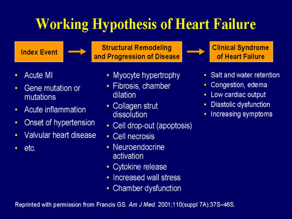
Neurohormonal effects of Heart Failure
SNS activity: “whipping a dying horse”-the catecholamine stimulation Stimulation of Alpha 1 & Beta 1&2 receptors Increases cardiac output by increasing heart rate& stroke volume Prolonged stimulation leads to myocyte hypertrophy, dilation, ischemia, arrhythmias, deterioration and death of cardiac cells Activates Renin-Angiotensin-Aldosterone System (RAAS)-> (further) vasoconstriction & sodium retention
-> (further) vasoconstriction & sodium retention.")
20
Prevalence of specific signs & symptoms in systolic & diastolic HF

21
Signs & Symptoms Dyspnea & fatigue Limitation of exercise tolerance
Fluid retention-may lead to pulmonary edema PND & orthopnea Cough, can be primarily nocturnal Poor appetite Early satiety & bloating Chest pain: not always in pts with CAD Palpitations Lightheadedness Especially positional Edema, ascites, anasarca Poor sleep quality Difficulty thinking clearly, or concentrating These abnormalities can impair functional capacity & quality of life

22
Clinical signs: Increased adrenergic activity
peripheral vasoconstriction (cool extremities) pallor &/or cyanosis of digits diaphoresis tachycardia loss of normal sinus rhythm distention (obvious) of peripheral veins due to vasoconstriction (prominent JVD) narrowed pulse pressure
 pallor &/or cyanosis of digits. diaphoresis. tachycardia. loss of normal sinus rhythm. distention (obvious) of peripheral veins due to vasoconstriction (prominent JVD) narrowed pulse pressure.")
23
Clinical signs Pulmonary rales & /or wheezes:
usually an acute heart failure finding Chronic HF pts mobilize pulm fluid into lymph nodes: are enlarged on x-ray & CT JVD: indication of right atrial pressure (preload) Normal JVD is under 10 cm HJR: compression over liver causes distention of JVD further Indicates congested abdomen/liver and/or inability of right heart to accept or eject the transiently increased volume
 Normal JVD is under 10 cm. HJR: compression over liver causes distention of JVD further. Indicates congested abdomen/liver and/or inability of right heart to accept or eject the transiently increased volume.")
24
LV systolic dysfunction
Ischemic: CAD is the cause of approximately 2/3 of pts LV dysfunction Remodeling: change in tissue & geometry of LV Neurohormonal changes: affect endothelium of vessels as well as cardiac muscle with deposition of fibrinous material alters contraction and electrical pathways of myocardium Neurohormonal activation of Renin-Angiotensin-Aldosterone system & Norepinephrine Down-regulation of alpha, beta 1 & beta 2 adrenergic receptors

25
Treatment of Systolic HF
Diuretics, ACE-Inhibitors, Beta blockers, Digoxin, Aldosterone blockers, Vasodilators & IV Inotropic agents Value of these agents shown in multiple clinical trials Shown to decrease symptoms & increase length/quality of life

27
Diastolic Dysfunction
Abstracted charts from 37,500 Medicare pts from National Heart Failure Project database Only 57% of pts had LV fxn assessed Of 19,710 pts with documented EF, 1/3 had EF> 50% This group was 79% women with mean age of 79.7 yrs

28
Characteristics of Outpatient Diastolic HF
Overwhelmingly female (85%) & elderly(70 yo) Marked increase in LV mass/volume ratio Higher BMI More likely to have HTN Exercise limitation similar to systolic HF pts
 & elderly(70 yo) Marked increase in LV mass/volume ratio. Higher BMI. More likely to have HTN. Exercise limitation similar to systolic HF pts.")
29
Diastolic Dysfunction
Increased myocardial stiffness Ventricular interaction or pericardial restraint Abbreviated LV filling time Multifactorial: thyrotoxicosis, AV fistula, beriberi Volume overload stress Obesity Impaired LV relaxation Myocardial ischemia Hypertrophy Systolic dysfunction DM Hypothyroidism

30
Diastolic Dysfunction Diagnostic Criteria
Required criteria: Normal EF (> 50%) Clinical Evidence of Heart Failure Framingham of Boston Criteria Plasma BNP &/or chest x-ray Cardiopulmonary exercise testing Confirmatory Criteria: LVH or Concentric Remodeling Left Atrial Enlargement (in absence of AF) Echo Doppler or Cath Evidence of Diastolic Dysfunction Exclusions: Non-Myocardial Disease
 Clinical Evidence of Heart Failure. Framingham of Boston Criteria. Plasma BNP &/or chest x-ray. Cardiopulmonary exercise testing. Confirmatory Criteria: LVH or Concentric Remodeling. Left Atrial Enlargement (in absence of AF) Echo Doppler or Cath Evidence of Diastolic Dysfunction. Exclusions: Non-Myocardial Disease.")
31
Treatment of Diastolic HF
Class I Control systolic & diastolic BP Control ventricular rate in AF Use diuretics to control edema Class IIA Coronary revascularization Class IIB Restore & maintain NSR Beta blocker, ACE-I, ARB, Calcium antagonists, digitalis

32
Treatment of DHF General approach: Symptom reduction
Control blood pressure Decrease circulating volume Salt & fluid restriction Diuretics Nitrates Neurohormonal blockade (ACEI/ARB) Treat tachycardia Increase duration of diastole (slow HR) in select pts Maintain synchronous atrial contraction
 Treat tachycardia. Increase duration of diastole (slow HR) in select pts. Maintain synchronous atrial contraction.")
33
Treatment DHF Lifestyle modification & education
Salt & fluid restriction Exercise program Other dietary concerns Weight control Target underlying mechanisms Drugs: that improve calcium homeostasis blunt hormonal activation prevent & regress fibrosis are in existence or development

34
Treatment of DHF Look at underlying disease processes contributing to DHF & choose drug combinations to combat them Eg: Diabetic elderly female Angiotensin blocking drug Diuretic beta blocker CCB if resting HR>70. Most often requires antihypertensive Lots of trial & error to finding right combination of meds that minimize symptoms & side effects

35
Patient self care at home: systolic & diastolic HF
Weigh daily same time same clothing amount Report daily gain of >3 lbs. or overall >5 lbs. Low salt diet (2 grams) Take all meds as prescribed Report any side-effects or problems with meds Know symptoms of HF & report worsening: SOB or decreased activity tolerance Increased fatigue, unable to sleep lying down Swelling of ankles or abdomen Frequent colds Decreased urination, increase in weight
 Take all meds as prescribed. Report any side-effects or problems with meds. Know symptoms of HF & report worsening: SOB or decreased activity tolerance. Increased fatigue, unable to sleep lying down. Swelling of ankles or abdomen. Frequent colds. Decreased urination, increase in weight.")
36
Pt. care at home: Participate in regular exercise & stress reduction
Plan daily activities in advance to conserve energy Plan strategies to help reduce fatigue: delegate jobs take naps or rest periods Withdrawal of meds known to affect clinical status: NSAIDS antiarrhythmic most calcium channel blockers

37
Lifestyle & monitoring
OTC medications to stay away from: NSAIDS: ASA (high dose) ibuprofen naproxen Decongestants: Sudafed anything with ephedrine Most diet pills
 ibuprofen. naproxen. Decongestants: Sudafed. anything with ephedrine. Most diet pills.")
38
Case Study 44 yo male, previous hx DM and DD renal tx ‘05, no known CAD (negative stress prior to tx) Presents with 4-6 wk hx progressive DOE, weight gain, decreased appetite, cough that worsens at night States he’s having hard time at work, carries 60 lb. bags & other equipment road construction Believes he has “bad cold”, treated by PCP for bronchitis/pneumonia without relief of symptoms

39
Case study Sx not improved after steroidsabx
History reveals change in appetite, bloating, PND-wakes up after 2 hrs coughing, sits on side of bed or walks around Reports chest “soreness”, mid sternal area, no radiation, no palpitations, no diaphoresis, n/v What would you suspect? What would you do next?

40
Case study CXR shows cardiomegaly, pulmonary congestion
12 lead ecg shows NSR no evidence of acute MI normal voltage TTE shows LV systolic dysfunction LVEF 25% moderately dilated RA & LA normal RV function LVEDD 65 mm (nl= 56) no thrombus Labs: lytes wnl, bun/cr:35/1.8, t bili 1.6, ast 66, alt 94, TSH 8.7 with normal fT4, TT3.
 no thrombus. Labs: lytes wnl, bun/cr:35/1.8, t bili 1.6, ast 66, alt 94, TSH 8.7 with normal fT4, TT3.")
41
Treatment If possible: Start or increase ACE inhibitor or ARB Diurese
Once near euvolemic Start or increase beta blocker (preferably carvedilol or metoprolol succinate)
")
42
6 months later… Calls with SOB without weight gain Appetite poor
Decreased activity tolerance Increased fatigue PND and orthopnea

43
What to do next? Assess over the phone: Labs:
What brings on SOB/activity limitations Assess for edema Other symptoms: chest discomfort palpitations Early satiety, bloating, nausea… Labs: Creatinine rise from 1.4 to 1.8 BUN rise from 28 to 36 Serum potassium 3.6, sodium 130

44
What do you do next? Most likely, increased renal indices from volume overload Diurese, based on symptoms and labs Leave ACE-I unchanged F/u labs in 3-7 days If equivocal, get pt to be assessed in clinic TTE can be done in pts difficult to assess volume status Can always schedule for RH cath (may need anticoag management in pts on warfarin)
")
45
Case study 2 78 yo female s/p OH Tx ‘02 presents with progressive SOB, weight gain longstanding hx htn obesity swelling of her legs bloating over past couple of months Hx reveals: poorly controlled BP for past 45 yrs sedentary lifestyle no DM hypothyroid States she’s been sleeping in her husband’s recliner for past 3 weeks sleep quality poor r/t fatigue poor quality of life “unable to do anything”

46
What do you suspect? What questions do you want to ask her?
What do you want to do with her?

47
Case Study 2 Physical exam: JVP elevated to 15 cm above RA
No scleral icterus Oropharynx pink, moist Lungs with few basilar crackles & wheezes Abdomen soft, obese; Liver WNL Extremities +2 to knees bilaterally Blood pressure: 178/92

48
Findings CXR shows pulmonary congestion TTE shows: BNP elevated to 528
concentric LV hypertrophy abnormal E/a ratio (diastolic filling) LVEF 75% BNP elevated to 528 lytes normal creatinine 2.4 LFT’s normal 12 lead ecg shows: increased voltage in precordial leads ( LV hypertrophy) NSR Left heart cath with minor luminal irregs RHC with elevated PCWP, RA,RV and PA pressures
 LVEF 75% BNP elevated to 528. lytes normal. creatinine 2.4. LFT’s normal. 12 lead ecg shows: increased voltage in precordial leads ( LV hypertrophy) NSR. Left heart cath with minor luminal irregs. RHC with elevated PCWP, RA,RV and PA pressures.")
49
CXR

50
What do you do next? Diurese Labs to evaluate lytes and renal function
Helps relieve symptoms Labs to evaluate lytes and renal function Can schedule for clinic visit Trend home monitoring data

51
Thank you!

Similar presentations


 Heart failure is the pathophysiological state in which an abnormality of cardiac function is responsible for failure of the heart to.>")


 Brunner, ch. 30, pp. 824-840.>")


 Class IV: symptoms at rest Class III: symptoms on less-than-ordinary exertion Class.>")


. “Heart (or cardiac) failure is the pathophysiological state in which the heart is unable to pump blood at a rate commensurate.>")
 failure is the state in which the heart is unable to pump blood.>")
