Download presentation
Presentation is loading. Please wait.

1
Anatomy & Physiology of the Female Reproductive Tract

4
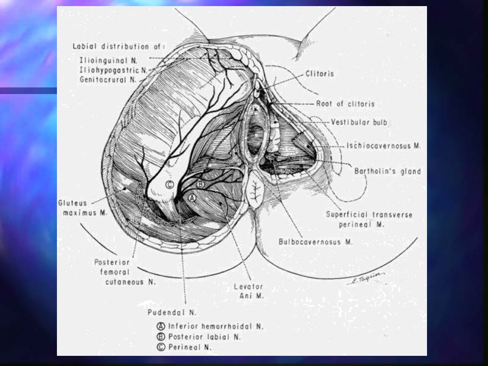
External Genital Organs
mons pubis labia majora labia minora prepuce (clitoral hood) frenulum of the labia minora = fourchette vestibule of the vagina external urethral orifice paraurethral glands (Skene’s glands) [prostate] Bartholin's gland
 frenulum of the labia minora = fourchette. vestibule of the vagina. external urethral orifice. paraurethral glands (Skene’s glands) [prostate] Bartholin s gland.")
6
Pubococcygeus Muscle main part of levator ani injuries
most likely muscle to be damaged during childbirth supports the bladder, urethra, vagina, and rectum injuries cystocele cystourethrocele or urethrocystocele rectocele urinary stress incontinence (weakening of pubovaginalis part of levator ani) => Kegel exercise
 => Kegel exercise.")
8
venous drainage: internal pudendal veins
vaginal orifice hymen greater vestibular glands Bartholin’s glands [bulbourethral glands] arterial supply two external pudendal arteries one internal pudendal artery venous drainage: internal pudendal veins

11
Lymph Drainage The external genitalia, anus, and anal canal drain to the superficial inguinal nodes. The lower one third of the vagina drains to the sacral nodes and the internal and common iliac nodes. The cervix drains to the external or internal iliac and sacral nodes

12
Lymph, cont’d The lower uterus drains to the external iliac nodes
The upper uterus drains into the ovarian lymphatics to the lumbar nodes. The lymphatics of the ovaries drain out of the pelvis to the lumbar nodes

14
Innervation ilioinguinal nerve
genital branch of the genitofemoral nerve perineal branch of the femoral cutaneous nerve of thigh perineal nerve

16
Pelvic Viscera Urogenital organs…
bladder, uterus, adnexa, and rectum Also have…the sigmoid colon, cecum, and ileum are components of the pelvic anatomy.

17
Pelvic Viscera urinary organs rectum ureters urinary bladder
pass medial to origin of uterine artery and continues to level of ischial spine, where is crossed superiorly by the uterine artery. Then passes close to lateral portion of vaginal fornix and enters posterosuperior angle of bladder urinary bladder hollow viscus with strong muscular walls trigone of bladder urethra - about 4 cm long, anterior to vagina rectum

18
Ligaments round ligament of uterus - attaches anterior-inferiorly to uterotubal junctions ligament of ovary - attached to uterus, posterior-inferior to uterotubal junctions broad ligament - encloses body of uterus, freely moveable transverse cervical ligaments - extend from cervix and lateral parts of vaginal fornix to lateral walls of pelvis uterosacral ligaments - pass superiorly and slightly posteriorly from sides of cervix to middle of sacrum, can be palpated through rectum as pass posteriorly at sides of rectum. Hold cervix in normal relationship to sacrum.

20
Broad Ligament Contains between its layers the fallopian tube; the ovary and the round ligament; the uterine and ovarian blood vessels, nerves, lymphatics, and fibromuscular tissue; and a portion of the ureter as it passes lateral to the uterosacral ligaments over the lateral angles of the vagina and into the base of the bladder

21
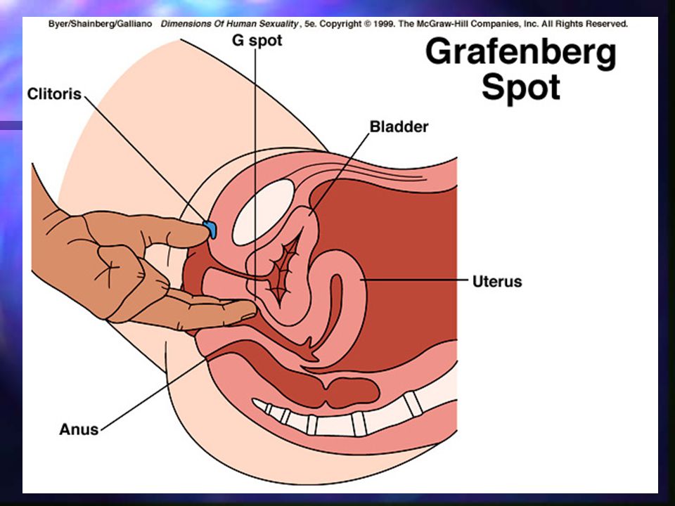
Internal Genital Organs
vagina fornix rectouterine pouch (pouch of Douglas) sphincters of vagina pubovaginalis muscle urogenital diaphragm bulbospongiosus muscle lymphatic drainage superior part into internal and external iliac lymph nodes middle part into the internal iliac lymph nodes vestibule into superficial inguinal lymph nodes
 sphincters of vagina. pubovaginalis muscle. urogenital diaphragm. bulbospongiosus muscle. lymphatic drainage. superior part into internal and external iliac lymph nodes. middle part into the internal iliac lymph nodes. vestibule into superficial inguinal lymph nodes.")
23
Uterus 7-8 cm long, 5-7 cm wide, 2-3 cm thick
projects superior-anteriorly over urinary bladder two major parts body (superior 2/3s) fundus cervix (inferior 1/3) internal os external os anterior lip posterior lip lined with columnar, mucus-secreting epithelium isthmus = a transitional zone between body and cervix
 fundus. cervix (inferior 1/3) internal os. external os. anterior lip. posterior lip. lined with columnar, mucus-secreting epithelium. isthmus = a transitional zone between body and cervix.")
25
wall of uterus consists of 3 layers:
Perimetrium/serosa - outer serous coat, peritoneum supported by thin layer of connective tissue myometrium mm smooth muscle, main branches of blood vessels and nerves of uterus are in this layer endometrium - inner mucous coat

26
uterine tubes 10-12 cm long, 1 cm diameter
extend laterally from cornua of uterus 4 parts infundibulum distal end abdominal ostium, about 2 mm in diameter 20-30 fimbriae ovarian fimbria is attached to ovary ampulla tortuous part widest and longest part, over 1/2 its length fertilization occurs here Most common site for ectopic

28
isthmus short 2.5 cm, narrow, thick-walled part of tube that enters the uterine cornu uterine part short segment that passes through thick myometrium of uterus uterine ostium (smaller than abdominal ostium)
")
30
Ovaries oval, almond-shaped, 3 cm long, 1.5 cm wide, 1 cm thick
ligaments superior (tubal) end of ovary is connected to lateral wall of pelvis by suspensory ligament of the ovary contains ovarian vessels and nerves ligament of ovary - connects inferior (uterine) end of ovary to lateral angle of uterus surface of ovary is not covered by peritoneum oocyte expelled into peritoneal cavity
 end of ovary is connected to lateral wall of pelvis by suspensory ligament of the ovary. contains ovarian vessels and nerves. ligament of ovary - connects inferior (uterine) end of ovary to lateral angle of uterus. surface of ovary is not covered by peritoneum. oocyte expelled into peritoneal cavity.")
31
Pelvis The bony and ligamentous pelvic mechanism is designed to…
protect the pelvic viscera support the vertebral column facilitate locomotion The pelvic girdle protects the viscera contained within its cavity from all ordinary trauma

32
Pelvis The bony pelvis is formed anteriorly and laterally by the innominate bones and posteriorly by the sacrum and coccyx The pelvic girdle is adapted for strength, support, and locomotion. In the erect position, the pelvic girdle is inclined forward.

35
Man vs. Woman The female pelvic inlet is oval; the male pelvic inlet is heart shaped. The female pelvis has a more regular outline than the male pelvis, in which the sacral promontory is more prominent and the sacrum is longer and more curved.

36
Female Bony Pelvis wider, shallower, and has larger superior and inferior pelvic apertures than male pelvis hip bones farther apart ischial tuberosities are farther apart because of wider pubic arch sacrum is less curved, which increases the size of the inferior pelvic aperture and the diameter of the birth canal obturator foramina is oval

37
Types of Bony Pelvis anthropoid = AP diameter > transverse diameter
23% females platypelloid uncommon android = wide transverse diameter, posterior part of aperture is narrow 32% females gynecoid = most spacious obstetrically 43% females

39
Superior Pelvic Aperture
AP diameter = measurement from the midpoint of the superior border of pubic symphysis to the midpoint of sacral promontory transverse diameter = greatest width, measured from linea terminalis on one side to this line on opposite side

40
determine prominence of ischial spines
oblique diameter = measurement from one iliopubic eminence to the opposite sacroiliac joint midplane diameter = interspinous diameter or distance between ischial spines and cannot be measured. Is estimated by palpating the scarospinous ligament through the vagina. The length of this ligament = about half the midplane diameter. determine prominence of ischial spines < 9.5 cm may prevent passage of fetus

41
Physiology Hypothalamus Anterior Pituitary Ovary
Endometrium & outflow tract

42
Hypothalamus Release of GnRH (gonadotropin-releasing hormone), also called LHRH, into the pituitary portal circulation via the pituitary stalk The menstrual cycle does not ‘begin’ here!! All are inter-related !

43
Hypothalamus What triggers the release of GnRH?
Unclear but in animal studies dopamine is inhibitory & norepinephrine is stimulatory For normal gonadotropin release, GnRH must be released in pulses. The pulse frequency & amplitude are critical for normal menses Decrease in pulse frequency will decrease LH release & increase FSH Increase pulse frequency will increase LH & decrease FSH

44
Anterior Pituitary Gonadotrophs respond to the GnRH by producing FSH (follicle stimulating hormone) & LH (Luteinizing hormone) into the general circulation Release at this level is also controlled by circulating levels of estrogen & progesterone (gonadal steroids)…positive & negative feedback
 & LH (Luteinizing hormone) into the general circulation. Release at this level is also controlled by circulating levels of estrogen & progesterone (gonadal steroids)…positive & negative feedback.")
45
Anterior Pituitary Stores & releases FSH & LH
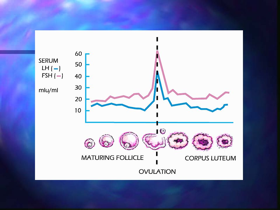
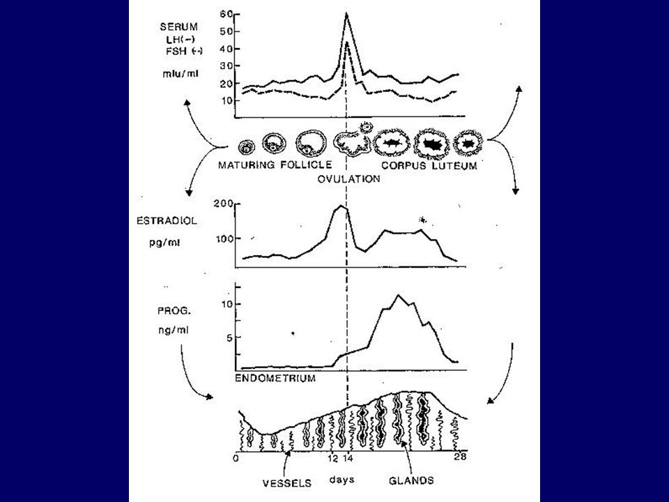
Day 1-7, follicular phase: estrogen from the ovary will stimulate storage of FSH & LH(in the pituitary)…also inhibits secretion Later in follicular phase with increasing estrogen levels (enlarging follicle) effect on gonadotrophs changes to stimulatory allowing for a secretion of LH which triggers ovulation
…also inhibits secretion. Later in follicular phase with increasing estrogen levels (enlarging follicle) effect on gonadotrophs changes to stimulatory allowing for a secretion of LH which triggers ovulation.")
47
Under the influence of LH, the follicle begins to secrete progesterone shortly before ovulation
Low level of progesterone will induce the FSH surge that occurs immediately prior to ovulation

48
FSH Surge matures the oocyte (stimulates gametogenesis
produces proteolytic enzymes needed for follicle rupture Increases the # of LH receptors(ovarian) required for optimal progesterone production in the luteal phase
 required for optimal progesterone production in the luteal phase.")
49
LH surge increase in intrafollicular proteolytic enzymes that destroy the basement membrane and allow follicular rupture luteinization of the granulosa cells and theca, resulting in increased progesterone production resumption of meiosis in the oocyte, thus preparing it for fertilization an influx of blood vessels into the follicle, preparing it to become a corpus luteum.

51
After ovulation, the secretion of estrogen & progesterone in high concentrations from the corpus luteum inhibits both gonadotrophs & GnRH As the corpus luteum dies off the hormone levels subside & FSH resumes the cycle

52
Ovary By the fifth week of embryonic life, germ cells have formed the ovary Maximum # of eggs the ovary is able to produce is at 20 weeks of gestation… 6-7 million! 1-2 million at birth 300,000 at the onset of puberty!

54
Ovary The functional unit is the FOLLICLE
Oocyte (frozen in the first stage of meiosis) surrounded by granulosa cells & adjacent stromal cells…Theca cells. FSH will target the granulosa cells LH will target the thecal & stromal cells
 surrounded by granulosa cells & adjacent stromal cells…Theca cells. FSH will target the granulosa cells. LH will target the thecal & stromal cells.")
55
Ovary, cont’d As the follicle matures, Antrum develops around the oocyte A bunch of follicles will develop around day 7 of cycle…a dominant follicle will win!

57
Ovary cont’d Rising estrogen levels from the maturing follicle itself will ‘prime’ the follicle for the LH surge. When estrogen levels reach 200pg/ml or greater for longer than 48 hours, the LH surge occurs The granulosa cells become luteinized just prior to ovulation & begin to produce progesterone

58
Progesterone rise is responsible for...
Facilitates the positive feedback action of estradiol in initiating the LH surge LH surge occurs about 36 hours prior to ovulation Responsible for the FSH peak

60
Ovary An avascular area will develop on the wall of the follicle & with the help of proteolytic enzymes ovulation occurs. The oocyte is picked up by the fimbriae of the tube If not met by a sperm will degenerate in hours!

61
Ovary After ovulation, luteinization will transform the ruptured follicle into a corpus luteum which produces estrogen & progesterone for the next days If not aided by secretion of hCG, the corpus luteum will become the corpus albicans

62
Androgens Androstenedione & testosterone are also secreted & can alter the ability of the ovary to respond to FSH & LH…may create atretic follicles early on

63
TWO CELL THEORY …of ovarian steroidogenesis
Theca cells produce androgens under the influence of LH Granulosa cells convert the androgens to estrogen under the influence of FSH

64
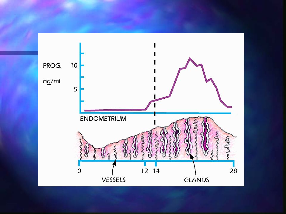
Endometrium Contains receptors for both estradiol & progesterone
Estradiol causes the proliferation, steady increase in thickness of lining When the corpus luteum starts producing progesterone; the proliferative effect of estradiol is neutralized & endometrial growth ceases

66
Endometrium The lining now becomes SECRETORY with the endometrial vessels coiling & preparing to shed If no baby… corpus luteum stops producing estrogen & progesterone. This withdrawal of steroid support from the endometrium causes endometrial breakdown

67
Why don’t women bleed to death every month??
Vascular spasm Thrombosis Resumption of endometrial proliferation under the influence of unopposed estrogen Myometrial ischemia - dysmenorrhea

Similar presentations