Download presentation
Presentation is loading. Please wait.

1
Anatomy and Physiology: The Digestive System
to accompany

2
Overview 1 Gastrointestinal (GI) Tract 2 Accessory Organs of the Head 3 Swallowing 4 Stomach 5 Accessory Organs of the Abdomen 6 Small Intestine 7 Large Intestine 8 Phases of Digestion 9 Food Molecules
 Tract 2 Accessory Organs of the Head 3 Swallowing 4 Stomach 5 Accessory Organs of the Abdomen 6 Small Intestine 7 Large Intestine 8 Phases of Digestion 9 Food Molecules")
3
Essential Terms digestion
process of mechanically or chemically breaking down food absorption passage of small molecules into blood and lymph digestive system organs which carry out process of digestion and absorption metabolism all the chemical reactions of the body

4
Introduction Digestive System
Composed of GI tract and accessory organs Breaks down ingested food for use by the body Digestion occurs by mechanical and chemical mechanisms Excretes waste products or feces through process of defecation

5
GI Tract / Alimentary Canal
Continuous tube from mouth to anus Mouth Pharynx Esophagus Stomach Small intestine Large intestine

6
Accessory Digestive Organs
Provide mechanical and chemical mechanisms to aid digestion Teeth Tongue Salivary glands Liver Gallbladder Pancreas

8
Functions of Digestive System
Ingestion Secretion Mixing and propulsion Motility Digestion Mechanical and chemical Absorption Defecation

9
Layers of GI Tract Same in all areas of GI tract
From deep to superficial: Mucosa Submucosa Muscularis Serosa

10
Figure 23.2

11
Layers of GI Tract Mucosa Submucosa Epithelium
Type varies Lamina propria – areolar connective tissue MALT – mucus-associated lymphatic tissue Muscularis mucosae – smooth muscle Submucosa Areolar connective tissue Blood and lymphatic vessels Neurons – submucosal plexus

12
Layers of GI Tract Muscularis Serosa Skeletal and smooth muscle
Neurons – myenteric plexus Serosa Areolar and simple squamous epithelium Visceral peritoneum

13
Peritoneum Mesothelium Parietal peritoneum Visceral peritoneum
Peritoneal cavity Retroperitoneal

14
Figure 23.3a

15
Figure 23.3b

16
Figure 23.3c

17
Figure 23.3d

18
Neural Innervation of GI Tract
Regulated by autonomic nervous system Enteric division Myenteric plexus / plexus of Auerbach Submucosal plexus / plexus of Meissner Able to function independently from rest of nervous system Linked to CNS by extrinsic sympathetic and parasympathetic nerves Sympathetic nerves decrease GI secretions & motility Parasympathetic nerves increase GI secretion and motility

19
Mouth Parts of Digestive System
Mouth formed by several parts: Cheeks Lips / labia Labial frenulum Orbicularis Vestibule Oral cavity proper Fauces Hard and soft palate Uvula Palatoglossal and palatopharyngeal arch

20
Figure 23.4

21
Tongue Skeletal muscle and mucous membrane
Helps form floor of oral cavity Extrinsic muscles Intrinsic muscles Lingual frenulum Papillae Fungiform Filiform Circumvallate Foliate Lingual glands Lingual lipase

22
Salivary Glands Release saliva to oral cavity
3 pairs of salivary glands Parotid Submandibular Sublingual

23
Composition of Saliva 99.5 % water 0.5% other solutes
Ions Mucus Immunoglobulin A Enzymes Salivation controlled by autonomic nervous system Stimulated by various mechanisms

24
Figure 23.5

25
Teeth External regions Internal components Crown Root Neck Enamel
Dentin Cementum Pulp cavity PulpRoot canals Apical foramen

26
Figure 23.6

27
Teeth Dentitions Deciduous teeth – first set
Permanent teeth – secondary Carry out mechanical digestion by mastication Creates bolus Salivary amylase Breakdown starch Lingual lipase Breakdown triglycerides

28
Figure 23.7

29
Pharynx Composed of skeletal muscle Lined by mucous membrane
Nasopharynx Oropharynx Laryngopharynx

30
Esophagus Collapsible muscular tube through esophageal hiatus of diaphragm Mucosa Submucosa contains areolar connective tissue Muscularis Skeletal muscle Upper and lower esophageal sphincter Adventitia Attaches esophagus to nearby structures Secrets mucus and transports food

31
Figure 23.8

32
Deglutition Stages of swallowing Voluntary Pharyngeal Esophageal
Mouth to oropharynx Pharyngeal Deglutition center in medulla oblongata and pons Closing of epiglottis Involuntary Esophageal Peristaltic contractions

33
Figure 23.9a,b

34
Figure 23.9c

35
Table 23.2

36
Stomach Serves as mixing chamber and storage area for ingested food
Rugae allow for increased volume 4 main regions Cardia Fundus Body Pylorus Pyloric antrum and canal Pyloric sphincter Lesser and greater curvatures

37
Figure 23.10a

38
Stomach Histology Mucosa Submucosa – areolar connective tissue
Surface mucous cells Lamina propria Muscularis mucosae Gastric glands and pits Parietal cells Chief cells G cells Submucosa – areolar connective tissue Muscularis 3 layers of smooth muscle Serosa

39
Figure 23.11a

40
Figure 23.11b

41
Mechanical and Chemical Digestion
Mixing waves caused by peristaltic movement Chyme released in process of gastric emptying Proton pumps bring H+ into the lumen Carbonic anhydrase forms carbonic acid to provide H+ and bicarbonate ions (HCO3-)
")
42
Figure 23.12

43
Mechanical and Chemical Digestion
Chemical digestion stimulated by nervous system Parasympathetic neurons release acetylcholine Works with gastrin HCl released in presence of histamine Pepsin begins digestion of proteins Stomach protected by alkaline mucus secretion Gastric lipase digests triglycerides Few molecules absorbed by stomach Water, ions, short-chain fatty acids, alcohol

44
Table 23.3 pt 1

45
Table 23.3 pt 2

46
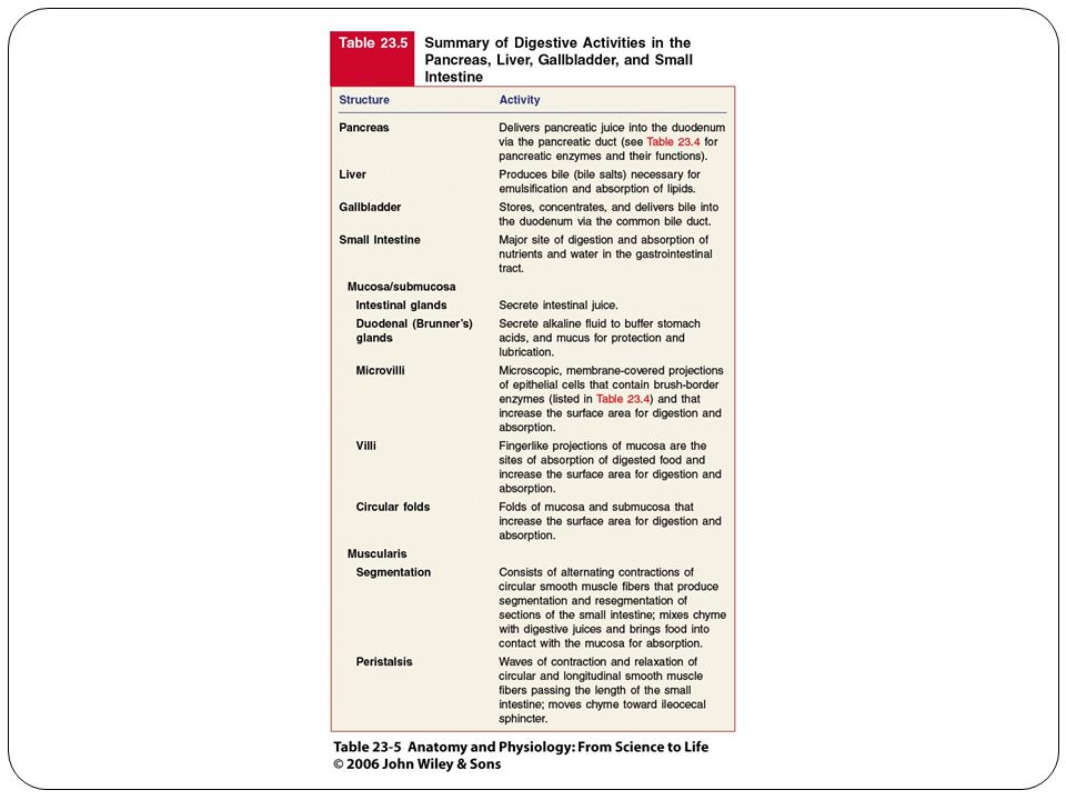
Pancreas Produces secretions to aid digestion Head Body Tail
Pancreatic duct /duct of Wirsung Hepatopancreatic ampulla Sphincter of the heatopancreatic ampulla (sphincter of (Oddi) Regulates passage of pancreatic juice and bile Accessory duct (duct of Santorini)
 Regulates passage of pancreatic juice and bile. Accessory duct (duct of Santorini)")
47
Figure 23.13a

48
Figure 23.13b

49
Figure 23.13c

50
Histology of Pancreas Glandular epithelial cells
99% exocrine clusters Secrete pancreatic juice Fluid and enzymes Pancreatic islets (islets of Langerhans) 1% endocrine cells Hormones Glucagon Insulin Somatostatin Pancreatic polypeptide
 1% endocrine cells. Hormones. Glucagon. Insulin. Somatostatin. Pancreatic polypeptide.")
51
Pancreatic Juice 1200-1500 mL/day pH 7.1-8.2 Water Salts
Sodium bicarbonate Enzymes Pancreatic amylase Trypsin Entereokinase Chymotrypsin Carboxypeptidase Elastase Pancreatic lipase Ribonuclease and deoxyribonuclease

52
Liver and Gallbladder Liver Largest gland at 1.4 kg (~3 lb)
Closely associated with liver

53
Anatomy of Liver Right and left lobe separated by falciform ligament
Quadrate lobe Caudate lobe Round ligament (ligamentum teres) Remnant of umbilical vein coronary ligaments
 Remnant of umbilical vein. coronary ligaments.")
54
Histology of Liver Lobule Reticuloendothelial (Kupffer) cells
Hepatocytes radiating from central vein Sinusoids Reticuloendothelial (Kupffer) cells Stationary phagocytes
 cells. Stationary phagocytes.")
55
Figure 23.14a

56
Figure 23.14b

57
Figure 23.14c

58
Figure 23.14d

59
Bile Duct System Bile secreted by hepatocytes Bile canaliculi
Right and left hepatic ducts Common hepatic duct Common bile duct Gallbladder for temporary storage of bile Cystic duct

60
Blood Supply of Liver Hepatic artery provides oxygenated blood
Hepatic portal vein provides deoxygenated blood Nutrients, drugs, toxins, microbes Hepatic artery and vein carry blood to sinusoids Substances exchanged by hepatocytes Blood drains to central vein and eventually hepatic vein Portal triad Hepatic portal vein Hepatic artery Bile duct

61
Figure 23.15

62
Bile 800-1000 mL/day pH 7.6 – 8.6 Water Bile acids Bile salts
Emulsification Cholesterol Lecithin Bile pigments Bilirubin Stercobilin

63
Liver Functions Metabolism of: Process drugs and hormones
Carbohydrates Lipids Proteins Process drugs and hormones Excrete bilirubin Synthesize bile salts Storage Glycogen Vtamins Minerals Phagocytosis Activate Vitamin D

64
Small Intestine Adapted for digestion and absorption
3 m (10 ft) living 6.5 m (21 ft) without muscle tone Duodenum Jejunum Ileum Ileocecal sphincter Connection to large intestine
 living. 6.5 m (21 ft) without muscle tone. Duodenum. Jejunum. Ileum. Ileocecal sphincter. Connection to large intestine.")
65
Figure 23.16a

66
Figure 23.16b

67
Histology of Small Intestine
Mucosa Cell types Absorptive Goblet Endocrine Paneth Lysozyme Intestinal glands (crypts of Lieberkühn) S cells Hormone secretin CCK cells Hormone – cholecystokinin (CCK)
 S cells. Hormone secretin. CCK cells. Hormone – cholecystokinin (CCK)")
68
Figure 23.17a

69
Figure 23.17b

70
Histology of Small Intestine
MALT – mucosa-associated lymphoid tissue Solitary lymphatic nodules Aggregated lymphatic follicles (Peyer’s patches) Submucosa Duodenal (Brunner’s glands) Alkaline secretion Muscularis Serosa
 Submucosa. Duodenal (Brunner’s glands) Alkaline secretion. Muscularis. Serosa.")
71
Adaptive Structures Small Intestine
Circular folds / plicae circulares Villi Lacteal Lymphatic capillary Microvilli Brush border Brush border enzymes Intestinal juice 1-2 liters / day pH 7.6

72
Figure 23.18a

73
Figure 23.18b

74
Mechanical Digestion in Small Intestine
Segmentation Localized Mix chyme with digestive juices Important for process of absorption Peristalsis Movement along the length of small intestine

75
Chemical Digestion in Small Intestine
Completes digestion of food from the stomach Carbohydrates Pancreatic amylase Glycogen and starch only -dextrinase Sucrase Lactase Maltase

76
Chemical Digestion in Small Intestine
Proteins Trypsin Chymotrypsin Elastase Carboxypeptidase Peptidases

77
Chemical Digestion in Small Intestine
Lipids Pancreatic lipase Emulsification Amphipathic bile salts Nucleic acids Nucleosidases Phosphatases

78
Table 23.4 pt 1

79
Table 23.4 pt 2

80
Absorption in Small Intestine
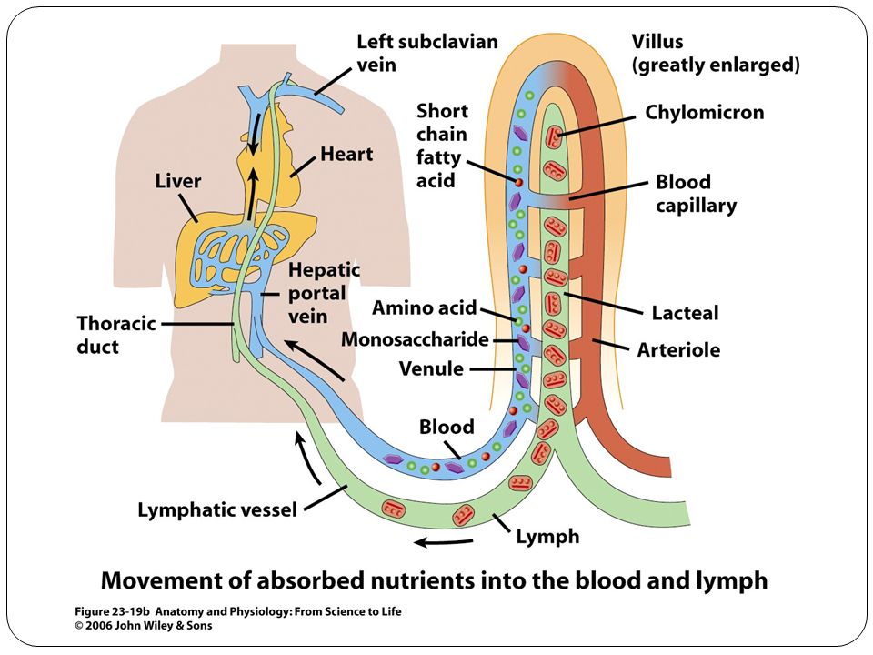
Passage of digested nutrients from gastointestinal tract into blood or lymph 90% of nutrients absorbed through small intestine Monosaccharides Facilitated diffusion Fructose Secondary active transport Glucose Galactose Enter blood through hepatic portal system

83
Absorption in Small Intestine
Amino acids Active transport Na+-dependent secondary active transport Dipeptides and tripeptides Symporter with H+

84
Absorption in Small Intestine
Lipids by simple diffusion Due to emulsification and digestion Micelles formed due to amphipathic nature of bile salts Chylomicrons Triglycerides coated with proteins Leave cells via exocytosis Enter blood vessels via lymphatic system Enterohepatic circulation

85
Absorption in Small Intestine
Electrolytes Diffusion Active transport Secondary active transport Vitamins Water Osmosis

88
Anatomy of Large Intestine
Mesocolon attaches to posterior abdominal wall Regions Cecum Colon Rectum Anal canal Ileocecal sphincter (valve) Allows passage into large intestine
 Allows passage into large intestine.")
89
Figure 23.21a

90
Figure 23.21b

91
Anatomy of Large Intestine
Cecum Pouch Attached appendix / veriform appendix Colon Ascending Transverse Descending Sigmoid Right and Left colic (splenic) flexures Rectum Anal canal Anal columns Anus Internal and external sphincter
 flexures. Rectum. Anal canal. Anal columns. Anus. Internal and external sphincter.")
92
Histology of Large Intestine
Mucosa Absorptive cells absorb mainly water Goblet cells secrete mucus Lymphatic nodules Submucosa Muscularis Haustra External longitudinal smooth muscle Teniae coli Internal circular smooth muscle Serosa Epiploic appendages

93
Figure 23.22a

94
Figure 23.22b

95
Figure 23.22c

96
Figure 23.22d

97
Mechanical digestion in Large Intestine
Gastroileal reflex Intensifies after a meal Occurs 3 or 4 times a day Haustral churning Distension and contraction of haustra Peristalsis Mass peristalsis

98
Chemical Digestion in Large Intestine
Bacteria: Ferment carbohydrates Gases produced are flatus or flatulence when excessive Break down proteins Decompose bilirubin Feces formed of dried chyme, inorganic salts, mucus, bacteria, undigested foods and other substances

99
Defecation Reflex Empties the rectum
Response to distention of rectal wall External anal sphincter voluntarily relaxed defecation occurs

101
Table 23.7

102
Phases of Digestion Cephalic Gastric Intestinal Cephalic Phase
Stimulation of the senses activates CNS Prepares mouth and stomach for food

104
Phases of Digestion Gastric Phase Begins with food in the stomach
Neural regulation Negative feedback system Stretch receptors Chemoreceptors Hormonal regulation Gastrin Released by G cells of gastric glands Controlled by negative feedback mechanism (pH)
")
105
Phases of Digestion Intestinal Phase Neural regulation
Begins with food in the small intestine Inhibitory effects to slow exit of chyme Neural regulation Enterogastric reflex Distension of duodenum

106
Phases of Digestion Intestinal Phase continued Hormonal regulation
Cholecystokinin (CCK) Stimulates release of pancreatic juice Contraction of gallbladder wall Relaxes sphincter of hepatopancreatic ampulla Secretin Response to acidic chyme Stimulates flow of pancreatic juice for buffering Inhibits secretion of gastric juice
 Stimulates release of pancreatic juice. Contraction of gallbladder wall. Relaxes sphincter of hepatopancreatic ampulla. Secretin. Response to acidic chyme. Stimulates flow of pancreatic juice for buffering. Inhibits secretion of gastric juice.")
107
Table 23.8

108
Six Main Types of Nutrients
Water Carbohydrates Lipids Proteins Minerals Vitamins Essential nutrients cannot be made in sufficient amounts by the body

109
Guidelines for Healthy Eating
Variety Maintain healthy weight Choose low fat foods Lots of vegetables, fruits, and grains Sugar in moderation Salt and sodium in moderation Alcohol in moderation Food Guide Pyramid

110
Figure 23.24

111
Nutrients Inorganic elements constitute 4% of body mass
Minerals Inorganic elements constitute 4% of body mass Regulate enzymatic reactions Serve as coenzymes Vitamins Organic molecules required in small amounts Most function as coenzymes Most cannot be synthesized by body Provitamins Fat-soluble vitamins A, D, E, and K Water soluble vitamins B and C Antioxidant vitamins

112
End

Similar presentations


 Turnover Require nutrients.>")


