Download presentation
Presentation is loading. Please wait.

1
Morphology and Differential Diagnosis

2
Welcome to Dermatology! No matter what area of medicine or surgery you pursue, you will get skin related questions from family, friends, and patients. The time frame is short, so make the best use of your time. Carry your book with you at all times and try to make it through all the photos.

3
Suggestions for a Successful Rotation Be on Time! Be attentive and helpful. Do not ask questions or make comments during the patient encounter. Please ask all questions outside the exam room. Please do not talk loudly in the hallway.

4
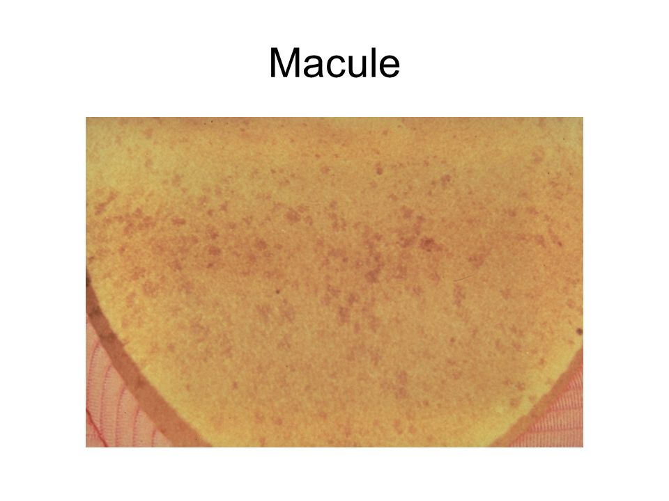
Macule

7
Patch

8
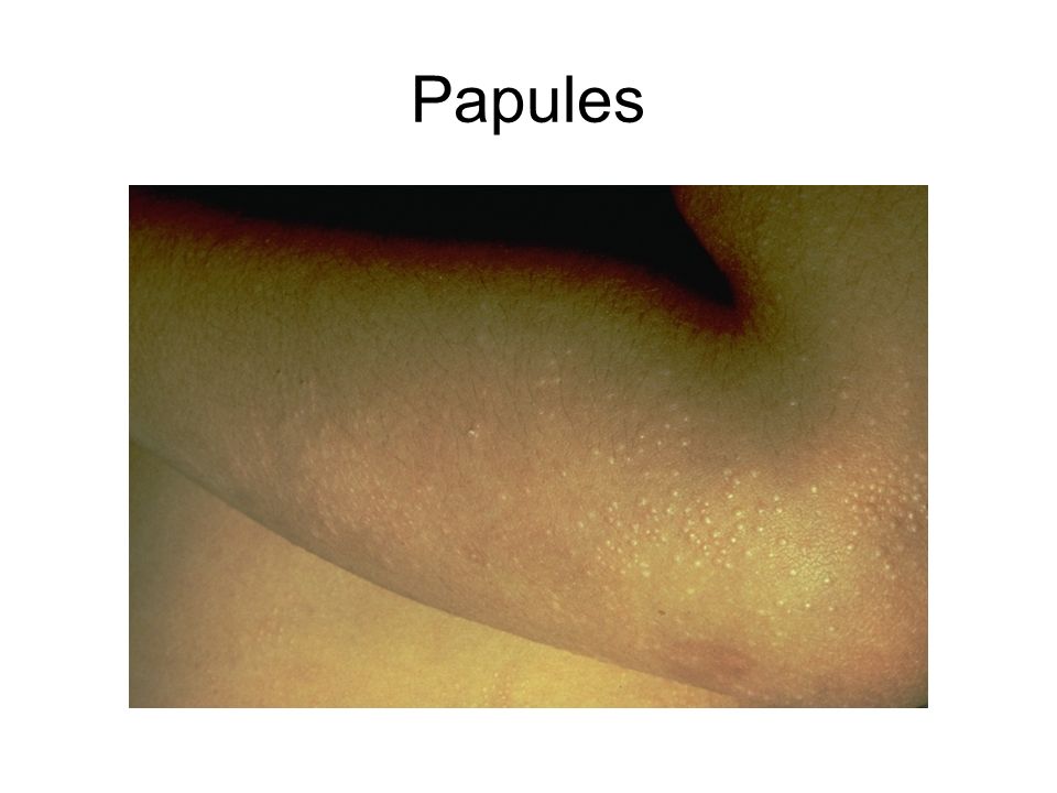
Papule

9
Papules

12
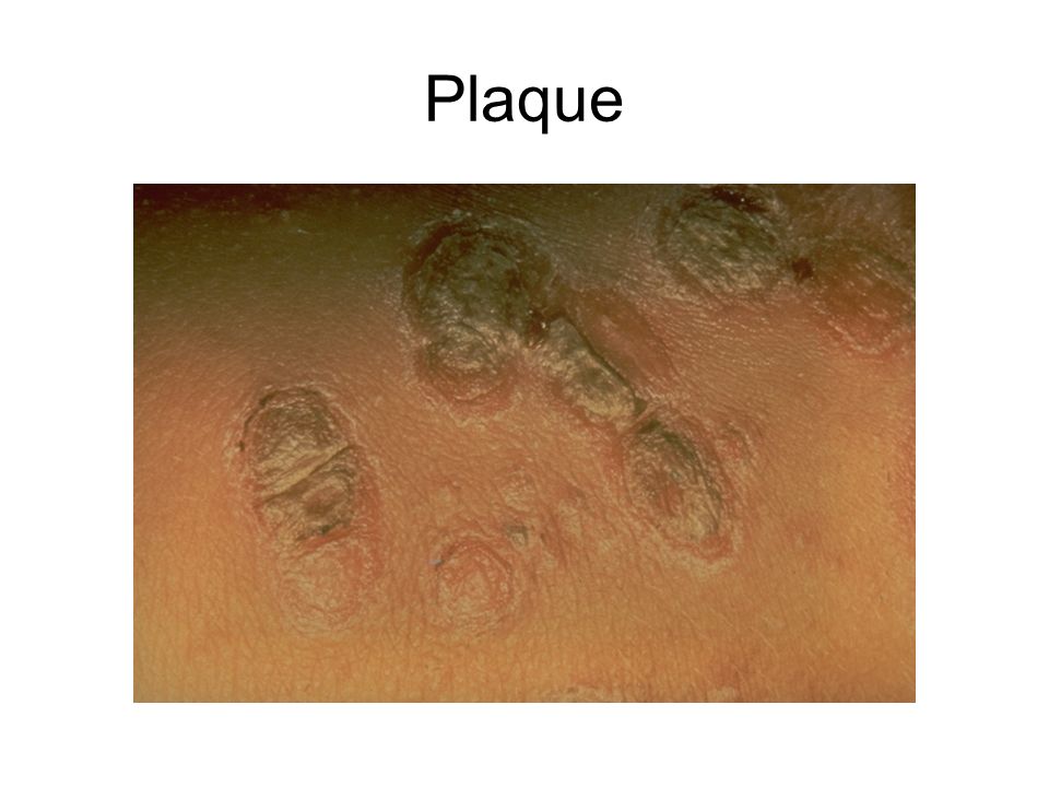
Plaque

15
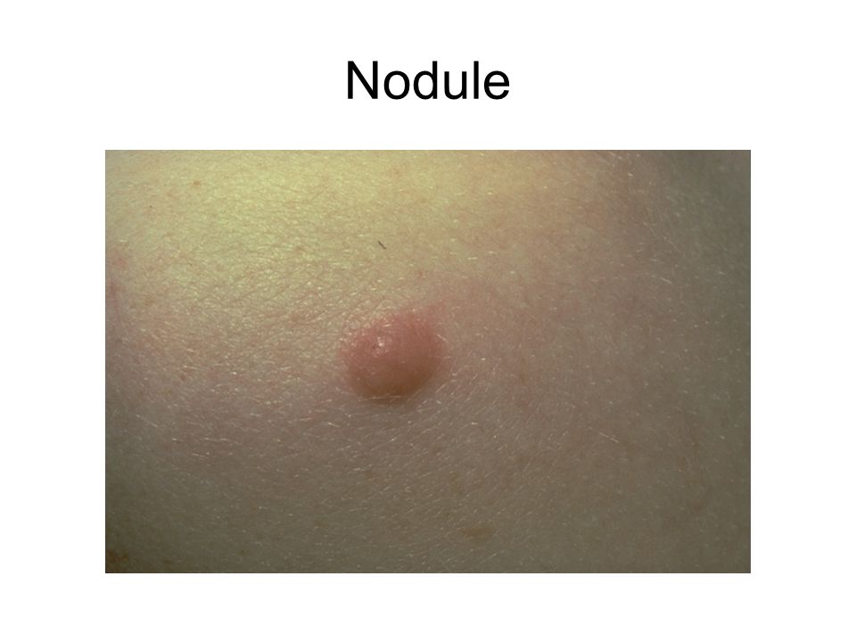
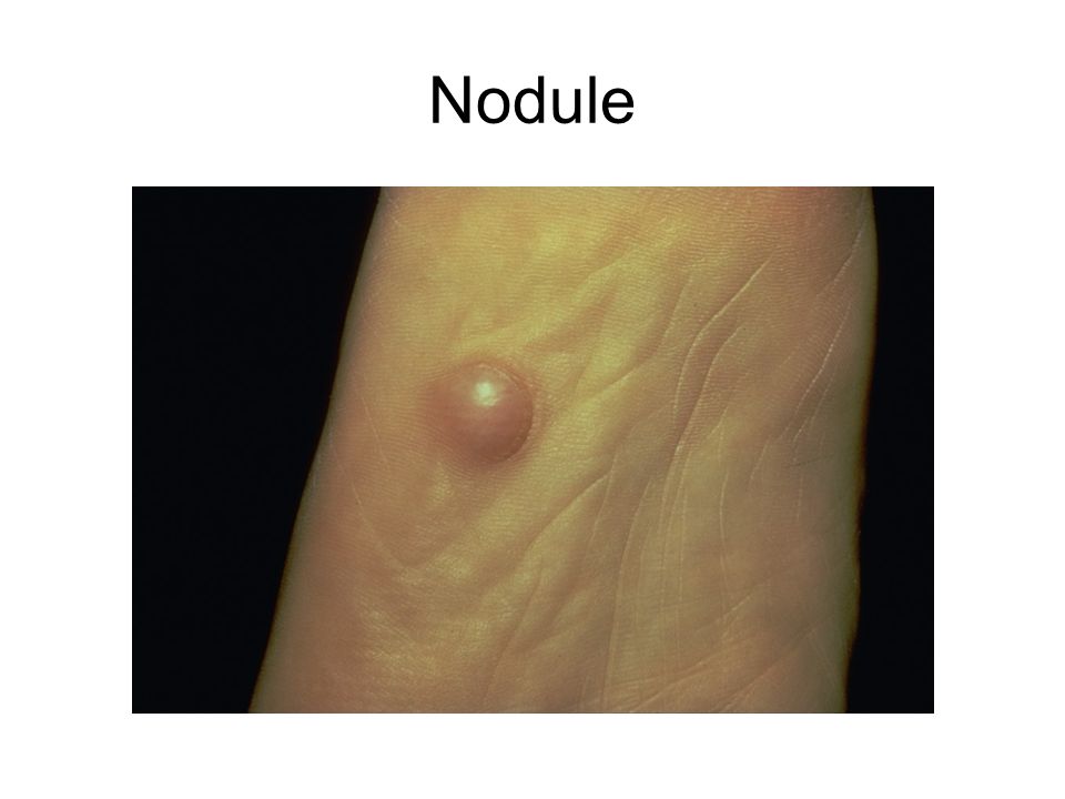
Nodule

18
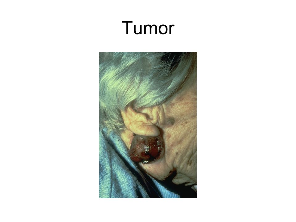
Tumor

21
Pustule

23
Vesicle

26
Bulla

31
Wheals

33
Special Skin Lesions Burrow: Thin linear papule or plaque Comedone: Follicular papule filled with keratinous plug which is open or closed Cyst: Papule or nodule filled with debris Telangiectasia: Dilated blood vessel less than 1 mm wide

34
Burrow

35
Comedone

36
Telangiectasia

37
Cyst

38
Secondary Lesions Scale Crust Erosions and ulcers Excoriations Fissures Scars Lichenification Atrophy

39
Scales

42
Crust

44
Excoriations

45
Erosion

47
Ulcer

50
Fissure

53
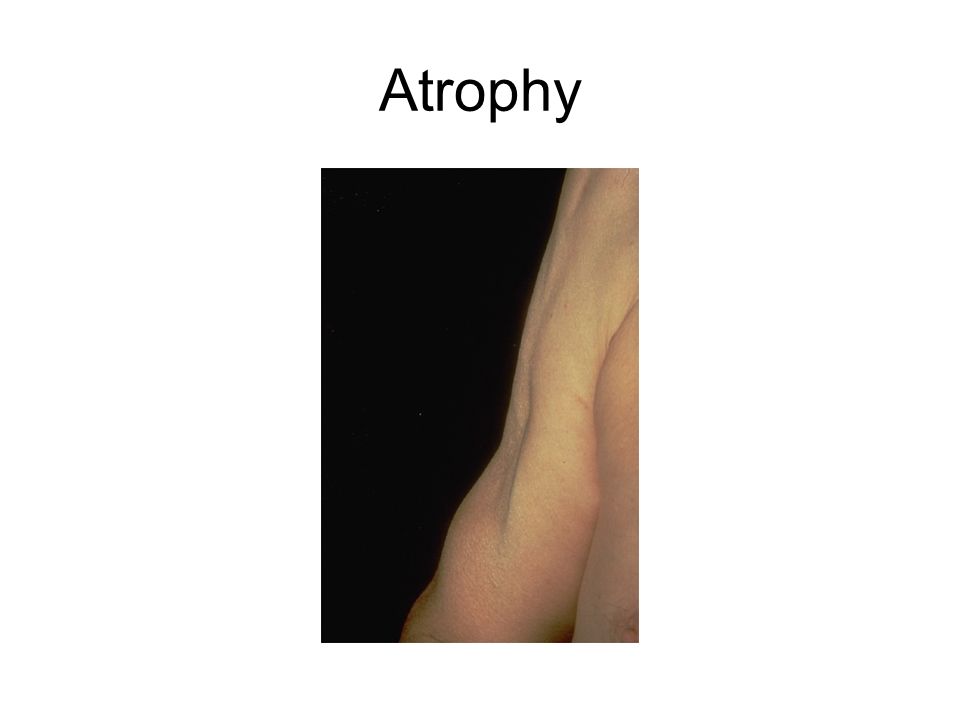
Atrophy

57
Scar

60
Lichenification

61
Configuration Annular Arcuate Geographic Discrete Confluent Serpiginous Linear Reticulated

62
Annular and arcuate

63
Linear

64
Erythema Subitum

65
Descriptors Punctate Lichenoid Umbilicated Scarletiniform, morbiliform Leonine

67
Color Pink Violet Orange Blue Green Yellow Black Brown

68
Color PinkPityriasis rosea VioletLichen planus OrangeJuvenile xanthogranuloma BlueAmioderone skin pigmentation GreenPseudomonas YellowXanthomas Blackeschar BrownCafé au lait spots

69
Color

70
Distribution

71
Morphologic categories Macular-Patch Papular Papulosquamous (scaly papules) Nodular Pustular Vesicular-bullous Urticarial Petechial Telangiectatis Burrow Poikiloderma Hyperkeratotic/scale Atrophic
 Nodular Pustular Vesicular-bullous Urticarial Petechial Telangiectatis Burrow Poikiloderma Hyperkeratotic/scale Atrophic")
72
More is missed by not looking than by not knowing M. McKay, M.D.

73
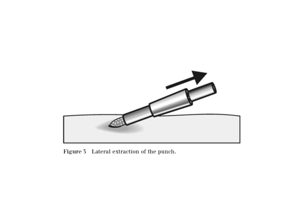
Procedures Liquid Nitrogen Electrodessication and curettage Biopsy –Punch –Shave –Excision

78
Seborrheic Keratosis Common Skin Tumor of unknown cause. Predilection for trunk, scalp, temples No malignant potential Increase incidence with age Easily treated with curettage or cryodestruction

79
Dermatosis Papulosa Nigra Most likely a subtype of seborrheic keratosis Malar areas, most commonly on African- American women

80
Acrocordons (Skin Tags) Common, occurring in about 25% of adults More common in obese individuals and often develop in pregnancy Frictional areas such as neck, axillae, inframammary and groin locations Can become irritated or infarcted because of torsion
 Common, occurring in about 25% of adults More common in obese individuals and often develop in pregnancy Frictional areas such as neck, axillae, inframammary and groin locations Can become irritated or infarcted because of torsion")
81
Dermatofibroma Firm papule often with brown pigmentation, most frequently seen on the anterior legs Dimple sign May be a reactive process to an insect bite reaction rather than a tumor If multiple, sometimes associated with systemic lupus erythematosis

82
Dermatofibroma

83
Keloids Hypertrophic scar which extends beyond the area of injury May have delayed onset, even up to years after injury Can be painful More common in African- Americans Treatment can be difficult and choices include intralesional steroids, radiation, careful excision, laser ablation

85
Epidermoid Cyst

86
Trichilemmal (Pilar) Cyst
 Cyst")
87
Actinic Keratosis

88
Keratosis Pilaris Follicular papules, commonly on extremities sandpaper feel 20% of the population affected Worsens in adolescence Common in Atopics and icthyosis May improve with keratolytics, retinoids, dermabrasion

89
Keratosis Pilaris

91
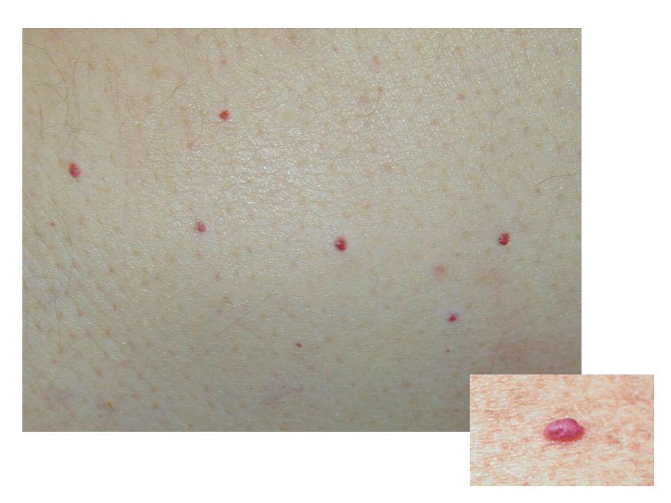
Cherry Angiomas Benign vascular proliferation senile hemangioma – dont use this term with patients Usually appear on trunk, start at age 30, increase with age Dilated capillaries Tx for cosmetic reasons only

Similar presentations


 ITEC VTCT Director of Aesthetics.>")


.>")