Download presentation
Presentation is loading. Please wait.

1
Skin as a protection against environmental threats Antti Lauerma, M.D., Ph.D. Chief Medical Officer FIOH Dermatology Figures: copyright Blackwell (Rook, Textbook of Dermatology)
.")
2
SKIN AS ORGAN Surface area 1.5 - 2 m2 Weight ~10% of body weight Purpose: To protect body against: –Mechanical stress –Physical damage –Pathogens –Foreign biologic material –Foreign nonbiologic material

4
SKIN: THREE LEVELS OF PROTECTION Mechanical barrier (stratum corneum) Innate immunity Acquired immunity
 Innate immunity Acquired immunity")
5
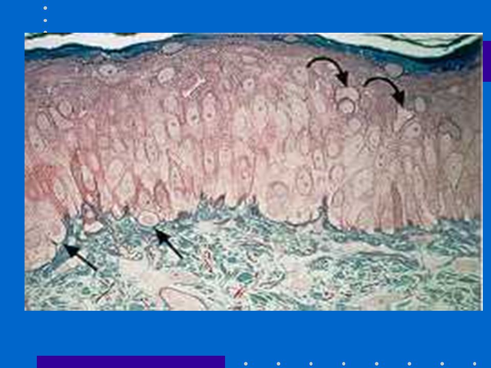
STRATUM CORNEUM The outer-most layer of skin Approximately 10 cell layers thick Consists of corneocytes and extracellular matrix Protective layer against water loss from body

6
STRATUM CORNEUM DAMAGE EXOGENOUS DAMAGE –excess washing (toxic hand dermatitis) ENDOGENOUS DAMAGE –inflammation STRUCTURAL DAMAGE –atopic skin
 ENDOGENOUS DAMAGE –inflammation STRUCTURAL DAMAGE –atopic skin")
7
STRATUM CORNEUM REPAIR Lipid synthesis in corneocytes Lipid synthesis in keratinocytes Basal keratinocyte proliferation

11
STRATUM CORNEUM REPAIR (2) Ointment/cream application UV light therapy Systemic retinoid use???
 Ointment/cream application UV light therapy Systemic retinoid use")
12
Coombs-Gell I Mast cells release histamine Vasodilatation Leakage of water to skin Intense pruritus 15 min - 2 hours (immediate hypersensitivity)
")
13
Coombs-Gell II Cytotoxic response Macrophage-mediated killing of unfit cells 24 hours Erythema multiforme

14
Coombs-Gell III Antibody-antigen complexes Complexes trapped at capillaries Exanthema 8-24 hours

15
Coombs-Gell IV APC presents antigen T-cell mediated cellular inflammation Allergic contact dermatitis 24-48 hours (delayed hypersensitivity)
")
17
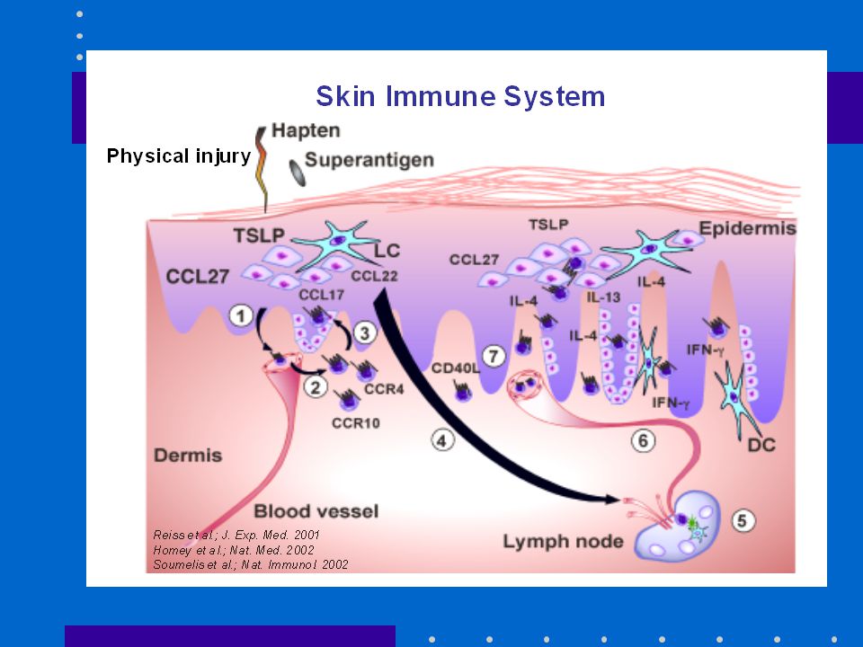
INNATE IMMUNITY IN SKIN - START Damage - danger signal Preformed IL-1a released from KC IL-1a stimulates KC to produce IL-1b, IL-6, TNFa and more IL-1a TOLL receptors have same effect as IL-1a, sharing NF-kappa-beta signalling

18
Arrival of Granulocytes Larger vessel - lower speed Attachment via P- and E-selectins Movement to dermis through CXC - chemokine gradient Proteases enable movement through ECM Entrance to epidermis, movement through epidermis (”zipper movement”)
")
19
Granulocytes in epidermis Presence of IL-1, IL-6, TNF-a, GM-CSF, IFNg induce a respiratory burst in granulocytes C3R, FCg receptors bind to microbes with opsonins (part of complement) to microbes 1 bacteria/min, total over 50 bacteria per granulocyte
 to microbes 1 bacteria/min, total over 50 bacteria per granulocyte")
20
Turn off the inflammation or call in the lymphocytes? Keratinocytes produce IL-10, IL1ra, aMSH. FB, MF, Lymphocytes produce TGF-beta: –IFN down –T cell anergy –Endoth. Cell Chk, adh mol down

21
Turn off the inflammation or call in the lymphocytes? Inflammation over 24- 35 hours starts acquired immunity Endothelial cells produce ICAM, VCAM T cells adhere to endothelial cells and enter skin via chemokine (CC, not CXC) gradient
 gradient.")
23
Lymphocytes in the skin Professional APC present antigen in the context of MHC II and B7.1/B7.2 to T cells. (If keratinocytes present antigen, anergy results (no B 7.1/7.2)) IFNg, IFNa, TNFa, and LPS, bacterial cell wall, CpG induce MF and DC to produce IL-12 IL-12 favors Th1 response Th1 T cells produce more IFNg that keeps up production of CC-chemokines
) IFNg, IFNa, TNFa, and LPS, bacterial cell wall, CpG induce MF and DC to produce IL-12 IL-12 favors Th1 response Th1 T cells produce more IFNg that keeps up production of CC-chemokines.")
24
What if ”danger” persists??? Inflammatory area will be isolated from surrounding tissue IL-4 and IL-10 induce giant cells from MF TGF beta stimulates action of giant cells and FB Granulomatous inflammation

Similar presentations


 BIOS 486A/586A>")

 It has made the self/nonself discrimination on an evolutionary time-scale It uses.>")


 Immune team. Cell-Mediated Immunity (CMI) Antigen T-lymphocytes Immune responses Immune responses.>")


 Prof. Dr. Zahid Shakoor MBBS, Ph D (London) College of Medicine King Saud University.>")

 System Nestor T. Hilvano, M.D., M.P.H. (Images Copyright Discover Biology, 5 th ed., Singh-Cundy and Cain, Textbook, 2012.)>")
