Download presentation
Presentation is loading. Please wait.

1
MYELOPROLIFERATIVE DISEASES By DR. FATIMA AL-QAHTANI CONSULTANT HAEMATOLOGIST

2
WHO Classification Chronic Myeloproliferative Disease --------------------------------------------------------------------------------------------------------------------------- Chronic Myelogenous Leukaemia [Ph chromosome, t(9;22)(q34;q11), BCR/ABL- positive ] Chronic Neutrophilic Leukaemia Chronic Eosinophilic Leukaemia (and the hypereosinophilic syndrome) Polycythaemia Vera Chronic Idiopathic Myelofibrosis (with extramedullary haematopoiesis) Essential Thrombocythaemia Chronic Myeloproliferative Disease, Unclassifiable
![WHO Classification Chronic Myeloproliferative Disease Chronic Myelogenous Leukaemia [Ph chromosome, t(9;22)(q34;q11), BCR/ABL- positive ] Chronic Neutrophilic Leukaemia Chronic Eosinophilic Leukaemia (and the hypereosinophilic syndrome) Polycythaemia Vera Chronic Idiopathic Myelofibrosis (with extramedullary haematopoiesis) Essential Thrombocythaemia Chronic Myeloproliferative Disease, Unclassifiable](http://images.slideplayer.com/9/2504235/slides/slide_2.jpg "WHO Classification Chronic Myeloproliferative Disease Chronic Myelogenous Leukaemia [Ph chromosome, t(9;22)(q34;q11), BCR/ABL- positive ] Chronic Neutrophilic Leukaemia Chronic Eosinophilic Leukaemia (and the hypereosinophilic syndrome) Polycythaemia Vera Chronic Idiopathic Myelofibrosis (with extramedullary haematopoiesis) Essential Thrombocythaemia Chronic Myeloproliferative Disease, Unclassifiable")
3
WHO Classification Myelodysplastic / Myeloproliferative Diseases ----------------------------------------------------------------- Chronic Myelomonocytic Leukaemia Atypical Chronic Myeloid Leukaemia Juvenile Myelomonocytic Leukaemia Myelodysplastic/Myeloproliferative Disease, Unclassifiable

5
Myeloid Disorders Usual Features at Diagnosis Disease BM cellularity % Marrow Blasts Maturation Morphology Haemato- poiesis Blood count (s) Organo- megaly Myeloproliferatie disorder Usually increased Normal or slightly increased (<10%) PresentRelatively normal EffectiveOne or more myeloid cell lines increased Common Myelodysplastic syndromes Usually increased, occasionally decreased Normal or increased (<20%) PresentDysplasia of one or more myeloid lineage IneffectiveCytopenia (S)Uncommon Myelodysplastic/ myeloproliferative disease Usually increased Normal or increased (<20%) PresentDysplasia of one or more myeloid lineages frequent Effective or ineffective; may vary among involved lineages VariableCommon Acute myeloid leukaemia Usually increased, occasionally decreased Increased (≥ 20%) Varies, frequently minimal May or may not be associated with dysplasia in one or more myeloid lines Ineffective or effective Variableuncommon
 Organo- megaly Myeloproliferatie disorder Usually increased Normal or slightly increased (<10%) PresentRelatively normal EffectiveOne or more myeloid cell lines increased Common Myelodysplastic syndromes Usually increased, occasionally decreased Normal or increased (<20%) PresentDysplasia of one or more myeloid lineage IneffectiveCytopenia (S)Uncommon Myelodysplastic/ myeloproliferative disease Usually increased Normal or increased (<20%) PresentDysplasia of one or more myeloid lineages frequent Effective or ineffective; may vary among involved lineages VariableCommon Acute myeloid leukaemia Usually increased, occasionally decreased Increased (≥ 20%) Varies, frequently minimal May or may not be associated with dysplasia in one or more myeloid lines Ineffective or effective Variableuncommon")
6
Myeloproliferative Disease Recurring Genetic Abnormalities and Their Frequency (%) at diagnosis DiseaseSpecific abnormalities(%)Recurring, nonspecific cytogenetic/genetic abnormalities (%) CML, CPt(9;22)(q34;q11), BCR/ABL100 CML, AP/BP t(9;22)(q34;q11), BCR/ABL100+8, +9Ph,+19,i(17q), t(3;21)(q26;q22)(EVI1/AML1)80 CNLNone+8, +9, del(20q), del(11q14)~10 CELNone+8, t(5;12)(q33;p13)(TEL/PDGFβR), dic(1;7), 8p11 (FGFR1) ? PVNone +8, +9, del(20q), del(13q), del(1p11)~15 CIMFNone+8, del(20q), -7/del(7q), del(11q), del(13q)~35 ETNone+8, del (13q)~5 CML, CP = Chronic myelogenous leukaemia, chronic phase; CML, AP/BP= Chronic myelogenous leukaemia, accelerated or blast phase; CNL = Chronic neutrophilic leukaemia; CEL = Chronic eosinophilic leukaemia; PV = Polycythaemia Vera; EVI-1 = ecotropic viral integration site 1 CIMF = Chronic idiopathic myelofibrosis; ET = Essential thrombocythaemia ? = Insufficient data available
, del(13q), del(1p11)~15 CIMFNone+8, del(20q), -7/del(7q), del(11q), del(13q)~35 ETNone+8, del (13q)~5 CML, CP = Chronic myelogenous leukaemia, chronic phase; CML, AP/BP= Chronic myelogenous leukaemia, accelerated or blast phase; CNL = Chronic neutrophilic leukaemia; CEL = Chronic eosinophilic leukaemia; PV = Polycythaemia Vera; EVI-1 = ecotropic viral integration site 1 CIMF = Chronic idiopathic myelofibrosis; ET = Essential thrombocythaemia . = Insufficient data available.")
7
Chronic Idiopathic Myelofibrosis Prefibrotic Stage Clinical findingsMorphological findings Spleen and liver: No or mild splenomegaly or hepatomegaly Blood: No or mild leukoerythroblastosis No or minimal red blood cell poikilocytosis; few if any dacrocytes Splenomegaly: Haematologic parameters variable, but often: Mild anaemia Mild to moderate leukocytosis Mild to marked thrombocytosis Bone marrow: Hypercellularity Neutrophilic proliferation Megakaryocytic proliferation and atypia (Clustering of megakaryocytes, abnormally lobulated megakaryocytic nuclei, naked megakaryocytic nuclei) Minimal or absent reticulin fibrosis
 Minimal or absent reticulin fibrosis")
8
Chronic Idiopathic Myelofibrosis Fibrotic Stage Clinical findingsMorphological findings Spleen and liver: Moderate to marked splenomegaly and hepatomegaly Blood: Leukoerythroblastosis Prominent red blood cell poikilocytosis with dacrocytes Haematology: Moderate to marked anaemia Low, normal or elevated WBC Platelet count decreased, normal or elevated Bone Marrow: Reticulin and/or collagen fibrosis Decreased cellularity Dilated marrow sinuses with intraluminal haematopoiesis Prominent megakaryocytic proliferation and atypia (clustering of megakaryocytes, abnormally lobulated megakaryocytic nuclei, naked nuclei) New bone formation (osteosclerosis)
 New bone formation (osteosclerosis)")
13
Essential Thrombocythaemia Diagnostic Criteria Positive Criteria 1.Sustained platelet count ≥600X10 9 /L 2.Bone marrow biopsy specimen showing proliferation mainly of the megakaryocytic lineage with increased numbers of enlarged, mature megakaryocytes Criteria of exclusion 1.No evidence of polycythaemia vera (PV) - Normal red cell mass or Hb <18.5g/dl in men, 16.5g/dl in women - Stainable iron in marrow, normal serum ferritin or normal MCV - If the former condition is not met, failure of iron trial to increase red cell mass or Hgb levels to the PV range 2.No evidence of CML - No Philadelphia chromosome and no BCR/ABL fusion gene
 - Normal red cell mass or Hb <18.5g/dl in men, 16.5g/dl in women - Stainable iron in marrow, normal serum ferritin or normal MCV - If the former condition is not met, failure of iron trial to increase red cell mass or Hgb levels to the PV range 2.No evidence of CML - No Philadelphia chromosome and no BCR/ABL fusion gene")
16
Essential Thrombocythaemia Diagnostic criteria (Continued) 3.No evidence of chronic idiopathic myelofibrosis - Collagen fibrosis absent - Reticulin fibrosis minimal or absent 4.No evidence of myelodysplastic syndrome - No del(5q), t(3;3)q21;q26), inv(3)(q21q26) - No significant granulocytic dysplasia, few if any micromegakaryocytes 5.No evidence that thrombocytosis is reactive due to: - Underlying inflammation or infection -Underlying neoplasm -Prior splenectomy
 3.No evidence of chronic idiopathic myelofibrosis - Collagen fibrosis absent - Reticulin fibrosis minimal or absent 4.No evidence of myelodysplastic syndrome - No del(5q), t(3;3)q21;q26), inv(3)(q21q26) - No significant granulocytic dysplasia, few if any micromegakaryocytes 5.No evidence that thrombocytosis is reactive due to: - Underlying inflammation or infection -Underlying neoplasm -Prior splenectomy")
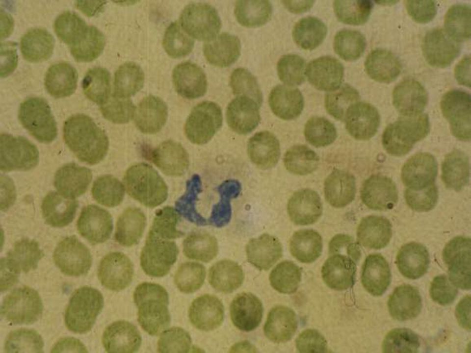
17
Giant Plat

18
Megakaryocytes in Clusters

19
Polycythaemia Vera ---------------------------------------------------------------------------------------------------------------------------------------------------------------------- - Hb: >17.5 gm/dl > 15.5 gm/dl -RBC: > 6.0 X 10 12 /L > 5.5X10 12 /L -PCV: > 51% > 48% -TRCV : > 36 ml/kg (25-35) > 32 ml/kg (22-32) -TPV: 40 – 50 ml/kg
 > 32 ml/kg (22-32) -TPV: 40 – 50 ml/kg")
20
Classification of Erythrocytosis Raised PCV (female >0.48; male>0.51) RCM (Interpreted using ICSH reference values) Increased RCMNormal RCM Absolute erythrocytosisApparent erythrocytosis Abbreviations: PCV = Packed Cell Volume; RCM = red cell mass; ICSH = International Council for Standardization in Haematology;
 RCM (Interpreted using ICSH reference values) Increased RCMNormal RCM Absolute erythrocytosisApparent erythrocytosis Abbreviations: PCV = Packed Cell Volume; RCM = red cell mass; ICSH = International Council for Standardization in Haematology;")
21
Primary Erythrocytosis Congenital # Truncation of the EPO receptor* Acquired Polycythaemia Vera* Secondary Erythrocytosis Congenital # e.g., high oxygen affinity Hb, autonomous high EPO production Acquired e.g., hypoxemia, renal disease # Sometimes familial * The only condition to be defined in this category at present EPO = erythropoietin

22
Polycythaemia Vera Causes ---------------------------------------------------------------------------------------------------------------------------------------------------------------------- Primary : Polycythaemia Vera Secondary: 1.Erythropoietin compensatory increase: High Altitude C.V. disease Pulmonary disease High Affinity Hb Heavy smoking Methaemoglobinaemia 2.Abnormal erythropoietin production: Renal diseases. Massive uterine fibromatosis Hepatocellular Carcinoma Cerebellar Haemangioblastoma Relative: Stress, Dehydration, Plasma Loss.

23
Polycythaemia Vera Clinical Features ---------------------------------------------------------------------------------------------------------------------------------------------------------------------- Headache, Lethargy, Dyspnea Weight Loss, Night Sweats Generalized pruritis (Increase after hot bath) Plethoric Appearance Haemorrhage & Thrombosis Hypertension (In about 1/3rd of the patients) Gout (Increased Uric Acid) Peptic Ulcers (In 5 – 10% of the patients) Splenomegaly (In 2/3rd of patients) Accidental Discovery (On Routine exam)
 Plethoric Appearance Haemorrhage & Thrombosis Hypertension (In about 1/3rd of the patients) Gout (Increased Uric Acid) Peptic Ulcers (In 5 – 10% of the patients) Splenomegaly (In 2/3rd of patients) Accidental Discovery (On Routine exam)")
24
Polycythaemia Vera Laboratory Investigations ---------------------------------------------------------------------------------------------------------------------------------------------------------------------- -C.B.C -Neutrophil Alkaline Phosphatase (N.A.P.) -Serum B 12 & B 12 binding capacity -Bone Marrow - Blood Viscosity -Uric Acid Level - Hb Electrophoresis -Arterial Oxygen Tension - T.R.C.V. -I.V. Pyelography, CT & US - JAK2: 74 – 97 % (PV) -Erythropoietin Assay 33 – 57 % (ET) 35 – 50 % (MF)
 -Erythropoietin Assay 33 – 57 % (ET) 35 – 50 % (MF).")
30
Polycythaemia Vera Proposed diagnostic criteria ------------------------------------------------------------------------------------------------------------------------------- A1 Raised red cell mass B1 Thrombocytosis (>25% above mean normal Platelet count>400X10 9 /1 predicted value) A2 Absence of a cause of B2 Neutrophil leukocytosis Secondary Polycythaemia neutrophil count >10X10 9 /1 A3 Palpable splenomegaly B3 Splenomegaly demonstrated by isotope/ultrasound scanning A4 Clonality marker B4 Characteristic BFU-E growth e.g., abnormal marrow karyotype or reduced serum erythropoietin ------------------------------------------------------------------------------------------------------------------------------------------------------------------------------------------------------------------------------------------------------------------ A1 + A2 + A3 or A4 establishes PV A1 + A2 + Two of B establishes PV
 A2 Absence of a cause of B2 Neutrophil leukocytosis Secondary Polycythaemia neutrophil count >10X10 9 /1 A3 Palpable splenomegaly B3 Splenomegaly demonstrated by isotope/ultrasound scanning A4 Clonality marker B4 Characteristic BFU-E growth e.g., abnormal marrow karyotype or reduced serum erythropoietin A1 + A2 + A3 or A4 establishes PV A1 + A2 + Two of B establishes PV")
31
Polycythaemia Vera Classic Polycythaemia Vera Study Group Diagnostic Criteria -------------------------------------------------------------------------------------------------------------------------------------------------------------------------- A1 ↑ Red Cell Mass B1 Thrombocytosis Male ≥36 ml/kg Platelet count >400,000/µl Female ≥32ml/kg B2 Leukocytosis >12,000/µl (No fever or infection) A2 Normal Arterial B3 ↑ Leukocyte Alkaline O 2 Saturation ≥92% Phosphatase score >100 (No fever or infection) A3 Splenomegaly ↑ Serum B 12 (>900pg/ml) or ↑ Unbound B 12 binding capacity (>2200pg/ml) -------------------------------------------------------------------------------------------------------------------------------------------------------------------------- Diagnosis is acceptable if the following combinations are present: A1 + A2 + A3 or A1 + A2 + any two from Category B.
 A2 Normal Arterial B3 ↑ Leukocyte Alkaline O 2 Saturation ≥92% Phosphatase score >100 (No fever or infection) A3 Splenomegaly ↑ Serum B 12 (>900pg/ml) or ↑ Unbound B 12 binding capacity (>2200pg/ml) Diagnosis is acceptable if the following combinations are present: A1 + A2 + A3 or A1 + A2 + any two from Category B.")
32
Polycythaemia Vera Treatment -------------------------------------------------------------- Venesecton Radioactive Phosphorus (P 32 ) Chemotherapy: e.g. Hydroxyurea

Similar presentations


 is defined as an increase in haemoglobin, PCV and red cell count. PCV is a more reliable indicator of polycythaemia.>")

II Dr. Ibrahim. A. Adam.>")

>")
 Myelodysplastic / myeloproliferative diseases (MDS/MPD) >")
:>")


 constitute one of five categories of myeloid malignancies, according to the World Health Organization (WHO) classification.>")

 - Essential Thrombocythemia - Myelofibrose Myeloid Methaplasia.>")
