Download presentation
Presentation is loading. Please wait.

1
Collected by Dr.Wala’a Gholam KAAU Jeddah, 2007
Skeletal X-ray Collected by Dr.Wala’a Gholam KAAU Jeddah, 2007

2
Skull (Lateral)
")
3
1. Frontal sinus 2. Ethmoidal sinus 3. Sphenoidal sinus 4
1 . Frontal sinus 2 . Ethmoidal sinus 3 . Sphenoidal sinus 4 . Maxillary sinus 5 . Anterior clinoid processes 6 . Hypophyseal fossa 7 . Posterior clinoid processes 8 . Clivus 9 . Great density of the petrous part of the temporal bone 10 . External acoustic meatus

4
11. Mastoid cells 12. Nasopharynx 13. Angle of mandible 14
11 . Mastoid cells 12 . Nasopharynx 13 . Angle of mandible 14 . Anterior arch of the atlas 15 . Dens of axis 16 . Posterior arch of the atlas 17 . Internal occipital protuberance A. Coronal suture B. Lambdoid suture C. The grooves for the branches of the middle meningeal vessels

5
Skull (Anteroposterior)
")
6
1. Frontal sinus 2. Crista galli 3. Cribriform plate 4
1 . Frontal sinus 2 . Crista galli 3 . Cribriform plate 4 . Lesser wing of sphenoid 5 . Superior orbital fissure 6 . Superior border of petrous part of temporal bone 7 . Dense shadow of petrous part of temporal bone 8 . Perpendicular plate of the ethmoid 9 . Vomer 10 . Maxillary sinus 11 . Inferior concha 12 . Ramus of mandible 13 . Body of mandible

7
Cervical Spine (Lateral)

8
1. Anterior arch of the atlas 2. Dens of axis 3
1 . Anterior arch of the atlas 2 . Dens of axis 3 . Posterior arch of the atlas 4 . Soft palate 5 . Root of the tongue 6 . Transverse process 7 . Intervertebral disc 8 . Inferior articular process 9 . Superior articular process 10 . Zygapophyseal (facet) joint 11 . Spinous process of C7 2nd-7th: The bodies of 2nd to 7th cervical vertebrae
 joint 11 . Spinous process of C7 2nd-7th: The bodies of 2nd to 7th cervical vertebrae.")
9
Cervical Spine (Anteroposterior)
")
10
1. Bifid spinous process of C3 2. Superimposed articular processes 3
1 . Bifid spinous process of C3 2 . Superimposed articular processes 3 . Uncinate processes 4 . Air filled trachea 5 . Transverse process of C7 6 . Transverse process of T1 7 . st rib 8 . Clavicle 4th-7th: The bodies of 4th to 7th cervical vertebrae

11
Shoulder Joint (Anteroposterior)
")
12
1. Clavicle 2. Acromioclavicular joint 3. Acromion 4
1 . Clavicle 2 . Acromioclavicular joint 3 . Acromion 4 . Greater tubercle of humerus 5 . Head of humerus 6 . Lesser tubercle of humerus 7 . Surgical neck of humerus 8 . Coracoid process 9 . Glenoid fossa 10 . Shoulder joint 11 . Lateral border of scapula

13
Elbow Joint (Anteroposterior)
")
14
1- Lateral supracondylar ridge 2- Medial supracondylar ridge 3- Olecranon fossa 4- Medial epicondyle 5- Lateral epicondyle 6- Capitulum 7- Olecranon 8- Trochlea 9- Coronoid process of ulna 10- Proximal radioulnar joint 11- Head of radius 12- Neck of radius 13- Tuberosity of radius 14- Ulna

15
Elbow Joint (Lateral)
")
16
6- Head of radius 7- Neck of radius 8- Tuberosity of radius 9- Ulna
1- Supracondylar ridge 2- Trochlea 3- Olecranon 4- Trochlear notch 5- Coronoid process of ulna

17
Forearm (Anteroposterior)
")
18
1- Scaphoid 2- Lunate 3- Styloid process of radius 4- Styloid process of ulna 5- Head of ulna 6- Radius 7- Ulna 8- Tuberosity of radius 9- Neck of radius 10- Head of radius 11- Proximal radioulnar joint

19
Hand (Carpal Tunnel)
")
20
1- Pisiform 2- Triquetrum 3- Hook of hamate 4- Capitate 5- Scaphoid 6- Trapezium 7- Ulna 8- Radius

21
Wrist Joint (Anteroposterior)
")
22
I-V: Metacarpals 1. Trapezium 2. Trapezoid 3. Capitate 4
I-V: Metacarpals 1 . Trapezium 2 . Trapezoid 3 . Capitate 4 . Head of capitate 5 . Hamate 6 . Hook of hamate 7 . Scaphoid 8 . Lunate 9 . Triquetrum 10 . Pisiform 11 . Styloid process of radius 12 . Head of ulna 13 . Styloid process of ulna 14 . Radiocarpal joint 15 . Distal radioulnar joint

23
Wrist Joint (Lateral)
")
24
1. 1st metacarpal 2. Metacarpals II-V 3. Trapezium 4
1. 1st metacarpal 2 . Metacarpals II-V 3 . Trapezium 4 . Tubercle of scaphoid 5 . Lunate 6 . Triquetrum 7 . Radiocarpal joint 8 . Distal end of radius 9 . Distal end of ulna

25
Hand (Dorsopalmar)
")
26
A. Thumb B. Index C. Middle finger D. Ring finger E. Little finger I-V
A. Thumb B. Index C. Middle finger D. Ring finger E. Little finger I-V. Metacarpal bones 1-4: Distal phalanx 2: Middle phalanx 3-5: Proximal phalanx 6: Sesamoid bones 7: Distal interphalangeal joint (DIP) 8: Proximal interphalangeal joint (PIP)
 8: Proximal interphalangeal joint (PIP)")
27
9- Metacarpophalangeal joint (V
9- Metacarpophalangeal joint (V.) 10- Carpometacarpal joints 11- Trapezium 12- Trapezoid 13- Capitate 14- Hamate 15- Scaphoid 16- Lunate 17- Triquetrum 18- Pisiform 19- Radius 20- Ulna
 10- Carpometacarpal joints 11- Trapezium 12- Trapezoid 13- Capitate 14- Hamate 15- Scaphoid 16- Lunate 17- Triquetrum 18- Pisiform 19- Radius 20- Ulna.")
28
Hand (Oblique)
")
29
A. Thumb B. Index C. Middle finger D. Ring finger
E. Little finger1,4. Distal phalanx 2- Middle phalanx 3-5- Proximal phalanx 6- Sesamoid bones 7- Distal interphalangeal joint (DIP) 8- Proximal interphalangeal joint (PIP) 9-Metacarpophalangeal joint (V.) 10- Carpometacarpal joints . 11-Trapezium 12- Trapezoid 13- Capitate 14- Hamate 15- Scaphoid 16- Lunate 17- Triquetrum 19- Radius 20- Ulna
 8- Proximal interphalangeal joint (PIP) 9-Metacarpophalangeal joint (V.) 10- Carpometacarpal joints Trapezium 12- Trapezoid 13- Capitate 14- Hamate 15- Scaphoid 16- Lunate 17- Triquetrum 19- Radius 20- Ulna.")
30
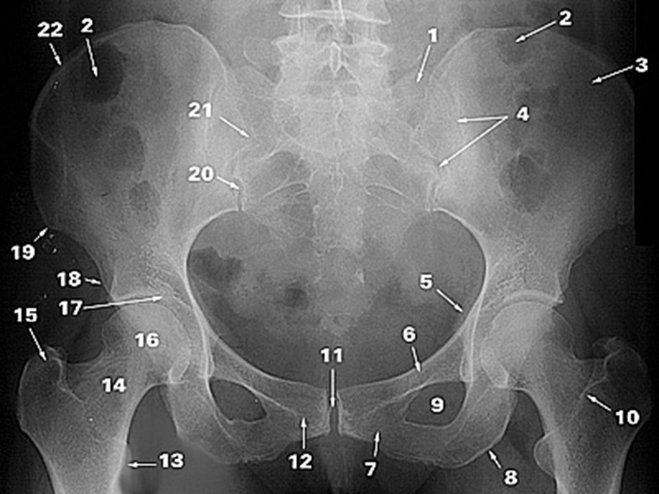
Pelvis (Anteroposterior)
")
32
1. Lateral part of the sacrum 2. Gas in colon 3. Ilium 4
1 . Lateral part of the sacrum 2 . Gas in colon 3 . Ilium 4 . Sacroiliac joint 5 . Ischial spine 6 . Superior ramus of pubis 7 . Inferior ramus of pubis 8 . Ischial tuberosity 9 . Obturator foramen 10 . Intertrochanteric crest 11 . Pubic symphysis 12 . Pubic tubercle 13 . Lesser trochanter 14 . Neck of femur 15 . Greater trochanter 16 . Head of femur 17 . Acetabular fossa 18 . Anterior inferior iliac spine 19 . Anterior superior iliac spine 20 . Posterior inferior iliac spine 21 . Posterior superior iliac spine 22 . Iliac crest

33
Hip Joint (Anteroposterior)

34
1. Anterior superior iliac spine 2. Ilium 3
1 . Anterior superior iliac spine 2 . Ilium 3 . Anterior inferior iliac spine 4 . Pelvic brim 5 . Acetabular fossa 6 . Head of femur 7 . Fovea 8 . Superior ramus of pubis 9 . Obturator foramen 10 . Inferior ramus of pubis 11 . Pubic symphysis 12 . Ischium 13 . Lesser trochanter 14 . Intertrochanteric crest 15 . Greater trochanter 16 . Neck of femur

35
Hip Joint (Frog Lateral)
")
36
1 .Greater trochanter 2 . Intertrochanteric crest 3 . Lesser trochanter 4 . Neck of femur 5 . Head of femur 6 .Acetabular fossa 7 . Superior ramus of pubis 8 . Obturator foramen 9 . Inferior ramus of pubis 10 . Ischium

37
Knee Joint (Anteroposterior)

38
1. Femur 2. Patella 3. Medial epicondyle of femur 4
1 . Femur 2 . Patella 3 . Medial epicondyle of femur 4 . Lateral epicondyle of femur 5 . Medial condyle of femur 6 . Lateral condyle of femur 7 . Intercondylar eminence 8 .Intercondylar notch 9 . Knee joint 10 . Lateral condyle of tibia 11 . Medial condyle of tibia 12 . Tibia 13 . Fibula

39
Knee Joint (Lateral)
")
40
1. Femur 2. Lateral condyle of femur 3. Medial condyle of femur 4
1 . Femur 2 . Lateral condyle of femur 3 . Medial condyle of femur 4 . Fabella 5 . Patella 6 . Base of patella 7 .Apex of patella 8 . Intercondylar eminence 9 . Apex of fibula 10 . Fibula 11 . Tibia 12 . Tibial tuberosity

41
Patella (Distal-proximal)

42
1. Patella 2. Medial part of patella 3. Lateral part of patella 4-5
1 . Patella 2 . Medial part of patella 3 . Lateral part of patella Patellofemoral joint 6 . Lateral femoral condyle 7 . Medial femoral condyle

43
Lower Leg (Anteroposterior)
")
44
1. Femur 2. Medial condyle of femur 3. Lateral condyle of femur 4
1 . Femur 2 . Medial condyle of femur 3 . Lateral condyle of femur 4 . Knee joint 5 . Intercondylar eminence 6 . Lateral condyle of tibia 7 . Medial condyle of tibia 8 . Fibula 9 . Tibia 10 . Head of fibula 11 . Neck of fibula

45
Lower Leg (Lateral)
")
46
1 . Femur 2 . Knee joint 3 . Intercondylar eminence 4 . Tibial tuberosity 5 . Fibula 6 . Tibia 7 . Ankle joint 8 . Talus 9 . Calcaneus

47
Ankle Joint (Lateral)
")
48
1- Fibula 2- Tibia 3- Ankle joint 4- Promontory of tibia 5- Trochlear surface of talus 6- Talus 7- Posterior tubercle of talus 8- Calcaneus 9- Sustentaculum tali 10- Tarsal tunnel 11- Navicular 12- Cuneiforms 13- Cuboid

49
Ankle Joint (Anteroposterior)
")
50
1- Fibula 2- Tibia 3- Distal tibiofibular joint 4- Malleolar fossa 5- Lateral malleolus 6- Ankle joint 7- Medial malleolus 8- Talus

51
Foot (Dorso-plantar)
")
52
A-E: Toes 1-5. (Great toe) I-V
A-E: Toes 1-5. (Great toe) I-V. Metatarsals 1-3: Distal phalax 4 Middle phalax 2-5 Proximal phalax 6- Interphalangeal joints 7- Metatarsophalangeal joints 8- bSesamoids 9- Head of metatarsal 10- Shaft (body) of metatarsal 11- Base of metatarsal 12- Cuneiforms 13- Navicular 14- Cuboid 15- Talus 16- Calcaneus 17- Tibia 18- Fibula 19- Tarsometatarsal joints 20- Transverse midtarsal joint
 I-V. Metatarsals 1-3: Distal phalax 4 Middle phalax 2-5 Proximal phalax. 6- Interphalangeal joints 7- Metatarsophalangeal joints 8- bSesamoids 9- Head of metatarsal 10- Shaft (body) of metatarsal 11- Base of metatarsal 12- Cuneiforms 13- Navicular 14- Cuboid 15- Talus 16- Calcaneus 17- Tibia 18- Fibula 19- Tarsometatarsal joints 20- Transverse midtarsal joint.")
53
Foot (Oblique)
")
54
6- Interphalangeal joints 7- Metatarsophalangeal joints 8- Sesamoids 9- Head of metatarsal 10- Shaft (body) of metatarsal 11- Base of metatarsal 12- Cuneiforms 13- Navicular 14- Cuboid 15- Talus 16- Calcaneus 17- Tibia 18- Fibula 19- Tarsometatarsal joints 20- Transverse midtarsal joint A-E: Toes 1-5. Great toe)) 1-3: Distal phalax 4- Middle phalax 2-5: Proximal phalax
) 1-3: Distal phalax 4- Middle phalax 2-5: Proximal phalax.")
55
collected by Dr.Wala’a Gholam KAAU Jeddah 2007
Source:

Similar presentations