Download presentation
Presentation is loading. Please wait.

1
LV Dssynchrony and Cardiac Resynchronization Therapy in Heart Failure Nisha I. Parikh MD MPH August 13 th 2008

2
Summary of Talk Background CRT Rational and Evidence for Benefit LV Dssynchrony by Echocardiography Evidence for Ability of Echo to Predict CRT Response

3
Hospital discharges for HF from 1979-2004 American Heart Association. Heart Disease and Stroke Statistics — 2007 Update 1,099,000

4
HF Total Expenditures: $27.9 Billion American Heart Association. Heart Disease and Stroke Statistics — 2007 Update

5
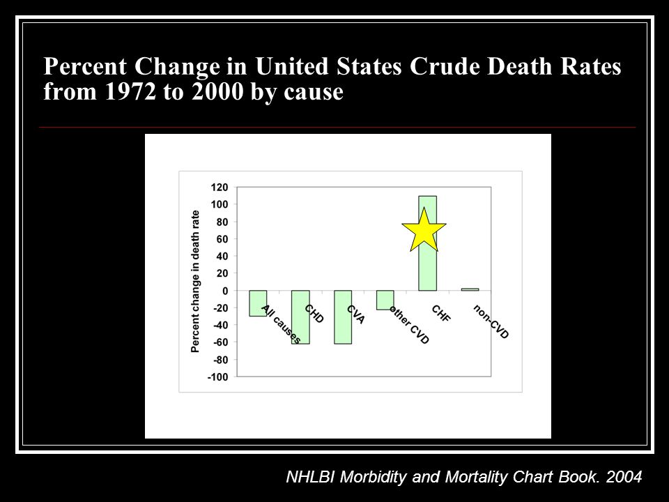
Percent Change in United States Crude Death Rates from 1972 to 2000 by cause NHLBI Morbidity and Mortality Chart Book. 2004
6
HF Therapy Jessup M, Brozena S. Medical Progress--Heart Failure. N Eng J Med 2003; 348: 2007-2018. Copyright 2002 Massachusetts Medical Society. All rights reserved.

7
Electrical dyssynchrony Abnormal ventricular depolarization, causing increased QRSd generates early and delayed ventricular contraction QRSd directly associated with EF BBB present in 20% of HF patients and 35% of patients with severely impaired EF BBB is an independent predictor of mortality especially QRSd > 120 ms

8
Mechanical dyssynchrony Intraventricular- refers to delayed activation of one LV region to another Interventricular- refers to delayed activation of LV relative to RV CRT aims to correct both

9
Achieving Cardiac Resynchronization Goal: Atrial synchronous biventricular pacing Transvenous approach for left ventricular lead via coronary sinus Back-up epicardial approach Right Atrial Lead Right Ventricular Lead Left Ventricular Lead From Dr. A. Goldman’s CRT Talk 2007

10
Cumulative Enrollment in Cardiac Resynchronization Randomized Trials 0 1000 2000 3000 4000 1999200020012002200320042005 Results Presented Cumulative Patients PATH CHF MUSTIC SR MUSTIC AF MIRACLE CONTAK CD MIRACLE ICD PATH CHF II COMPANION MIRACLE ICD II CARE HF

11
CRT benefits Reduced mitral regurgitation Increased 6-minute hall walk distance Improved NYHA functional class ranking Increased peak VO2 and treadmill exercise time Reduced QRS duration Reversal of maladaptive remodeling Fewer days in hospital over 6 months Improved clinical composite response Reduced morbidity and mortality

12
Improvement with CRT - MR

13
Regional Wall Motion With CRT: Improved LVEF Septum Lateral Pacing Off Pacing On Regional Fractional Area Change Seconds0.40 Seconds0.40 Adapted from Kass DA. Rev Cardiovasc Med. 2003;4(suppl 2):S3-S13. Adapted from Kawaguchi M, et al. J Am Coll Cardiol. 2002;39:2052-2058.
:S3-S13. Adapted from Kawaguchi M, et al. J Am Coll Cardiol. 2002;39:")
14
CRT Promotes Reverse Remodeling in Class II CHF Control (n=85) CRT (n=69) Abraham et al., Circulation 2004; 110:2864-2868 P=0.04P=0.01P=0.02
 CRT (n=69) Abraham et al., Circulation 2004; 110: P=0.04P=0.01P=0.02")
15
CRT Improves Quality of Life and NYHA Functional Class * P < 0.05 Abraham et al., 2003

16
CRT Improves Exercise Capacity * P < 0.05 Abraham et al., 2003

17
Progressive Heart Failure Mortality 51% Relative Reduction with CRT Favors CRTFavors No CRT CONTAK CD (n=490) MIRACLE ICD (n=554) MIRACLE (n=532) MUSTIC (n=58) Overall (n=1634) Bradley DJ, et al. JAMA 2003;289:730-740 Overall odds ratio (95% CI) of 0.49 (0.25 - 0.93)
 of 0.49 ( ).")
18
Summary of Major Trials Significant clinical benefit of CRT in patients with class III-IV HF, low EF, and QRS > 120 Improvement in symptoms Improvement in objective standards of HF Meta-analysis 29% decrease in HF hospitalization (13% vs. 17.4%) 51% decrease in deaths from HF (1.7% vs. 3.5%) Trend toward decrease in overall mortality (4.9% vs 6.3%) BUT: >30% non-responders consistent through most trials Bradley et al. JAMA 2003;289:730
 51% decrease in deaths from HF (1.7% vs. 3.5%) Trend toward decrease in overall mortality (4.9% vs 6.3%) BUT: >30% non-responders consistent through most trials Bradley et al. JAMA 2003;289:730.")
19
How to best predict who will respond to CRT? ?Use of Echo/ imaging parameters

20
Intraventricular Dyssynchrony M-Mode Echo Tissue Velocity Strain Imaging Three Dimensional Echo

21
M-Mode Septal to posterior wall delay Measures time between maximal displacement of septum and posterior wall (SPWMD) ≥ 130 ms considered significant Easy to perform No specific equipment needed
 ≥ 130 ms considered significant Easy to perform No specific equipment needed")
22
Copyright ©2008 American Heart Association Anderson, L. J. et al. Circulation 2008;117:2009-2023 M-mode echocardiography with color-coded tissue velocity. a, Timing of ventricular septal (VS) wall motion is difficult to define because of its severe hypokinesis and the lack of distinct peaks. b, Color coding of tissue velocity helps to identify the exact wall motion timing as transition point of blue to red color for septal wall (arrows) and red to blue color for posterior wall (arrowheads) (right)
 wall motion is difficult to define because of its severe hypokinesis and the lack of distinct peaks. b, Color coding of tissue velocity helps to identify the exact wall motion timing as transition point of blue to red color for septal wall (arrows) and red to blue color for posterior wall (arrowheads) (right).")
23
M-Mode- SPWMD Disadvantages Can only be quantified in regions perpendicular to U/S beam Only feasible in half of patients studied In several reports, septal-posterior wall delay didn’t predict outcome after CRT Only assesses motion of septal and posterior walls

24
Tissue Velocity Measurement of either longitudinal tissue velocity or deformation (strain) - Opposing wall peak delay of > 60-65 ms 1-2 - Yu index: global 12 segment Asynchrony Index ≥ 33 ms 3 High temporal resolution Color-coded TDI- allows simultaneous processing of multiple samples from the same image Susceptible to translational motion or tethering effect Bax et al, Am J Card 2003 Bax et al, Am J Card 2004
 - Opposing wall peak delay of > ms Yu index: global 12 segment Asynchrony Index ≥ 33 ms 3 High temporal resolution Color-coded TDI- allows simultaneous processing of multiple samples from the same image Susceptible to translational motion or tethering effect Bax et al, Am J Card 2003 Bax et al, Am J Card 2004")
25
Copyright ©2008 American Heart Association Anderson, L. J. et al. Circulation 2008;117:2009-2023 Tissue velocity waveforms in a normal subject from 4-chamber (left), apical long-axis (middle), and 2-chamber views (right )
, apical long-axis (middle), and 2-chamber views (right ).")
26
Copyright ©2008 American Heart Association Anderson, L. J. et al. Circulation 2008;117:2009-2023 Color-coded tissue velocity recordings from 12 LV segments before (a) and after (b) CRT in 65-year-old patient with nonischemic cardiomyopathy whose LVEF improved by 17% at 6 months after CRT Before CRT After CRT Apical 4 ChLong axis2 Chamber
 and after (b) CRT in 65-year-old patient with nonischemic cardiomyopathy whose LVEF improved by 17% at 6 months after CRT Before CRT After CRT Apical 4 ChLong axis2 Chamber.")
27
Tissue Velocity- Disadvantages Susceptible to translational motion or tethering effect Color coding can vary with time window setting Requires specific equipment

28
Strain Imaging TDI-derived and Speckle tracking Abnormal strain pattern- premature early systolic shortening of septum accompanied by lateral prestretch and followed by postsystolic lateral wall shortening Less affected by tethering / translational motion

29
Copyright ©2008 American Heart Association Anderson, L. J. et al. Circulation 2008;117:2009-2023 Radial strain curves from short-axis view of speckle tracking Echocardiography: Significant timing difference was found among time to peak radial strain before CRT (a), and it was reduced after CRT (b).
, and it was reduced after CRT (b)..")
30
Strain imaging Dependent on image quality; not feasible in all patients Mixed results with respects to predicting success after CRT

31
3-D Echo Only one image allows entire assessment Short-term improvements in 3-D dyssynchrony index noted after CRT

32
Three Dimensional Echocardiography

33
3-D Echo No study to date shows 3D Echo predicts response to CRT Highly dependent on image quality Incomplete inclusion of the apex Can’t perform in a-fib or rhythm with several ectopic beats

34
Interventricular Dyssynchrony Difference in preejection period between PW doppler in Ao and PA - Correlates with QRSd - Exceeds 40s in patients with QRDs>150 ms - Shown to be predictive of response post-CRT in SCART and CARE-HF trials TV delay between RV and LV free wall not predictive of effect of CRT

35
Evidence for echo in predicting CRT outcomes Limited echo-CRT studies with hard endpoints Thus far, trials have enrolled 4000 patients based on ECG versus ~500 by echocardiogram PROSPECT Study- largest study

36
Copyright ©2008 American Heart Association Chung, E. S. et al. Circulation 2008;117:2608-2616 Enrollment and follow-up of patients in PROSPECT

37
PROSPECT patient population Mean age 68 years Male71% NYHA class III96% Mean LVEF23% Prior MI48% Beta-blockers85% Ace-I92%

38
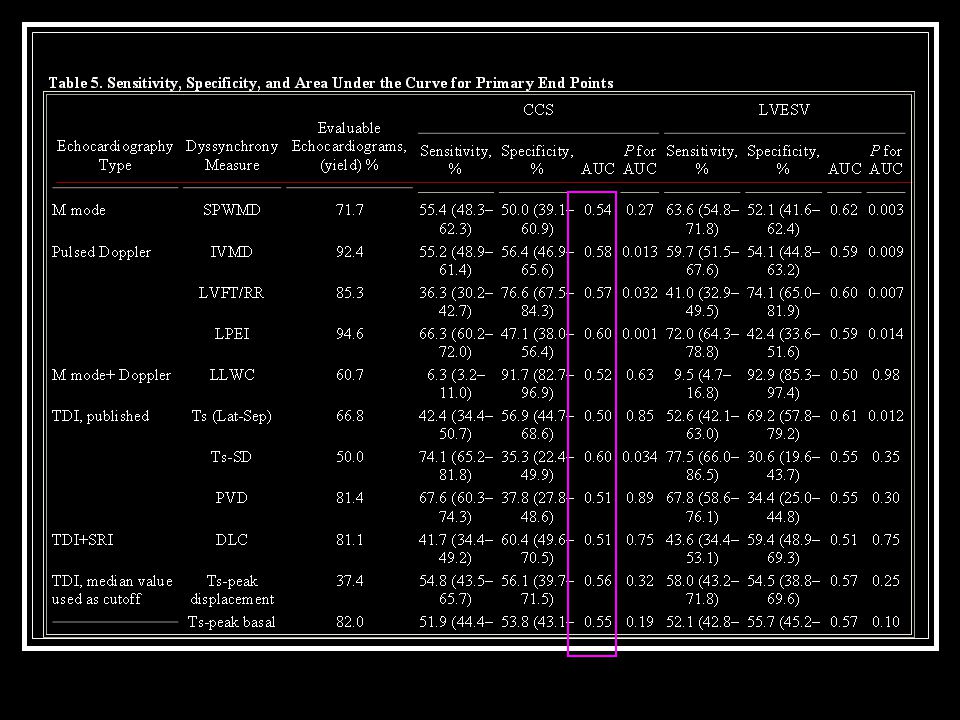
Endpoints- Composite clinical score Worsened (died, hospitalized, worsened heart failure, demonstrated worsening in NYHA class at last observation carried forward, moderate or marked worsening of patient global assessment score at last observation carried forward, or permanently discontinued CRT because of or associated with worsening heart failure Improved (not worsened as defined above and demonstrated improvement in NYHA class at last observation carried forward or had moderate or marked improvement in patient global assessment score at last observation carried forward) Unchanged (the patient was neither improved nor worsened)
 Unchanged (the patient was neither improved nor worsened)")
39
Copyright ©2008 American Heart Association Chung, E. S. et al. Circulation 2008;117:2608-2616 PROSPECT RESULTS: CCS and LVESV response rates

41
Area Under the Curve

42
PROSPECT Conclusions Echocardiographic measures of dyssynchrony aimed at improving patient selection criteria for CRT did not have a clinically relevant impact on improving response rates Echocardiographic parameters assessing dyssynchrony do not have enough predictive value to be recommended as selection criteria for CRT beyond current indications

43
Current ACC/AHA/NASPE 2005 Guideline Update Patients with LVEF 35%, sinus rhythm, and New York Heart Association functional class III or ambulatory class IV symptoms despite recommended optimal medical therapy and who have cardiac dyssynchrony, which is currently defined as a QRS duration >120 ms, should receive CRT unless contraindicated (Class: I, Level of Evidence: A).
.")
44
Other roles for Echo in CRT Assess LVEF Assess pre- and post-valvular regurgitation Assess best location of lead placement

45
Future directions >30% non-responders consistent through most trials Studies should aim to characterize the non- responders

Similar presentations