Download presentation
Presentation is loading. Please wait.

1
MULTICELLULAR ORGANISMS
Cell-Cell Adhesion Cell-Matrix Adhesion The Extracellular Matrix, ECM M. Habibi-Rezaei

3
Cell-Cell Interactions

4
Cleavage

5
MULTICELLULAR ORGANISMS
The appearance of multicellular organisms allows specialization of cells and formation of organs Vertebrates have more than 100 specialized cell types (plants have more than 15) A special matrix, the extracellular matrix, ECM, fills out the space between cells
 A special matrix, the extracellular matrix, ECM, fills out the space between cells.")
6
Cell Signals Direct contact Paracrine signaling Endocrine signaling
hormones Synaptic signaling neurotransmitters

7
Cell Signaling

8
Cell Surface Receptors

9
MULTICELLULAR ORGANISMS
By means of cell adhesion molecules, CAMs, cells are capable of recognizing each other Plasma membrane receptors take care of cell-ECM interactions

12
CELL-CELL ADHESION MOLECULES
Cadherins Ig superfamily CAMs Selectins Integrins

13
Septate Junctions (Invertebrates) Plasmodesmata (Plants)
Functional Categories of Cell Junctions Occluding Anchoring Communication Adherens Junctions Tight Junctions Gap Junctions Cadherin Desomosomes Septate Junctions (Invertebrates) Chemical Synapses Focal Adhesions Plasmodesmata (Plants) Integrin Hemi-desmosomes
 Chemical Synapses. Focal Adhesions. Plasmodesmata (Plants) Integrin. Hemi-desmosomes.")
14
Adhesion Molecules and Extra Junctional Adhesion:
Types of cell-cell adhesion

15
Cell adhesion Emphasis on cell migration: Embryogenesis Immune cell chemotaxis Tumor cell metastasis Types of adhesion molecules involved in these processes

16
Major Families of Cell Adhesion Molecules (CAMs)
Cadherins: participate in adherens junctions (adhesion belts) & desmosomes Immunoglobulin-like CAMs (ICAMs): only extrajunctional Integrins: cell matrix adhesion, hemidesmosomes, focal contacts Selectins: transient adhesion of leucocytes to blood vessels Integral membrane proteoglycans
 & desmosomes. Immunoglobulin-like CAMs (ICAMs): only extrajunctional. Integrins: cell matrix adhesion, hemidesmosomes, focal contacts. Selectins: transient adhesion of leucocytes to blood vessels. Integral membrane proteoglycans.")
17
Cell-Cell Recognition and Adhesion
What are the different categories of adhesion receptors? What is the difference between homophilic and heterophilic interactions? immunoglobulin superfamily proteins (e.g. N-CAM), cadherins, selectins, integrins homophilic – adhesion occurs when 2 identical molecules bind, one from each cell heterophilic – cell adhesion receptor on one cell interacts with different molecule on other cell
, cadherins, selectins, integrins. homophilic – adhesion occurs when 2 identical molecules bind, one from each cell. heterophilic – cell adhesion receptor on one cell interacts with different molecule on other cell.")
20
CADHERINS A family of Ca2+-dependent CAMs
Ca2+ causes dimerization of Cadherins The binding is homophilic

21
Cadherins – Ca++ dependent
Homophilic binding Cadherins – Ca++ dependent

22
Cadherins are Responsible for Cell-Cell Adhesion

23
Cadherins are homophilic, calcium dependent adhesion molecules

24
Primary tumor suppressor function of E- cadherin
Sequesteration of cytoplasmatic pools of ß-catenin Which prevents ß-catenin Of entering the nucleus and starting transcription programm

25
Cadherins Mediate Cell Sorting Due to Homophilic Binding
Cells in culture sort themselves based upon: 1) The type of cadherins they express 2) The level of cadherins they express
 The type of cadherins they express. 2) The level of cadherins they express.")
26
Adherens Junctions Help Fold Epithelial Cells
Fig

27
Embryogenesis & Cadherins
Expression of specific cadherins accompanies morphogenetic movements during embryogenesis

30
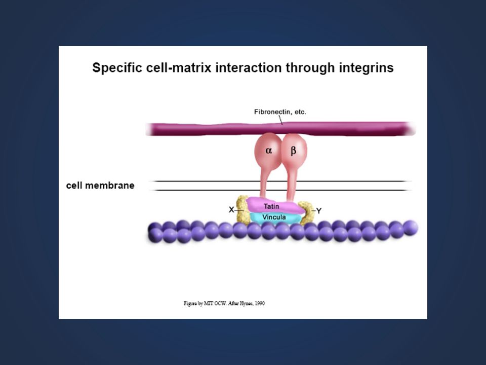
Integrins - Binding to extracellular matrix
Tripeptide binding sequence Protease cleaves Talin; binding/ uncoupling with actin Integrins - Binding to extracellular matrix

32
Immunoglobulin Superfamily CAMs (ICAMs) Important during neural development Binding is calcium-independent
 Important during neural development Binding is calcium-independent")
34
SELECTINS Selectins are involved in extravasation
Inflammatory signals activate endothelial cells making P-Selectin undergo exocytosis P-Selectin on the surface of endothelial cells binds a specific carbohydrate ligand (Sialyl Lewis -x) on leukocytes The leukocytes attach to the endothelial wall and roll slowly on it PAF and integrins are then activated and the leukocytes start to extravasate
 on leukocytes. The leukocytes attach to the endothelial wall and roll slowly on it. PAF and integrins are then activated and the leukocytes start to extravasate.")
35
Selectins mediate adhesions of white blood cells (leukocytes) during extravasation (migration of cells out of blood vessels) Adhesion is weak & transient Involved in other process, including adhesion of early embryo to uterine wall

36
Model of Extravasation

38
Integrins are Heterodimers

39
fibronectin, vitronectin, laminin, collagen etc (18 a-subunit members
dimerized single-transmembrane proteins consist of alpha and beta subunits, (18 a-subunit members and 8 b-subunit members) combine to form at least 25 different integrin receptors fibronectin, vitronectin, laminin, collagen etc . ECM molecules focal adhesion complex.
 combine to form at least. 25 different integrin receptors. fibronectin, vitronectin, laminin, collagen etc. . ECM. molecules. focal adhesion. complex.")
40
INTEGRINS serve as a velcro for cell migration
The cell moves by actin-driven "ruffling" it's membrane. In moving cell, integrins are turned on in “front” of cell, griping to ECM and pulling. At back, integrins are off. Internalised and recycled In resting cell, most of the integrins are inactive (Not ligand-binded)
")
41
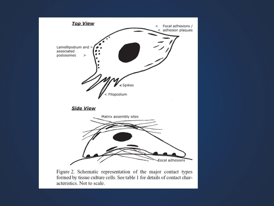
Focal Adhesions: Connect Cells to the EC Matrix
Adhesion Proteins Integrins Attach actin filaments from cell to matrix Connect to cytoplasmic anchor proteins Anchor Proteins Fig

42
Circ Res 89:

43
Integrins and their ligands
J Biol Chem 275:21785, 2000

44
Integrin Clustering Mediates Intracellular Signaling

46
Forms diverse structures
Extracellular Matrix Forms diverse structures Bone Ligament Tendon Vessel Connective tissue Skin Biological processes Adhesion Mechanical support Migration Proliferation Signalling Diseases Arthritis Atherosclerosis Cancer Asthma

47
Extracellular Matrix Prominent in Connective Tissues

48
Figure 4.1

49
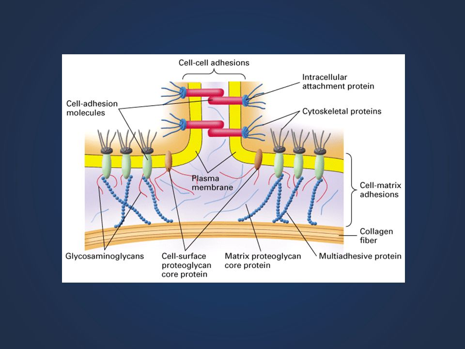
Extracellular Matrix is Contains of 3 Main Components:
Perlecan Large ( aggrecan, versican) Small ( decorin, Biglycan, Fibromodulin, Lumicin) Proteoglycans Collagens Multi Adhesive Matrix Proteins At least 12 types Fibronectin Laminin Nidogen Entactin
 Small ( decorin, Biglycan, Fibromodulin, Lumicin) Proteoglycans. Collagens. Multi Adhesive Matrix Proteins. At least 12 types. Fibronectin. Laminin. Nidogen. Entactin.")
50
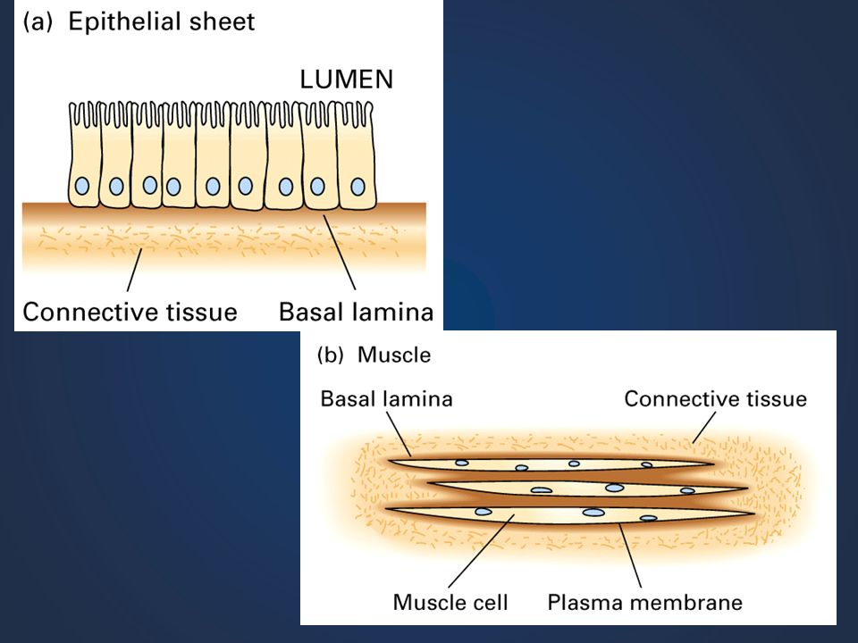
ECM Basement Membrane thin extracellular layer made up of basal lamina
closest to epithelial cells secreted by epithelial cells Components reticular lamina deep to basal lamina part of connective tissue layer produced by fibroblasts

51
Composition of The Extracellular Matrix
Bone Cornea

52
MAJOR ECM CONSTITUENTS
Hyaluronan Proteoglycans Collagens Elastin Fibronectin Laminin Enactin/nidogen Protein in green, glycosaminoglycan in red.

53
PLASMA MEMBRANE PROTEOGLYCANS
Annu Rev Biochem 68:729,’99

54
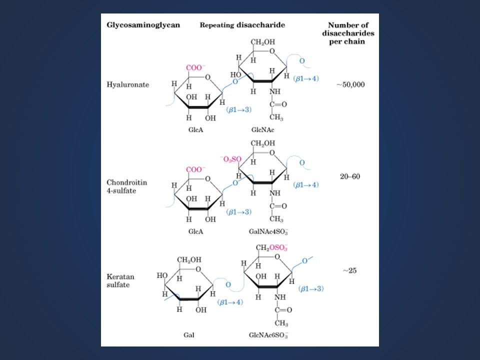
Glycosaminoglycan (GAG) Chains are Composed of a Repeating Disaccharide Sequence
Contains carboxyl & sulfate groups (-) Charge
 Charge.")
55
Hyaluronan is a GAG Chain Composed of a Repeating Disaccharide Sequence
Does not form proteoglycans; Contains only carboxyl groups (-) Charge 19-38
 Charge")
56
HYALURONAN Relative volumes

57
Some Common Proteoglycans

58
Proteoglycans = GAG Chain + Core Protein
19-39

59
Proteoglycans & Hyaluronan Associate to Form Large Complexes in the ECM
19-41

61
Basal Lamina Some functions:
Provide structural definition & integrity to tissues Acts as selective filter of small compounds Determines cell polarity Organize cell surface proteins on adjacent cell membranes Promotes cell survival, proliferation, differentiation Serves as “pathway” for cell migration

62
Basil Lamina: a specialized sheet of ECM

63
Scanning Electron Micrograph of an Epithelium
21_019.jpg

64
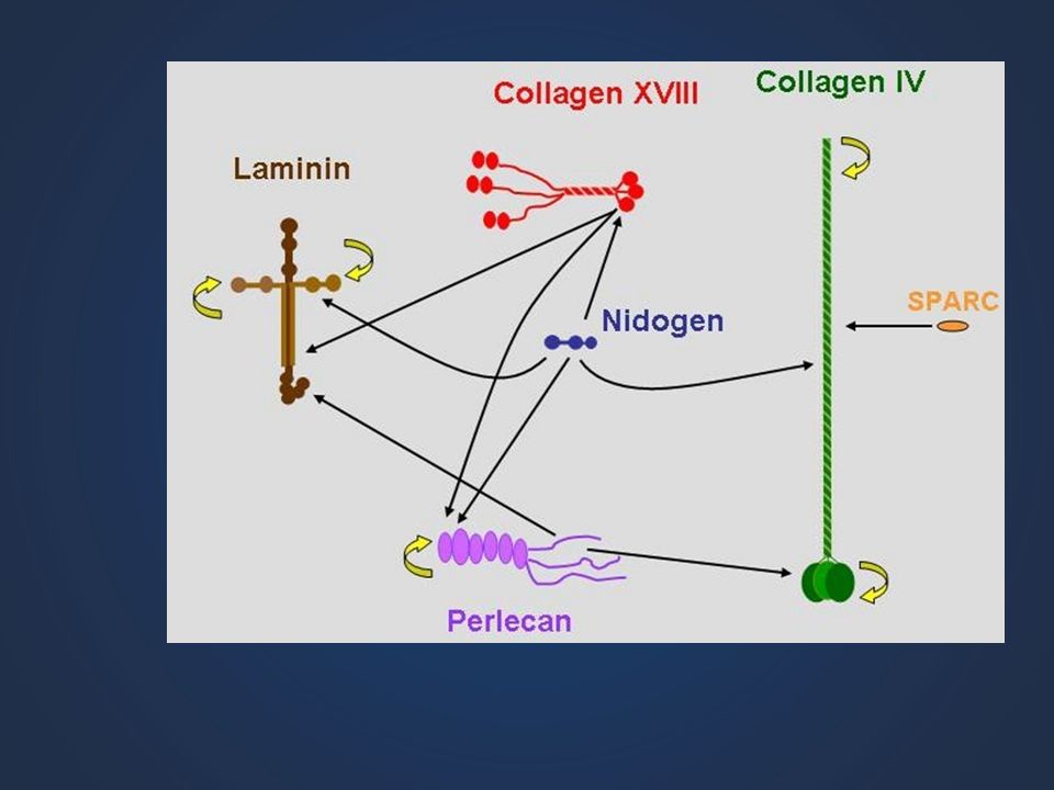
Composition of Basal lamina

66
Collagen

67
COLLAGENS A helix comprised of homotrimer & heterotrimer polypeptides (alpha chains) Major proteins of ECMs Many different alpha chains Multiple structures (involves cross-linking of chains) fibrils fibril-associated network forming Fig
 fibrils. fibril-associated. network forming. Fig")
68
Some types of collagen & their properties

69
Formation of Collagen FIBRILS and FIBERS

70
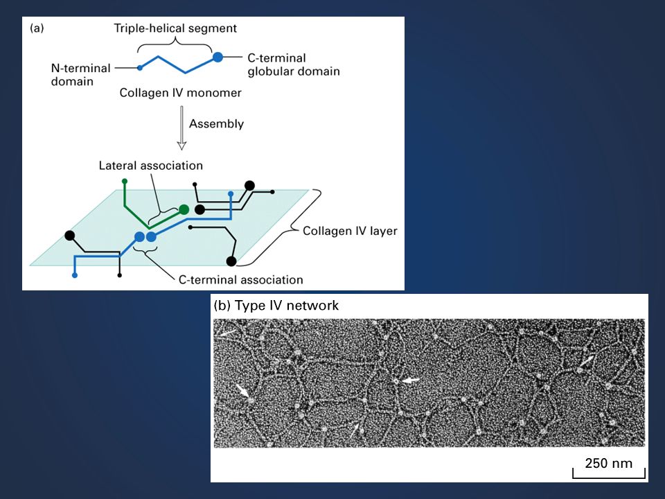
Formation of Collagen Networks

71
COLLAGEN ASSEMBLIES Ann Med 33:7, 2001

73
Heterotrimeric glycoprotein Basal lamina constituent
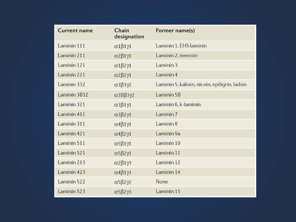
LAMININ Heterotrimeric glycoprotein Basal lamina constituent Multiple binding domains

74
2. Laminin i. a cross-shaped-protein with four binding sites for: i. cells trans-membrane proteins including integrins ii. other laminins, iii. proteoglycans and iv. collagen. : thus forming networks of extracellular fibers, including an inter-laced web in the basement membrane 3. entactin. another web-forming protein found in the basement membrane. Lin

75
Binding Domains of Laminin
Self assembly Type IV collagen Heparan sulfate Enactin/nidogen Cell Surface integrin nonintegrin Cell Suface Binding Sites J. Anat. 193:1, ‘98

77
FIBRONECTIN (FN) Extracellular dimeric glycoprotein
Differential splicing Multiple functional domains cell binding RGD sequence of FN other specificities heparin binding collagen binding fibrin binding Organized into a matrix Fig A,C

78
5. fibronectin. Important multi-valent linker
5. fibronectin. Important multi-valent linker. Multiple recognition sites on each of two peptides. constructed of two similar (not identical) peptides joined by disulfide bridges. A family of proteins. Each one has modular construction, with multiple binding sites for components of the ECM: i. collagen, ii. proteoglycans, integrins an arg-gly-asp amino acid (RGD) sequence functions to make a web of proteins, proteoglycans and cells by cross linking them Especially important as binding sites for cells within the ECM due to integrin binding. Collagen proteoglycan laminin integrin H2N COOH s s s s COOH H2N
 peptides joined by disulfide bridges. A family of proteins. Each one has modular construction, with multiple binding sites for components of the ECM: i. collagen, ii. proteoglycans, integrins an arg-gly-asp amino acid (RGD) sequence. functions to make a web of proteins, proteoglycans and cells by cross linking them. Especially important as binding sites for cells within the ECM due to integrin binding. Collagen proteoglycan laminin integrin. H2N. COOH. s. s. s. s. COOH. H2N.")
80
Matrix Metalloproteases MMPs

81
Genes Dev 14:2123,’00

82
Matrix Metalloproteases
Genes Dev 14:2123,’00

83
ADAM A Disintegrin And Metalloprotease
Distintegrin and Metalloproteinase (ADAM) ADAM proteins are members of the same superfamily as MMPs, namely the Metzincins, named for their zinc binding domains and their structurally important C-terminal conserved methionine residue. The name ADAM stands for “A Disintegrin And Metalloprotease” and like the name suggests, ADAM proteins are cell surface proteins that possess both an adhesion domain as well as a protease domain (Wolfsberg, TG et al. J Cell Biol 1995; 131:275–278). There are more than 35 members of the ADAM family of proteins; the precise function of many the ADAM family members are unknown, but some, such as ADAM17 (a.k.a. tumor necrosis factor–a converting enzyme) have known biological functions. An additional class of ADAM related proteins are known as the ADAMTS proteins. ADAMTS proteins are structurally homologous to ADAM proteins, but they contain at least one C-terminal thrombospondin type 1 (TSP1) repeat and are secreted rather than membrane bound. ADAMTS1 and ADAMTS8 are inhibitors of angiogenesis, and others, such as ADAMTS5, cleave extracellular proteoglycans such as aggrecan.
 ADAM proteins are members of the same superfamily as MMPs, namely the Metzincins, named for their zinc binding domains and their structurally important C-terminal conserved methionine residue. The name ADAM stands for A Disintegrin And Metalloprotease and like the name suggests, ADAM proteins are cell surface proteins that possess both an adhesion domain as well as a protease domain (Wolfsberg, TG et al. J Cell Biol 1995; 131:275–278). There are more than 35 members of the ADAM family of proteins; the precise function of many the ADAM family members are unknown, but some, such as ADAM17 (a.k.a. tumor necrosis factor–a converting enzyme) have known biological functions. An additional class of ADAM related proteins are known as the ADAMTS proteins. ADAMTS proteins are structurally homologous to ADAM proteins, but they contain at least one C-terminal thrombospondin type 1 (TSP1) repeat and are secreted rather than membrane bound. ADAMTS1 and ADAMTS8 are inhibitors of angiogenesis, and others, such as ADAMTS5, cleave extracellular proteoglycans such as aggrecan.")
84
ADAM A Disintegrin And Metalloprotease
transmembrane domain A disintegrin is a molecule that binds to an integrin. Trends Genet. 16:83, ‘00

85
Trends Genet. 16:83, ‘00

87
Tumor Cell Metastasis

88
Proteases allow cells to move through ECM, basal lamina
White blood cells Tumor cells

89
CELL JUNCTIONS Adherens junctions Gap junctions Tight junctions
Desmosomes/Hemidesmosomes Focal adhesions

90
Cell-Matrix Interactions

97
Summary of lectin families Lectin family Typical saccharide ligands
Lectin family Typical saccharide ligands Subcellular location Examples of functions Calnexin Glc1Man9 ER Protein sorting in the endoplasmic reticulum. M-type lectins Man8 ER-associated degradation of glycoproteins. L-type lectins Various ER, ERGIC, Golgi P-type lectins Man 6-phosphate, others Secretory pathway Protein sorting post-Golgi, glycoprotein trafficking, ER-associated degradation of glycoproteins, enzyme targeting. C-type lectins Cell membrane, extracellular Cell adhesion (selectins), glycoprotein clearance, innate immunity (collectins). Galectins -Galactosides Cytoplasm, extracellular Glycan crosslinking in the extracellular matrix. I-type lectins (siglecs) Sialic acid Cell membrane Cell adhesion. R-type lectins Golgi, Cell membrane Enzyme targeting, glycoprotein hormone turnover. F-box lectins GlcNAc2 Cytoplasm Degradation of misfolded glycoproteins. Ficolins GlcNAc, GalNAc Innate immunity. Chitinase-like lectins Chito-oligosaccharides Extracellular Collagen metabolism (YKL-40). F-type lectins Fuc-terminating oligosaccharides Intelectins Gal, galactofuranose, pentoses Extracellular/cell membrane Innate immunity. Fertilization and embryogenesis.
, glycoprotein clearance, innate immunity (collectins). Galectins. -Galactosides. Cytoplasm, extracellular. Glycan crosslinking in the extracellular matrix. I-type lectins (siglecs) Sialic acid. Cell membrane. Cell adhesion. R-type lectins. Golgi, Cell membrane. Enzyme targeting, glycoprotein hormone turnover. F-box lectins. GlcNAc2. Cytoplasm. Degradation of misfolded glycoproteins. Ficolins. GlcNAc, GalNAc. Innate immunity. Chitinase-like lectins. Chito-oligosaccharides. Extracellular. Collagen metabolism (YKL-40). F-type lectins. Fuc-terminating oligosaccharides. Intelectins. Gal, galactofuranose, pentoses. Extracellular/cell membrane. Innate immunity. Fertilization and embryogenesis.")
98
Hesselson et al., 2004; Kubota et al., 2004
Common name Gene Isoforms[a] LOF phenotype(s)[b] References Collagen IV α1 emb-9 1 Emb (2-3X) Guo et al., 1991; Gupta et al, 1997 Collagen IV α2 let-2 2 Sibley et al., 1993, 1994 Collagen XVIII cle-1 4 Neuro, gonad morph Ackley et al., 2001, 2003 Fibulin-1 fbl-1 Gonad morph Hesselson et al., 2004; Kubota et al., 2004 Hemicentin him-4 Tissue adhesion, aneuploidy Vogel and Hedgecock, 2001 Laminin αA lam-3 Emb, Acc Huang et al., 2003 Laminin αB epi-1 Emb, Acc, Ste Laminin β lam-1 N.D. Laminin γ lam-2 Nidogen nid-1 3 Neuro Kang and Kramer, 2000; Kim and Wadsworth, 2000 Osteonectin/SPARC ost-1 Acc (L1-L2) Fitzgerald and Schwarzbauer, 1998 Papilin ppn-1 Emb (hyp enclosure) Kramerova et al., 2000 Perlecan unc-52 >5 Pat Rogalski et al., 1993, 1995
[b] References. Collagen IV α1. emb Emb (2-3X) Guo et al., 1991; Gupta et al, Collagen IV α2. let Sibley et al., 1993, Collagen XVIII. cle Neuro, gonad morph. Ackley et al., 2001, Fibulin-1. fbl-1. Gonad morph. Hesselson et al., 2004; Kubota et al., Hemicentin. him-4. Tissue adhesion, aneuploidy. Vogel and Hedgecock, Laminin αA. lam-3. Emb, Acc. Huang et al., Laminin αB. epi-1. Emb, Acc, Ste. Laminin β. lam-1. N.D. Laminin γ. lam-2. Nidogen. nid Neuro. Kang and Kramer, 2000; Kim and Wadsworth, Osteonectin/SPARC. ost-1. Acc (L1-L2) Fitzgerald and Schwarzbauer, Papilin. ppn-1. Emb (hyp enclosure) Kramerova et al., Perlecan. unc-52. >5. Pat. Rogalski et al., 1993,")
100
COLLAGENS The most abundant animal protein At least 16 types exist
The structural unit is composed of three 300 nm long coiled subunits in a triple helix The helical structure depends on the abundant presence of glycin, proline (and hydroxyproline) making a motif gly-pro-x, which is necessary for twisting together the three strands
 making a motif gly-pro-x, which is necessary for twisting together the three strands.")
102
COLLAGENS 2 Collagens are synthesized as precursors called procollagens They are glycosylated in ER and Golgi adding Gal and Gly to hydroxy-lysine residues and long oligosaccharides to selected asparagine residues Proline and lysine are hydroxylated Disulphide bonds are made between the N- and C-terminal parts of the propeptides After exocytosis the N- and C-terminals are “trimmed”, only then can the fibrils be formed

103
COLLAGENS 3 Lack of vitamin C prevents hydroxylation impaired fibrils Mutations or deletions of -chains in Collagen I can lead to the disease Osteogenesis imperfecta

106
LAMININ Laminin is a key component of the basal lamina

113
DISEASES OF THE BASAL LAMINA
Alport’s syndrome appears as impaired ultrafiltration in the kidney resulting in renal failure and hearing loss. Mutations in collagen IV -chains result in this syndrome. Antibodies against 3-chains of collagen IV lead to pulmonary hemorrhage and renal failure (Goodpasture’s syndrome)
")
114
FIBRONECTIN Fibronectins attach cells to collagens
Fibronectins are dimers Fibronectins express the RGD sequence recognized by integrins

116
PROTEOGLYCANS 1 The Polysaccharides in proteoglycans are long repeating polymers of dissacharides called Glucosaminoglycans (GAGs) One sugar of the dissacharides is a uronic acid and the other is an aminosugar (e.g. N-acetylglucosamine) One or both sugars contain one or two sulphate residues
 One or both sugars contain one or two sulphate residues.")
120
HYALURONAN (HA) HA is a GAG found in ECM
HA is also a key component of complex proteoglycans HA consists of approx. 50,000 disaccharides in a random coil. It can be bound to the surface receptor CD44 HA gives strength, flexibility and smoothness to the ECM and forms a viscous hydrated gel in which cells can migrate HA makes the ECM able to resist compression

121
PROTEOGLYCANS 2 Heparin sulphate and chondroitin sulphate are added to a 3-sugar “linker” (Xyl-Gal-Gal) added to a Serine in the core protein Proteoglycans are found both in ECM and attached to the plasma membrane

124
PROTEOGLYCANS IN THE ECM
In cartilage the key proteoglycan is aggrecan The central component of aggrecan is a carbohydrate, hyaluronan At 40 nm intervals aggrecan core proteins are attached (assisted by a linker protein) to a decasaccharide sequence in hyaluronan Attached to the aggrecan core protein are multiple GAGs (via the trisaccharide linker) The GAGs in aggrecan are chondroitinsulphate and keratin sulphate MW of an aggrecan 2 x 10⁸
 to a decasaccharide sequence in hyaluronan. Attached to the aggrecan core protein are multiple GAGs (via the trisaccharide linker) The GAGs in aggrecan are chondroitinsulphate and keratin sulphate. MW of an aggrecan 2 x 10⁸.")
127
PROTEOGLYCANS ON THE CELL SURFACE
A typical example is syndecan The core protein spans the membrane with a short cytosolic domain The GAGs are attached via the trisaccharide linker to serine residues The GAGs in syndecan are heparan sulphate chains Syndecan binds extracellularly to collagens and fibronectin and intracellularly to the cytoskeleton

131
DISEASES OF GAG Rare genetic defects in enzymes required for the synthesis of Dermatan sulfate lead to defects in bones, joints, muscles, and skin. The individuals do not grow to normal hight and appear prematurely aged.

134
NEURONAL CELL ADHESION MOLECULES LEARNING AND MEMORY
Male humans with L1-mutations develop Mental retardation Hydrocephalos Adducted thumbs NCAM knock-out animals develop Morphological changes in bulbus olfactorius and hippocampus Impaired learning Emotional disturbances Modulation of NCAM and L1-function interferes with LTP and learning and memory

135
CELL-MATRIX ADHESION Integrins Collagens Laminin and Fibronectin
Proteoglycans and Glucosaminoglycans

136
CELL MATRIX ADHESION Integrins on the cell surface mediate cell-ECM binding Integrins are composed of an- and a -chain There are 3 different -chains and more than 10 types of -chains The chain composition determines the ligand specificity The affinity is generally low (Kd )
")
137
INTEGRINS Integrins can be activated through a signal from the interior of the cell Activation involves conformational changes of the integrin Various integrins recognize specific sequences in their ligands. E.g. 41 recognizes EILDV (in VCAM-1 and in fibronectin) and 51 recognizes RGD in many ECM proteins
 and 51 recognizes RGD in many ECM proteins.")
138
INTEGRIN CONTAINING JUNCTIONS
A junction consists of an exterior ligand, a transmem-brane protein, a linker, and a cytoskeletal component An adherence junction connects an ECM component with an integrin linked to an adapter (e.g. vinculin) and F-actin A hemidesmosome connects an ECM-component to integrin and via an adapter (e.g. plectin) to intermediate filaments (keratins)
 and F-actin. A hemidesmosome connects an ECM-component to integrin and via an adapter (e.g. plectin) to intermediate filaments (keratins)")
139
INTEGRIN DISEASES Genetic defects in integrin 2 lead to leucocyte-adhesion deficiency. The patient becomes susceptible to bacterial infections

140
DISINTEGRINS Disintegrins contain the RGD sequence and interfere with integrin-ECM adhesion allowing deadhesion and cell migration The ADAMs (A Disintegrin And a Metalloprotease) “remodel” surface proteins; f.x. at the fusion of sperm and egg, the fusion of myoblasts during myogenesis, release of TNF from the surface
 remodel surface proteins; f.x. at the fusion of sperm and egg, the fusion of myoblasts during myogenesis, release of TNF from the surface.")
Similar presentations


 through specialized integral membrane.>")

 –soluble procollagen excreted –cross linked –‘insoluble’ tropocollagen’ –assembled into a fibril (details.>")


? Something that is made by virtually all multi-cellular organisms. Elaborate.>")


