Download presentation
Presentation is loading. Please wait.

1
Using tests to help with diagnosis Preparation for Clinical Skills 1

2
Tests that you might hear discussed during clinical skills 1 3 patients Each one illustrates how a clinician can use tests to help them manage a patient Some simple questions Use the information we have already covered Think about what is going on Think about how can the test help you as a doctor to manage a patient Discussed in greater detail during CS1

3
From patient to diagnosis 44% Referring GP diagnosis unchanged 41% Diagnosis changed after history 7% Diagnosis changed after examination 8% Diagnosis changed after investigation

4
Cost of Tests Chest X-ray £16.00 CT chest £56.00 Full blood count £5.00

5
Cost to the patient Equivalent Dose (Sv) Dose required to sterilise medical products 25 000 Typical total radiotherapy dose to cancer tumour 60 50% survival probability, whole body dose 4 Legal worker dose limit (whole body) 0.02 Average annual dose from all sources in Cornwall 0.008 Average annual dose from natural radiation 0.002 Typical chest X-ray dose 0.00002 Average dose from a flight from UK to Spain 0.00001 Ct scan to abdomen or pelvis delivers about 500 x radiation as a single CXR
 Dose required to sterilise medical products Typical total radiotherapy dose to cancer tumour 60 50% survival probability, whole body dose 4 Legal worker dose limit (whole body) 0.02 Average annual dose from all sources in Cornwall Average annual dose from natural radiation Typical chest X-ray dose Average dose from a flight from UK to Spain Ct scan to abdomen or pelvis delivers about 500 x radiation as a single CXR")
6
Blood tests Why do we do them? 1.Screen asymptomatic patients for a disease 2.To confirm or refute a diagnosis 3.As marker of disease progression / response to therapy

7
What do you get?

8
Blood tests Blood count Us and Es Glucose HbA1c Liver function Cholesterol Subtypes of cholesterol Special blood tests PSA Hormones Thyroid Virus screens Cultures for infection Simple blood testsAlso available to GPs Radiology ECG 24 hour ECG Echocardiogram

10
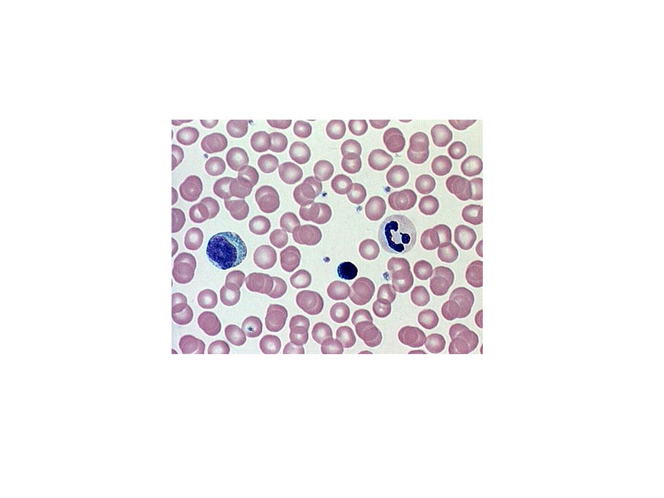
Full blood count Blood count Hb Red cell size White blood cells Different types of white blood cells Platelets counts the number of red cells, white cells, and platelets per ml of blood. measures the size of the red blood cells and calculates their average (mean) size. calculates the proportion of blood made up from red blood cells (the haematocrit). measures the amount of haemoglobin in the red blood cells. http://www.patient.co.uk/health/B lood-Test-Blood-Count-and- Smear.htm
 size. calculates the proportion of blood made up from red blood cells (the haematocrit). measures the amount of haemoglobin in the red blood cells. lood-Test-Blood-Count-and- Smear.htm.")
11
Erythrocyte sedimentation rate (ESR) Indirect measure of acute phase response Measures rate of fall of erythrocytes through plasma Depends on how red cells aggregate May be disparity between ESR and CRP in certain conditions
 Indirect measure of acute phase response Measures rate of fall of erythrocytes through plasma Depends on how red cells aggregate May be disparity between ESR and CRP in certain conditions")
12
Blood biochemistry Urea Creatinine eGFR Sodium Pottasium Glucose Protein waste Muscle breakdown indicates kidney function Estimated Glomerular filtration Blood electrolytes vital for life and cardiac function Transportable energy

13
Factors affecting urea and creatinine

14
C-reactive protein (CRP) Acute phase protein Increases within 6 hours inflammatory stimulus Half life 19 hours
 Acute phase protein Increases within 6 hours inflammatory stimulus Half life 19 hours")
15
Some examples

16
A patient coming for a check up Mr Smith Aged 55 Visits his GP for routine check up What can the GP offer Why does he do this?

17
Cholesterol The total circulating cholesterol Low density ( bad) High density (“good”) Circulating fats Total cholesterol – made up of: LDL HDL Triglycerides
 High density ( good ) Circulating fats Total cholesterol – made up of: LDL HDL Triglycerides")
19
Other tests Liver function HbA1c PSA Thyroid function Hormones Tests liver function and when it is not working Glycosylated Hb – Released from prostate Thyroid hormones Test endocrine function

20
Prevention What is Primary prevention and what is secondary prevention? Can you suggest any examples?

21
Reducing risk factors What are the risk factors for people with vascular disease? How can these be changed and what will the effect be? How does a doctor decide what to do and how does this get explained to a patient?

23
Mr White – Aged 62 with a history of chest tightness and shortness of breath Which symptoms suggest that a patient has Angina? What is happening in an anginal attack? What can be done to prevent it ? How do we investigate it?

24
Initial tests Blood tests X ray of chest Plain ECG

25
Chest x ray Aortic knuckle Left ventricle Pulmonary arteries Diaphragm Air in stomach

26
Resting ECG

27
Uses of ECG Looking at rhythm of the heart Information about conduction of electrical impulse Information about damage to heart muscle Specific patterns of appearance used to help find out about heart, electrolyte changes

29
Hyperkalaemia

30
The patient is referred to the cardiac clinic What can a hospital doctor do? Exercise ECG 24 hour ECG for heart rythm Isotope scans looking at perfusion of heart muscle Angiogram to look at blood flow Echocardiogram

31
Exercise ECG – Stress test

32
24 hour ECG

33
Isotope scans

34
Angiography

35
Therapeutic intervention Balloon angioplastyCoronary stent

36
Echocardiogram

37
Mr Green 63 SUDDEN ACUTE CHEST PAIN Which factors on a history suggest that the pain is coming from his heart? What is the cause of the pain and what is the process called? What are the risks to the patient without prompt treatment? What actions can the GP or paramedic at the scene do to reduce the extent of heart muscle damage?

38
Cardiac chest pain?

39
Acute coronary syndrome ( ACS)
")
40
Pathology in a heart attack

42
The patient is seen in the emergency department What tests will help to confirm the diagnosis of a heart attack? What actions are used to limit the damage to the patients heart How are tests used to monitor the patient after the initial event?

43
Blood tests Troponins – proteins released by damaged muscle into the blood stream Levels elevated after 12 hours May remain elevated for up to 2 weeks

44
Chemical markers in Acute coronary syndromes Blood markers Troponins – breakdown products of cardiac muscle cells – suggest cellular damage Enzymes – some specific for Cardiac muscle CK MB fraction Some non specific

45
ECG in MI There are characteristic changes ECG can record these Site of changes suggests location of the damage The shape of the changes may suggest severity of the damage

47
Complications of MI....... Changes to heart rhythm – slow, fast, very fast....... Loss of pump function Leaking of valves Blood clots Drop in blood pressure Failure of heart pump ECG monitoring.......

48
A medical emergency

49
Thrombolysis and primary angioplasty

Similar presentations


 CAD is most common form of heart disease and causes premature death. In UK, 1 in 3 men and.>")

