Download presentation
Presentation is loading. Please wait.

1
Pathology Dept/ Sulaimani School of Medicine /University of Sulaimani
Hemopoiesis Dr. Sana D. Jalal M.B.Ch.B/F.I.B.M.S.Path Pathology Dept/ Sulaimani School of Medicine /University of Sulaimani

2
Haematology: is the study of the blood and its diseases
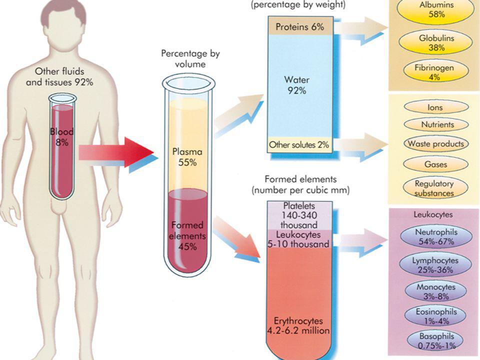
Blood : is a suspension of non-dividing end-stage cells of three types namely, red cells (erythrocytes), White cells (leucocytes) and platelets (thrombocytes). The suspending fluid is the Plasma.
, White cells (leucocytes) and platelets (thrombocytes). The suspending fluid is the Plasma.")
4
Function of Blood Delivery of substances needed for cellular metabolism. Removal of waste. Defense against microorganisms & injury. Maintenance of acid base balance.

5
Hemopoietic System Hemopoiesis is the process of production of all blood cells. Hemopoietic System is comprised of two major cell lines : Myeloid Lymphoid

7
Simplified chart of hemopoeisis from Stem cell to end stage cells
Stem cell COMMITMENT Precursors Multipotential Stem cells Erythroid precursors Proerythroblasts RBC Pluripotential Stem cells Myeloid precursors Myeloblasts Neutrophil Megak-aryocyte Precursors for megak. Platelets Lymph nodes Courtesy of Professor Nasir Allawi B or T Lymphocytes Lymphoid Stem cells

11
The Concept of STEM CELLS
Stem cells are cells from which all Hemopoietic elements originate. They are characterized by their ability of Self-renewal & Differentiation. Stem cells require for their proliferation and differentiation : - Certain regulatory factors (Hemopoietic Growth Factors). - Suitable microenvironment, provided by the marrow space. Stem cells Precursors End-stage cells The End stage cells have a restricted life span, ranging from 4 months for RBC, days for platelets, hours for granulocytes. These cells have to be continuously replaced by bone marrow, and BM should produce daily some 2.5 billion red cells/kg, 2.5 billion red cells/kg platelets, and 1 billion granulocytes/kg. Actually any normal individual will produce a no. of hemopoietic cells equivalent to his body weight /year, to maintain his peripheral blood counts.
. - Suitable microenvironment, provided by the marrow space. Stem cells Precursors End-stage cells. The End stage cells have a restricted life span, ranging from 4 months for RBC, days for platelets, hours for granulocytes. These cells have to be continuously replaced by bone marrow, and BM should produce daily some 2.5 billion red cells/kg, 2.5 billion red cells/kg platelets, and 1 billion granulocytes/kg. Actually any normal individual will produce a no. of hemopoietic cells equivalent to his body weight /year, to maintain his peripheral blood counts.")
12
Myeloid Hemopoietic System
The earliest evidence of myelopoiesis ( mesoblastic phase) occur in the yolk sac of the embryo. In this stage the hemopoietic system consists primarily of mesenchymal derived primitive erythroblast. The hepatic phase begins in the second month of fetal life. ( megakaryocytes and granulocytes appears in the sinusoids of liver) At wks gestation , the liver is the principle site of hemopoiesis till approximately 24 wks. Bone marrow function starts from the 4th – 5th months of gestation and is the major site of Hemopoiesis till birth.
 occur in the yolk sac of the embryo. In this stage the hemopoietic system consists primarily of mesenchymal derived primitive erythroblast. The hepatic phase begins in the second month of fetal life. ( megakaryocytes and granulocytes appears in the sinusoids of liver) At wks gestation , the liver is the principle site of hemopoiesis till approximately 24 wks. Bone marrow function starts from the 4th – 5th months of gestation and is the major site of Hemopoiesis till birth.")
13
Myeloid Hemopoietic System
Myeloid hemopoietic system in normal individual in the first yr. of postnatal life is in both axial and radial skeleton. Thereafter, there is a gradual regression of the hemopoiesis in the long bones until about the age of 15 , when the flat bones of the central skeleton ((pelvis, vertebral column, cranium, ribs ,sternum), and epiphysis of long proximal bones are the exclusive sites of hemopoiesis. The BM in postnatal life comprises about 3.5%-6% of total body weight and in aggregate mass approximates the size of liver.
, and epiphysis of long proximal bones are the exclusive sites of hemopoiesis. The BM in postnatal life comprises about 3.5%-6% of total body weight and in aggregate mass approximates the size of liver.")
14
Structure of Bone Marrow
Bone Marrow Consist of Hemopoietic tissue, adipose tissue and stroma. The stroma consist of delicate framework of connective tissue. The vascular supply is derived from the nutrient artery which ramify through the marrow space. The arterioles of the nutrient artery branch into capillaries which are continuous within a system of thin walled of sinusoids. The adventitial reticular cells synthesize the extravascular collagen and adhesive protein.

16
Microscopical Features:
Erythroblasts or normoblasts Microscopical Features: Reduction in the cell size. Loss of nucleoli. Clumping of nuclear chromatin. Hemoglobinization of the cytoplasm. Loss of nuclei. Proerythoblast Reticulocyte Early intermediate late Mature RBC Proerythoblast The earliest Erythroid recognizable cell is the Proerythroblast, which is large cell with high N/C ratio, basophilic cytoplasm, and open nuclear chromatin with prominent nucleolus. The next stage is early normoblast, in which the cell loses its nucleolus, but cytoplasm remains blue. The next stage is the intermediate normoblast in which the N/C ratio gets smaller, while the cytoplasm becomes less basophilic, as Hb increases.while the nuclear chromatin becomes more condensed and clumped The next stage is the late normoblast in which the cytoplasm become more pink due to Hbinization, and nuclear chromatin more condensed. The nucleus is then extruded to form a reticulocyte, which remains normally about two days in the marrow and is then released to circulation and matures finally into a mature anucleated erythrocyte. Normoblasts Intermediate Late Red Cell precursors (in marrow)
")
17
Microscopical Features:
Reduction in the cell size. Loss of nucleoli. Granulation of cytoplasm ( primary and secondary). Nuclear segmentation. Stages of Maturation of the Granulocytic Series In the bone marrow Blood Bone marrow
. Nuclear segmentation. Stages of Maturation of the Granulocytic Series. In the bone marrow. Blood. Bone marrow.")
18
Neutrophils

19
Blood leucocyte morphology

20
Leucocytes in Blood Leucocyte counts range in a normal adult between 4-10 x 109/L. Normally majority of the cells seen are neutrophils, followed by lymphocytes, monocytes, eosinophils and basophils. Determination of the proportion of various leucocyte types in the blood is called Differential leucocyte count.

21
Terms used to denote changes in leucocytes numbers:
Leucocytosis : increased no. of leucocytes above 10.0 x 10⁹/L. Leucopenia : Reduced total leucocyte count below 4.0 x 10⁹/L.

22
Megakaryocyte : the precursor of Platelets in the marrow
Microscopical Features: Increased cell size. Lobulation of the nuclei. Cytoplasmic granulation.

23
Platelets in blood Platelets count normally ranges between
150 to 450 x 109/L. Increased Platelets above 450 is called thrombocytosis. Reduced Platelets below 150 is called thrombocytopenia.

24
Life span of blood cells in peripheral circulation
- Red cells : 120 days. - Granulocytes : ~ 1day. - Platelets : 7-10 days.

25
Normal Blood Film

26
Bone marrow examination
In certain conditions a bone marrow examination is required for diagnosis or follow-up of patients. There are two types of marrow procedures : 1. Bone marrow aspirate : done from iliac crest or sternum, in which a specimen is aspirated using a wide bore needle from the active marrow, smeared, stained and then examined for any abnormalities. 2. Bone marrow biopsy : here a core of bone marrow tissue is taken, and processed and stained as in histopathological specimens (H&E stain)
")
27
Bone marrow sets Aspirate Set Biopsy Set

28
Common sites for Bone marrow procedures in adults
Best x x Manubrium Sternii Post. Superior Iliac spine

29
Bone marrow aspiration
Bone marrow aspirate smear Fragment Higher magnification of trail. Trail

30
Bone marrow section stained with H&E stain
Bone Marrow Biopsy Bone marrow biopsy slide-core of BM Bone marrow section stained with H&E stain

Similar presentations


. Blood functions to transport oxygen, carbon dioxide,>")


, MPhil (NUST) Asst Prof. Physiology.>")
