Download presentation
Presentation is loading. Please wait.

1
Histology The study of tissues

2
What are tissues? Tissues are collections of specialized cells and cell products that perform a limited number of functions.

3
Four Basic Tissue Types
Epithelial – lining and secretory tissue Connective – supportive and nutritive tissue Muscular – contracts to produce movement Nervous – integration and control

4
Characteristics of Epithelial Tissue
Cellularity– epithelia are composed almost entirely of cells bound closely together by interconnections called cell junctions. Polarity-epithelial tissue have an exposed surface (apical cells) that either faces the exterior of the body or some internal space and a base (basal cells). Attachment—the base of the epithelium is bound to a thin basal lamina or basement membrane.
 that either faces the exterior of the body or some internal space and a base (basal cells). Attachment—the base of the epithelium is bound to a thin basal lamina or basement membrane.")
5
More characteristics of Epithelial Tissue
Avascularity—epithelia are avascular, which means that they lack blood vessels. They have to get their nutrients by diffusion or absorption Regeneration—epithelial cells that are damaged or lost are continuously replaced by the stem cells in the epithelium

6
Functions of Epithelial Tissue
Four major functions: Provide physical protection Control permeability Provide sensation (They have a large sensory nerve supply.) Produce specialized secretions (Glands)
 Produce specialized secretions (Glands)")
7
Naming Epithelial Tissue based on shape
Epithelium is named according to shape, structure, and arrangement of cells. Shapes of Epithelium •squamous - thin and flat cells •cuboidal - cube shaped cells •columnar - column shaped cells

8
Naming Epithelial Tissue based on layers
simple - single layer of cells stratified - multilayered cells

9
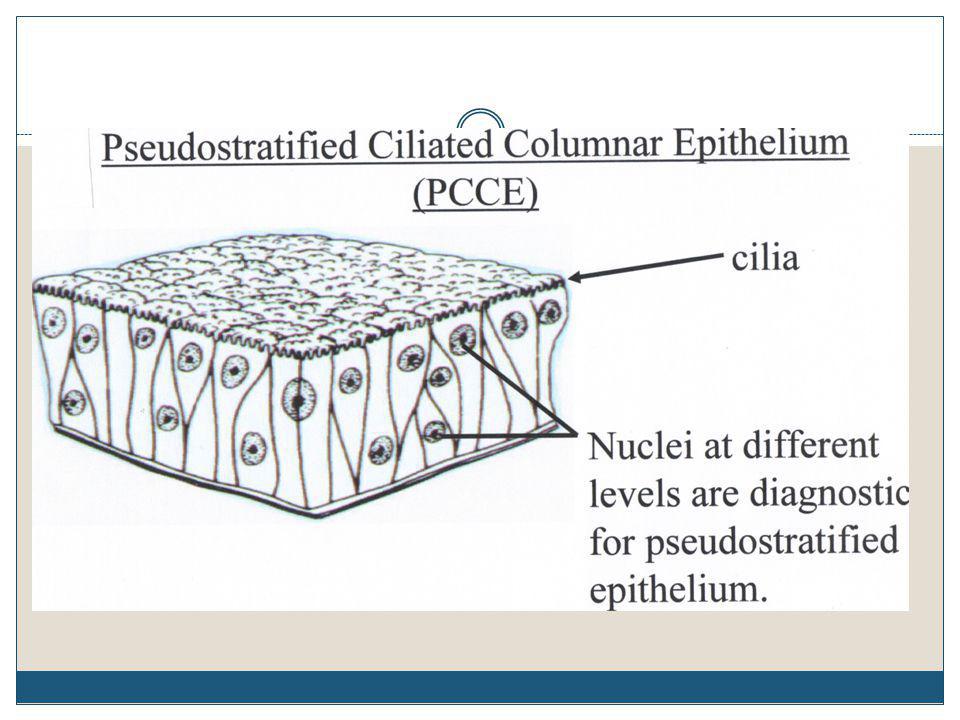
pseudostratified - false stratified
ciliated - cells possess cilia

10
What do you HAVE to know? -Structure, function, and location of seven epithelial tissue types -what each tissue type looks like enough to be able to identify the type from a picture

11
The 7 Epithelial Tissue Types that you have to know are . . .
Simple Squamous Simple Cuboidal Simple Columnar Stratified Squamous Stratified Cuboidal (sometimes referred to as Transitional) Pseudostratified Ciliated Columnar Keratinized (or Cornified) Stratified Squamous
 Pseudostratified Ciliated Columnar. Keratinized (or Cornified) Stratified Squamous.")
12
Simple Squamous Epithelium
Structure-The thinnest tissue of the body. One layer, flattened (squashed) nucleus. Function-Allows transport across membranes in lungs and capillaries. Secretes fluid in serous membranes (e.g. pericardial and pleural membranes, mesenteries). Location-Lines cardiovascular system, covers organs, forms glomerular capsules in kidney.
 nucleus. Function-Allows transport across membranes in lungs and capillaries. Secretes fluid in serous membranes (e.g. pericardial and pleural membranes, mesenteries). Location-Lines cardiovascular system, covers organs, forms glomerular capsules in kidney.")
16
Simple Cuboidal Structure-single layer, cube-like cells with spherical (round) nuclei. Function-secretion, absorption Location-Kidney tubules, small ducts of glands

19
Simple Columnar Structure Function Location

26
Stratified Cuboidal (sometimes referred to as “Transitional”)
Structure- many layered (usually 2), cube like cells, spherical (round) nuclei Function-absorption, secretion Location- mammary and some sweat glands.
, cube like cells, spherical (round) nuclei. Function-absorption, secretion. Location- mammary and some sweat glands.")
28
Keratinized Stratified Squamous
Structure- there is a layer of dead cells above the stratified squamous cells (nearest the apical surface). Function-protection of areas of external, sometimes extreme abrasion Location- Palms of hands and soles of feet.
. Function-protection of areas of external, sometimes extreme abrasion. Location- Palms of hands and soles of feet.")
30
Summary of Epithelial Tissue
Look for basement membrane and apical surface (could be a lumen). Determine how many layers there are between the basement membrane and the free or apical surface. Simple=one layer, stratified=multiple layers, pseudostratified=looks like stratified, but are all connected to the basement membrane Look at the shape of the cells nearest the apical surface. Name the cells according to the shapes.
. Determine how many layers there are between the basement membrane and the free or apical surface. Simple=one layer, stratified=multiple layers, pseudostratified=looks like stratified, but are all connected to the basement membrane. Look at the shape of the cells nearest the apical surface. Name the cells according to the shapes.")
31
Name this type

32
Quiz Time Name this tissue type

33
Name this tissue type

34
Tissue Notes Part 2 Muscle and Connective Tissue

35
Muscle Tissue Is specialized for contraction
Produces all body movement

36
3 Types of Muscle Tissue Smooth Muscle: Skeletal muscle:
found in walls of hollow, contracting organs (blood vessels; urinary bladder; respiratory, digestive and reproductive tracts) Skeletal muscle: body muscles responsible for movement Cardiac muscle: only in the heart
 Skeletal muscle: body muscles responsible for movement. Cardiac muscle: only in the heart.")
37
Skeletal Muscle Cells Skeletal muscle cells: are long and thin
are usually called muscle fibers do not divide new fibers are produced by stem cells (satellite cells)
")
38
Skeletal Muscle Striated, voluntary, and multinucleated Figure 4–18a

39
MUSCLE TISSUE SKELETAL Voluntary movement Long and cylindrical
Transverse striation Each fiber is multi-nuclear

40
Smooth Muscle Cells Smooth muscle cells: are small and tapered
can divide and regenerate

41
Smooth Muscle Tissue Nonstriated, involuntary, and single nucleus
Figure 4–18c

42
MUSCLE TISSUE SMOOTH Involuntary movement Predominant
Long, spindle shape Single nucleus Internal organs

43
MUSCLE TISSUE CARDIAC Striations Involuntary One nucleus Deep center
Heart muscle

44
Cardiac Muscle Cells Cardiac muscle cells: are called cardiocytes
form branching networks connected at intercalated disks Where they branch is called a bifurcation. are regulated by pacemaker cells

45
Cardiac Muscle Tissue Striated, involuntary, and single nucleus
Figure 4–18b

46
Connective Tissue

47
Connective Tissue The essential characteristic that distinguishes connective tissue from the other three tissue types is that it consists of cells separated from each other by an extracellular MATRIX.

48
Cell types in a Matrix -blast: create the matrix
-cytes: maintain the matrix -clasts: breakdown the matrix

49
Major Components of the Matrix
Protein fibers Collagen-very strong and flexible but not elastic Reticular-thin and “fillers” of space Elastin-elastic Ground Substance with non-fibrous proteins and other molecules. Bone Cartilage Fluid Blood

50
Classification of Connective Tissue
Fibrous Connective Tissue Loose Connective Tissue Areolar Dense Connective Tissue Regular-very strong in one direction (tendons and ligaments) Irregular-less strength but in many directions (dermis of skin)
 Irregular-less strength but in many directions (dermis of skin)")
51
Areolar Structure- has a viscous matrix with an irregular arrangement of fibers Function-loose packing, support, and nourishment for the surrounding structures Location-widely distributed throughout the body; fascia, which attaches the skin to underlying tissue

52
Areolar

53
Adipose Structure-Little extracellular material. The adipocytes, or fat cells, are so full of lipid that the cytoplasm is pushed to the periphery of the cell. Function-Insulation, packing material, energy storage, and protection of organs Location-mesenteries, subcutanous area (below the skin) and surrounding organs
 and surrounding organs.")
54
Adipose

55
Fibrous (Dense or Collagenous)
Structure-Matrix composed of collagen fibers running in (somewhat) the same direction. Function-Withstand great pulling forces with great tensile strength and stretch resistance. Location-Tendons and Ligaments
 the same direction. Function-Withstand great pulling forces with great tensile strength and stretch resistance. Location-Tendons and Ligaments.")
56
Dense Fibrous Tendon (connects muscle to bone—muscle stretches)
Ligament (connects bone to bone—ligament stretches)
")
57
Cartilage (Hyaline) Structure-Collagen fibers are small and evenly dispersed in the matrix. The cartilage cells, or chondrocytes, are found in spaces, or lacunae, within the rigid matrix. Function-Allows growth of long bones. Provides rigidity with some flexibility. Location-Growing long bones, costal cartilage of ribs, nasal cartilage, articulating surface of bones.

58
Cartilage (Hyaline)
")
59
Bone (Compact) Structure-Hard matrix with osteocytes within lacunae that are distributed around the central canal Function-Provide strength and support. Forms the outer shell of bones Location-Shafts of long bones, outer shell on all bones

60
Bone (Compact)
")
61
Key to Connective Tissue
Always found in a MATRIX. For American High School Anatomy and Physiology: A Adipose A Areolar B Bone C Cartilage D/F Dense Fibrous

62
Nervous Tissue

63
Neuron (Multipolar) Structure-axon, cell body and dendrite
Function-conduct impulses, store information, process thought Location-brain, spinal cord, peripheral nerves

64
Neuron

65
General Review

Similar presentations