Download presentation
Presentation is loading. Please wait.

1
ED Approach to the Dyspneic Patient
University of Utah Medical Center Division of Emergency Medicine Medical Student Orientation

2
Dyspnea Subjective feeling of shortness of breath
Difficult Labored Uncomfortable Ventilatory demands exceed respiratory function Alterations in: Gas exchange Pulmonary circulation Respiratory mechanics O2-carrying capacity of blood Cardiovascular function

3
Differential Diagnosis
Upper Airway Obstruction Angioedema Epiglottitis Foreign Body Vocal cord paralysis/spasm Pulmonary Aspiration Asthma COPD exacerbation Pneumonia Pneumothorax Pleural Effusion ARDS Toxic Inhalation Metabolic/Systemic Anaphylaxis Anemia Hyperthyroidism Sepsis Acidosis Salicylate intoxication Obesity Cardiovascular CHF Pulmonary edema Cardiac tamponade Acute MI Dysrhythmia Pulmonary Embolus Neuromuscular Guillain-Barre Syndrome Myasthenia gravis Psychogenic Hyperventilation syndrome

4
Cases…

5
Case 1 59 yo female CC: HPI PMHx left upper chest pain
shortness of breath HPI Sudden onset while watching television Increased pain with inspiration Non productive cough No fevers or chills Tried acetaminophen without relief PMHx Hypertension hypercholesterolemia

6
Case 1 Surgical Hx Social Hx Family Hx
2 wks s/p partial colectomy for diverticulitis Social Hx No tobacco, EtOH or drug use Married Works in the food industries Family Hx hypertension

7
Case 1 ROS: negative Vitals: T:37 HR: 62 RR: 20 BP: 120/64 SpO2: 98% room air Physical Exam: essentially normal Assessment?? Plan?

8
Pulmonary Embolism Occurs a lot more than we think it does!
1.5 million DVT 30% symptomatic PE, 30% asymptomatic PE 50k deaths/year 2.5% mortality if dx’d 30% mortality if not dx’d High index of suspicion 1.5 million DVTs per year. 30% of those go onto symptomatic PE, 30% asymptomatic, the other 40% no PE sequelae Dx’d = diagnosed

9
Symptoms of Acute Pulmonary Embolism
Massive Emboli Submassive Emboli (n=197) (n=130) Chest Pain 85% 82% Pleuritic 64% Non Pleuritic 6% 8% Dyspnea Apprehension 65% 50% Cough 53% 52% Hemoptysis 23% 40% Sweats 29% Syncope 20% 4% Classic findings are not so sensitive…
 (n=130) Chest Pain. 85% 82% Pleuritic. 64% Non Pleuritic. 6% 8% Dyspnea. Apprehension. 65% 50% Cough. 53% 52% Hemoptysis. 23% 40% Sweats. 29% Syncope. 20% 4% Classic findings are not so sensitive…")
10
Pulmonary Embolism Risk factors Post-op Inactivity Chronic disease
casts Chronic disease Hypercoagulable states Malignancies Protein C&S deficiency Lupus anticoagulants Estrogen therapy Factor V Leiden

11
Signs of Acute Pulmonary Embolism
Massive PE Submassive PE RR > 16/min 95% 87% Rales 57% 60% Increased S2 58% 45% HR >100/min 48% 38% Temp > 37.8 43% 42% Phlebitis 36% 26% Gallop 39% 25% Diaphoresis 27% Edema 23% Murmur 16% Cyanosis 9% 97% one of the following: Chest pain, RR > 20, Dyspnea

12
Pulmonary Embolism ECG findings S1Q3T3 Tachycardia 25 % of the time
RV strain Tachycardia Most common

13
When to test?!? Everyone? High risk only?
Who is safe to clinically rule out PE?

14
PERC/Well’s Criteria Clinical rules to limit testing
Low risk pts have false positive rates and morbidity/mortality with treatment Directs when to work-up

15
Wells et al. Ann Int Med 2001; 135:98-107
Pulmonary Embolus Wells Criteria – What is the pre-test probability? 3.0 Signs/Symptoms of DVT 1.5 HR>100 1.5 Immobilization >3d or surgery in past 4 wks. 1.5 Prior DVT or PE 1.0 Hemoptysis 1.0 Malignancy 2.0 PE as likely or more likely than alternative diagnosis High Probability > 6.0 Moderate Probability 2.0 – 6.0 Low Probability < 2.0 Wells et al. Ann Int Med 2001; 135:98-107

16
PERC Rule Age <50 HR <100 RA SpO2 >94% No prior PE/DVT
No recent surgery No estrogen No DVT findings No hemoptysis Will have a PTP <2% and therefore will not benefit from an evaluation for PE Kline JA et al. J. Thrombosis Haemostasis 2004; 2:

17
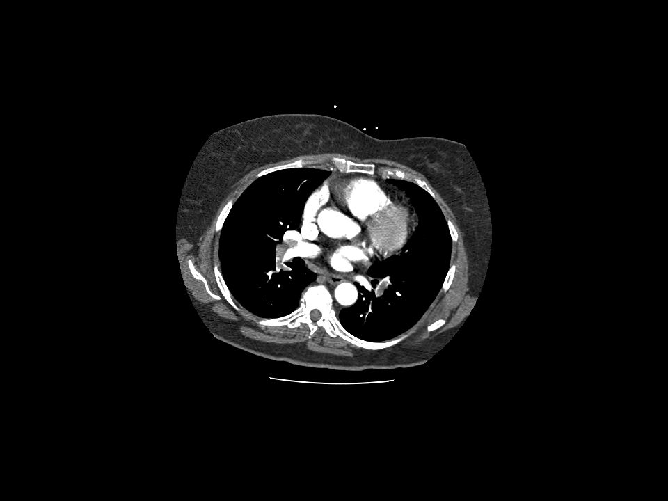
Imaging CXR V/Q Scan CT chest Angiography

18
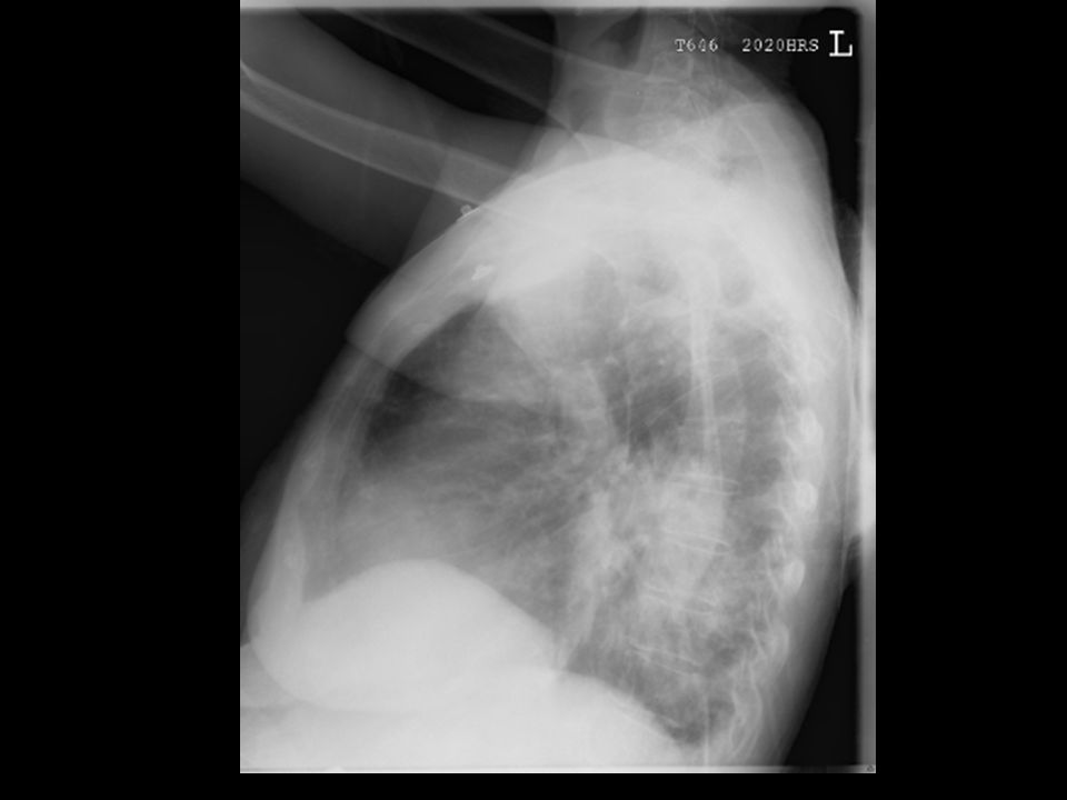
CXR

19
Normal excludes PE, otherwise in context of patient
VQ Scan Normal excludes PE, otherwise in context of patient

20
90% sensitive, 95% specific

24
Konstantinides et al NEJM 2002;347(15):1143-1150
Pulmonary Embolism Treatment High suspicion prior to imaging = heparin Proven with imaging = heparin (LMW or UFH) Thrombolytics in select cases Perimortem RV dysfunction on echo Pulmonary HTN on echo Pulmonary HTN on R heart cath New ECG signs of RV strain Konstantinides et al NEJM 2002;347(15):
 Thrombolytics in select cases. Perimortem. RV dysfunction on echo. Pulmonary HTN on echo. Pulmonary HTN on R heart cath. New ECG signs of RV strain. Konstantinides et al NEJM 2002;347(15):")
25
Case 1 Summary Risk: age, post-op Pleuritic chest pain
Mild tachypnea but vital signs otherwise normal = don’t be fooled! High index of suspicion!

26
Case 2 85 yo male CC: Cough, fever HPI:
3 days of progressive cough with green sputum production. Fevers and chills Pleuritic R sided chest pain PMHx: CAD, HTN, hypercholesterolemia

27
Case 2 Surg Hx: TURP, Coronary stent x 2, appy
Soc Hx: remote tobacco, occasional EtOH, no drug use. Widowed. Retired fisherman. FHx: Coronary disease ROS: no HA, abdominal pain, N/V/D, urinary symptoms

28
Case 2 Vitals: T 38.5 HR 95 RR 20 BP 105/62 SpO2 94% room air
Physical: HEENT: dry mucous membranes Cor: RRR no murmurs Lungs: LLL crackles & occ wheeze Abd: soft NT/ND Assessment?? Plan?

29
Pneumonia #1 infectious mortality
#6 overall 1% as outpt, 25% when needing admission #1 cause nosocomial infectious mortality Up to 50% mortality 25-50% of all ICU pts get pneumonia

30
Pathogens Typical S pneumoniae, H Flu, Staphylococcus
AtypicalLegionella, Mycoplasma, Chlamydia EtohKlebsiella pneumoniae DM/DKAS pneumoniae/S aureus HIVbased on CD4 count COPDHaemophilus influenzae/Moraxella catarrhalis Sickle CellS pneumoniae/H influenzae

31
Diagnosis History/Physical CXR CBC Blood Cx Urine Cx

32
Treatment Ceftriaxone + Macrolide or Fluroquinolone (moxi/levo)
Typical and Atypical coverage May to Cefepime for better G- Hospital/Nursing Home Health care associated (includes dialysis pts) Add Vanco Admit or outpt therapy?
 Add Vanco. Admit or outpt therapy")
33
PNA Severity Score Physical examination Age: Nursing home : +10
AMS: RR >30/minute: SBP <90mmHg: Temp <35, >40C: Pulse >125/minute: Laboratory findings pH <7.35: BUN >30: Sodium <130 mEq/L: Glucose >250: Hct <30%t: PO2 <60 mmHg: Pleural effusion: Age: Males: Age Females: Age -10 Nursing home : Comorbid illnesses Neoplastic disease: Liver disease: CHF: CVA disease: Renal disease:

34
PSS 30d Mortality Prediciton
Total Score Rank Site or Rx Mortality (%) None I Outpt 0.1 <70 II 0.6 71-90 III 90-130 IV Inpt >130 V 27-29
 None. I. Outpt <70. II III IV. Inpt >130. V")
35
CURB-65 Confusion? BUN > 19 mg/dL (7 mmol/L)?
Respiratory Rate ≥ 30? Systolic BP < 90 mmHg orDiastolic BP ≤ 60 mmHg? Age ≥ 65? For each yes answer pt gets 1 point

36
CURB-65 Score 30 day mortality
1 = 2.7%, outpt treatment 2 = 6.8%, consider inpt vs close outpt tx 3 = 14%, inpt tx, poss ICU 4 = 27.8%, inpt, prob ICU 5 = 27.8%, prob ICU tx CAVEAT: notice the score does not take into account hypoxia.

37
Atypical Pneumonia

38
RLL Pneumonia

40
RUL Pneumonia

41
LUL Pneumonia

42
Case 3 24 yo female CC: Shortness of breath, wheezing HPI:
2 days of gradual increased shortness of breath Worse today without relief with albuterol MDI Non productive cough No fevers Recently got a new kitten

43
Case 3 PMHx: asthma All/Meds: none/albuterol MDI Surgical Hx: none
No prior hospitalizations All/Meds: none/albuterol MDI Surgical Hx: none Social Hx: ½ ppd tobacco, no EtOH or drugs. Single. Waitress FHx: COPD ROS: negative

44
Case 3 Vitals: T 37.8 HR 105 RR 22 BP 140/90 SpO2 91% RA
Exam: +accessory muscle use, decreased air movement and very little wheezing Assessment?? Plan?

45
Asthma chronic, nonprogressive lung disorder characterized by:
Increased airway responsiveness Airway inflammation Reversible airway obstruction

46
Physical Exam Tachypnea Tachycardia Cough Prolonged expiratory phase
Wheezing NOT an accurate indicator of the severity of an attack BEWARE of the silent chest!!! Wheezing may be ABSENT or only barely audible in patients with severe obstruction

47
Physical Examination Severe obstruction: Inability to speak
Use of accessory muscles Altered mental status Diaphoresis The ‘silent chest’

48
Can we accurately risk stratify asthma patients with our exam alone?
No… clinicians & patients are notoriously inaccurate when assessing severity. Checking an objective measure of lung function is considered the standard.

49
Assessment Tools Clinical scoring systems Peak expiratory flow rates
Pulse oximetry Arterial blood gases Chest radiography CBC

50
Peak Expiratory Flow Rates
Should be measured before and after each treatment Easiest test to perform in the ED

51
Peak Expiratory Flow Rates
Provides an objective measure Based on height, age, gender Is effort-dependent Useful to assess the response to Rx <25% Severe 25%-50% Moderate 50%-70% Mild >70% Discharge Goal

52
Pulse Oximetry Used to assess and follow oxygenation
O2 sats < 90% indicate a severe asthma attack and significant hypoxemia May have near-normal pulse-ox with impending hypercapneic respiratory failure

53
Arterial Blood Gases Respiratory alkalosis typical
Inaccurate predictor of outcome Will seldom alter your treatment plan Painful and not free

54
Chest Radiography Adds little to decision making in most patients
The presence of ‘abnormal’ findings on CXR seldom alters management Should not be ordered routinely

55
Indications for CXR First episode of wheezing Unclear diagnosis
Patients refractory to therapy Respiratory failure Clinical evidence of infection, pneumothorax, or pneumomediastinum

56
Complete Blood Count Often elevated from stress of acute asthma attack or chronic steroid use Mild eosinophilia is common NOT routinely ordered Indications: infectious work-up

57
Pharmacotherapy Beta-agonists Corticosteroids Anticholinergics

58
Beta Agonists Mainstay of acute therapy
Promote bronchodilation by increasing cAMP Primary effect is small airways Onset of action < 5 min

59
β-Agonists: MDI vs. Nebulizer?
Both are equally effective, even in severe asthma MDI is substantially cheaper 6 puffs = 2.5 mg via a holding chamber nebulizer

60
Anticholinergic Agents
Produce bronchodilation by inhibition of vagally-mediated bronchoconstriction Decrease cGMP Primarily affect large, central airways Onset of action up to 30 min and peak in 1-2 hrs Use in combination with beta-agonists as first-line therapy

61
Steroids Administer early
Used to treat the inflammatory component of asthma Reduce the rate of relapse and the rate of hospital admission

62
Oral Versus IV? Both routes equally effective Oral route preferred
Methylprednisolone mg IV Prednisone 1-2mg/kg PO Oral route preferred Easier and faster Decreases pain/anxiety of IV Cheaper Indications for IV steroids: Severe asthma attack, ie dyspnea, use of accessory muscles Nausea/vomiting

63
Inhaled Steroids In chronic asthma the regular use of inhaled steroids has been shown to: Suppress airway inflammation Decrease beta-agonists use Decrease the frequency of acute exacerbations Decrease mortality related to acute asthma

64
Evidence Supporting the Role of Inhaled
Corticosteroids In Controlling Asthma The emergency physician can use the “rule of two” to determine if a patient’s asthma is well controlled: Use of a rescue inhaler >2 times a week Awakening with an asthma attack > 2 times a month Use of >2 quick-relief β-agonist canisters/year Singer A. Acad Emerg Med 2005; 45:

65
Inhaled Steroids After Discharge?
Use BID Always use a spacer Rinse mouth after use to reduce complications (dysphonia, S/T, oropharyngeal candidiasis)
")
66
Case 4 69 yo male CC: difficulty breathing HPI
Recent cold symptoms x 4 days Now with cough, increased shortness of breath Poor exercise tolerance Cough is productive with yellow sputum No fevers, N/V/D, or other complaints

67
Case 4 PMHx: HTN, COPD, hypercholesterolemia All: PCN
Meds: combivent, lipitor, HCTZ Surgical Hx: cholecystectomy Social Hx: 70 pk-yr tobacco, +EtOH, no drug use; married, retired ship builder FHx: emphysema ROS: negative

68
Case 4 Vitals: T 37.6 HR 100 RR 20 BP 150/94 SpO2 89% room air
Physical: pursed-lip breathing, barrel chest, using accessory muscles. Distant heart and lung sounds, occasional wheeze. +clubbing Assessment?? Plan?

69
COPD Definition Chronic bronchitis: Chronic, productive cough x 3 months in each of 2 successive years in which other causes of chronic cough have been eliminated (Blue bloaters) Emphysema: abnormal permanent enlargement of the air spaces distal to the terminal bronchioles, accompanied by destruction of bronchiolar walls but without obvious fibrosis (Pink puffers)
 Emphysema: abnormal permanent enlargement of the air spaces distal to the terminal bronchioles, accompanied by destruction of bronchiolar walls but without obvious fibrosis (Pink puffers)")
70
COPD Exacerbations Worsening airflow obstruction due to Bronchospasm
Sputum production (infectious, environmental irritants) Cardiovascular deterioration
 Cardiovascular deterioration.")
71
COPD History Physical exam Progressive shortness of breath
Increased sputum production Audible wheezing Physical exam Tachypnea Hypoxemia Cyanosis Agitation Hypercarbia (confusion, stupor, inadequate respiratory effort) Sitting up, pursed-lip breathing (PEEP) Diminished breath sounds, prolonged expiratory phase, wheezing
 Sitting up, pursed-lip breathing (PEEP) Diminished breath sounds, prolonged expiratory phase, wheezing.")
72
COPD Work-up CBC (r/o anemia) CXR (r/o infection, ptx, CHF) ECG
Other labs Lytes Cardiac enzymes BNP Theophylline level (if on med, uncommon these days)
")
73
COPD Treatment Oxygen Bronchodilation Decrease mucous production
Most have baseline sats of 88-91% with mod/severe disease Hypoxic drive Bronchodilation Beta-agonists i.e. albuterol Decrease mucous production Anticholinergic i.e. atrovent Decrease inflammation Steroid therapy Treat infection or underlying cause Similar to asthma treatment Combivent or Duoneb

74
Summary Dyspnea = Subjective Large differential to consider…
Pulmonary Embolus Pneumonia Asthma COPD AMI, CHF, Anemia, Tox, pneumothorax, airway obstruction etc. Discussed more of the common complaints

Similar presentations


 cecal carcinoma Past History: –Anemia, chronic stable angina, GERD Medications: –Diltiazem, Losec.>")


. In 1995, asthma hospitalizations.>")


Asthma 2)COPD 3)Comparison.>")
 Ch.Bronchitis Emphysema (C.O.P.D) Ch.Bronchitis Emphysema AISHA M SIDDIQUI.>")


