Download presentation
Presentation is loading. Please wait.

1
Single Complete Denture
SDS 421 Rahn, A; Heartwell, C: Text book of complete dentures. 5th ed. Pages 481 – Sharry, JJ: Complete Denture Prosthodontics. 5th ed. Pages 310 –

2
Edentulous Maxilla Vs Class I Mandibular Arch
A ' Typical ' Presentation Edentulous Maxilla Vs Class I Mandibular Arch

3
General Considerations in Construction & Delivery
The main problem in treating patients who need a CD to occlude with a natural dentition (without any restoration or restored with a partial prosthesis – FPD or RPD) is that the natural teeth can transmit large forces to a denture whose supporting structures are unable to resist them adequately. ,,,,
 is that the natural teeth can transmit large forces to a denture whose supporting structures are unable to resist them adequately. ,,,,")
4
Thus damage to the edentulous ridge or inability to wear the denture may easily occur.

5
To avoid these sequelae, the basic ingredients of good prosthetic treatment – an adequate denture base, correct jaw relations, good occlusion, and free articulation – must be provided.

6
Single Complete Denture Syndrome
Definition ‘the characteristic features that occur when an edentulous maxilla is opposed by natural mandibular anterior teeth, including loss of bone from anterior portion of the maxillary ridge, overgrowth of the tuberosities, papillary hyperplasia of the hard palatal mucosa, extrusion of mandibular anterior teeth, and the loss of alveolar bone and height beneath the mandibular removable partial denture bases, also called an anterior hyperfunction syndrome’. ‘The Glossary of Prosthodontic Terms, 1999’

7
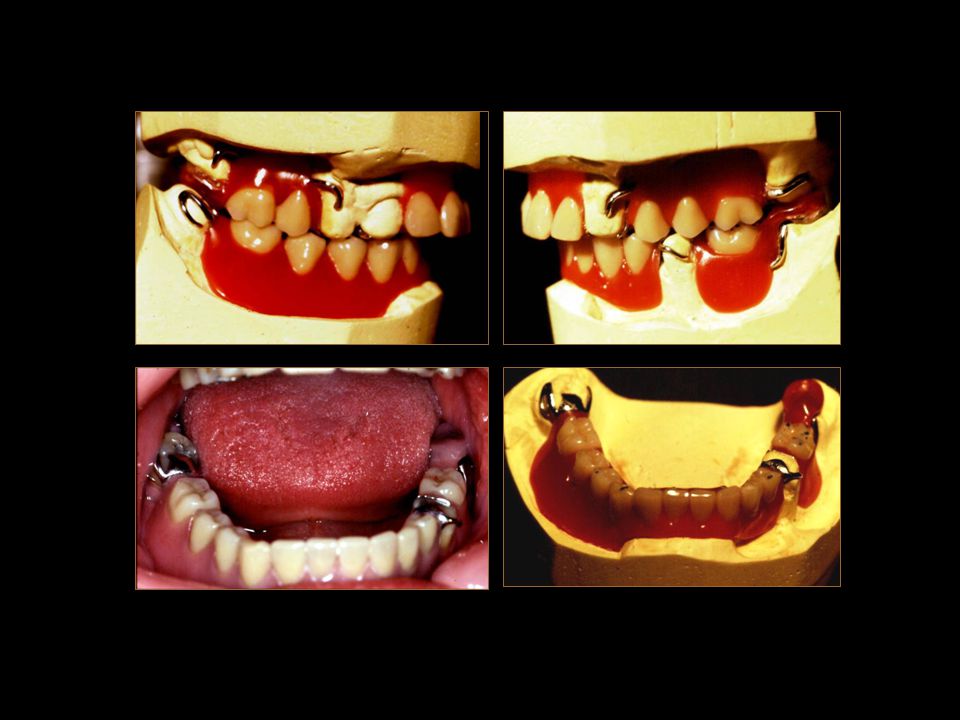
Single Complete Denture Syndrome
Characteristics 1. Loss of bone from anterior portion of the maxillary ridge 2. Overgrowth or enlargement of the maxillary tuberosities 3. Papillary hyperplasia of the hard palatal mucosa 4. Extrusion of the mandibular anterior teeth and 5. Loss of alveolar bone and height beneath the mandibular removable partial denture bases.

8
Loss of bone from anterior portion of the maxillary ridge
Single Complete Denture Syndrome

9
Overgrowth or enlargement of the maxillary tuberosities
Single Complete Denture Syndrome

10
Papillary hyperplasia of the hard palatal mucosa
Single Complete Denture Syndrome

11
Extrusion of the mandibular anterior teeth
Single Complete Denture Syndrome

12
Single Complete Denture Syndrome
Preventive Measures Preventing the degenerative changes produced by a maxillary complete denture opposed by a class I mandibular RPD is by treatment planning to avoid this combination of prostheses - causing combination syndrome. The options are, 1. Extraction of remaining anterior mandibular teeth – CD/CD. 2. Retain the week posterior teeth as abutments by means of endodontic treatment and periodontic recalls. 3. Overlay dentures in the mandible. 4. Implants placed in the posterior region. 5. Maximum coverage of the distal extension ridges in RPD.

13
Single Complete Denture Syndrome
Retained roots can be used as OD abutments that will preserve the alveolar ridge – hence prevent the ‘SCD Syndrome’ Single Complete Denture Syndrome

14
Single Complete Denture Syndrome
Application of basic prosthetic principles ‘maximum area of coverage’ ‘proper OP orientation’ and ‘balanced occlusion’ ensure preservation of the alveolar bone – prevention of SCD syndrome Single Complete Denture Syndrome

15
Single Complete Denture Syndrome
An attempt to equalize the support gained from the teeth and the ridge by ‘altered cast impression’, will minimize denture rotation – preservation of the alveolar ridge. Single Complete Denture Syndrome

16
Minimizing Occlusal Forces on the Denture Foundation
There are two reasons for the difficulty associated with the provision of a successful complete denture for these patients. 1. The biting forces applied by the natural teeth are very high (198 lb by a natural molar tooth Vs 26 lbs by a CD). 2. Disrupted occlusal plane of the remaining natural teeth applies horizontally directed forces to the opposing denture. Solution 1. Maximum denture base extension and precise jaw relation records with proper articulation of the teeth. 2. Reduce the magnitude of damaging forces by correcting the occlusal plane orientation – occlusal grinding, extraction and restoration of the tilted or over-erupted teeth.
. 2. Disrupted occlusal plane of the remaining natural teeth applies horizontally directed forces to the opposing denture. Solution. 1. Maximum denture base extension and precise jaw relation records with proper articulation of the teeth. 2. Reduce the magnitude of damaging forces by correcting the occlusal plane orientation – occlusal grinding, extraction and restoration of the tilted or over-erupted teeth.")
17
Single Mandibular Complete Denture The Complicating Factors
Very rarely the mandibular arch is the edentulous one. It usually happens due to surgical or accidental trauma, i.e., radiation therapy of the jaw, fall, vehicle accident or gunshot. Three factors should be considered while treating such patients. 1. Preservation of Residual Ridge: Opposing natural teeth would apply greater force and tongue activity can lead to denture movement – hence rate of bone resorption could be high. 2. Necessity for Retaining Maxillary teeth: These may be needed to retain a prosthesis, e.g., cleft palate, or these may be esthetically attractive and periodontally healthy. 3. Mental Trauma: Loss of mandibular teeth is already traumatic to the patient and advising the removal of remaining maxillary teeth will undoubtedly be traumatic that may lead to depression.

18
Single Mandibular Complete Denture The Favorable Situations
Occasionally, constructing a single mandibular complete denture is not potentially harmful, e.g., 1. When the maxillary arch has already been restored with a complete denture. 2. When all the maxillary posterior teeth are also missing and the patient needs a bilateral distal extension maxillary RPD. The biting forces applied by the RPD will be lass is magnitude. 3. Very old and frail patient – the biting forces applied by the natural teeth would be small, hence less damage to the mandible.

19
Single Mandibular Complete Denture

20
Various Combinations for Constructing a SCD Opposed by: Natural dentition, FPDs …….

21
Various Combinations for Constructing a SCD Opposed by: Class I or II RPD, or an existing CD

22
Special considerations for obtaining the Impression
Maximum tissue surface coverage is essential. Special impression technique for recording the impression of flabby ridge without displacement.

23
The Impression Technique
Impression can be obtained with ‘impression plaster’ that will cause minimal tissue displacement of the flabby ridge. The Impression Technique

24
The Impression Technique
Two stage Impression Technique Special design of custom tray - ‘window’ for the flabby ridge The Impression Technique

25
The Impression Technique
After completing border molding & impression of the firm tissues, impression plaster is painted with a soft brush to record the flabby ridge without displacement. The Impression Technique

26
The Impression Technique
Two stage Impression can be obtained with a combination of silicon impression material and impression plaster. The Impression Technique

27
Special considerations for recording the Maxillo-mandibular relations
The basic principles of recording vertical and centric jaw relations for edentulous patients must be followed. Following two difficulties can be met with while constructing a single complete denture, 1. An error may be made while recording the OVD if the upper wax rim is trimmed to represent the incisal level below the upper lip and parallel to the ala – tragal line. The labio-lingual thickness of the wax rim may have to be reduced to accommodate the lower natural teeth behind the upper wax rim.

28
Maxillo - mandibular relations
2. If a ‘gothic arch tracing’ is to be used for recoding the centric relation, then attaching the recording plate to mandibular natural teeth could pose a problem. Maxillo - mandibular relations

29
Maxillo - mandibular relations
Alternate methods of recording CR (check bites) can be used to avoid this difficulty. Maxillo - mandibular relations
 can be used to avoid this difficulty. Maxillo - mandibular relations.")
30
Modifying the Opposing Occlusion ‘Indications’
Not infrequently, the opposing dentition of the mandibular arch (natural teeth or prosthetically restored teeth with RPD & FPD) has to be modified prior to the fabrication of a maxillary complete denture. 1. Malposed or tilted mandibular posterior teeth. 2. Over-erupted mandibular anterior teeth. 3. Over-erupted mandibular posterior teeth. These conditions will result in an irregular occlusal plane that will result in unacceptable occlusal function and esthetics. 4. Too large occlusal table (bucco-lingual width) of the natural teeth may have to be narrowed by grinding of the buccal & lingual surfaces to accommodate the denture teeth.
 has to be modified prior to the fabrication of a maxillary complete denture. 1. Malposed or tilted mandibular posterior teeth. 2. Over-erupted mandibular anterior teeth. 3. Over-erupted mandibular posterior teeth. These conditions will result in an irregular occlusal plane that will result in unacceptable occlusal function and esthetics. 4. Too large occlusal table (bucco-lingual width) of the natural teeth may have to be narrowed by grinding of the buccal & lingual surfaces to accommodate the denture teeth.")
31
Modifying the Opposing Occlusion by grinding the incisal edges.

32
The aim of grinding the opposing natural teeth is to establish a regular occlusal plane.

33
Perfecting the Occlusal Plane
An irregular occlusal plane of the opposing natural / artificial teeth (picket fence arrangement) is unacceptable, hence their irregularity must be altered by, a. orthodontic means – intrusion, extrusion, etc. b. placing restorations – crowns, onlay prosthesis, etc. c. occlusal grinding – to reshape the teeth and to create a suitable occlusal surface with low cusp height. If this correction is not made, the finished prosthesis may have balanced contact of teeth in centric relation position only and not in the eccentric occlusion – hence lack of stability will result.
 is unacceptable, hence their irregularity must be altered by, a. orthodontic means – intrusion, extrusion, etc. b. placing restorations – crowns, onlay prosthesis, etc. c. occlusal grinding – to reshape the teeth and to create a suitable occlusal surface with low cusp height. If this correction is not made, the finished prosthesis may have balanced contact of teeth in centric relation position only and not in the eccentric occlusion – hence lack of stability will result.")
34
Perfecting the Occlusal Plane

35
Occlusal Adjustment ‘1st Method’
Pre-requisite to any occlusal grinding of the natural teeth is mounted study casts. In the event of edentulous maxilla, a metal occlusal template can be placed on the occlusal surface of the mandibular study cast to evaluate the existing occlusal plane.

36
This curved template when positioned on the cast will guide the operator for determining the areas and the amount of occlusal grinding needed on all the teeth (natural and artificial, if present). The cast tooth surfaces are painted with a spray paint before grinding – the altered areas become unpainted after grinding and serve as a guide for intra oral adjustment. The template is then sterilized and used as a guide in the oral procedures.

37
Occlusal Adjustment ‘2nd Method’
This method is time consuming but accurate and enables one (specially the beginner) to appreciate why these adjustments are necessary. It also gives immediate picture of the improvements made. 1. Proceed to the stage of recording CR record and mount the casts. 2. Start the set up of the artificial teeth – as the interferences in the natural occlusion become apparent, they are adjusted on the cast and marked with a pencil for future reference. 3. Once a favorable occlusion has been achieved on the articulator, similar adjustments are carried out on the natural teeth intra-orally using markings on the cast and the denture set up as a guide. Although due to the presence of restorations and sensitive dentine areas, it is not always possible to carry out all the adjustments needed, a substantial amount can usually be done.
 to appreciate why these adjustments are necessary. It also gives immediate picture of the improvements made. 1. Proceed to the stage of recording CR record and mount the casts. 2. Start the set up of the artificial teeth – as the interferences in the natural occlusion become apparent, they are adjusted on the cast and marked with a pencil for future reference. 3. Once a favorable occlusion has been achieved on the articulator, similar adjustments are carried out on the natural teeth intra-orally using markings on the cast and the denture set up as a guide. Although due to the presence of restorations and sensitive dentine areas, it is not always possible to carry out all the adjustments needed, a substantial amount can usually be done.")
38
Mesially tilted Opposing ‘last’ Molar tooth
The over-erupted or mesially tilted remaining molar teeth are a hazard to the success of a denture as the steeply inclined occlusal surface would tend to drive the opposing denture forward in CO as well as in eccentric occlusions.

39
Mesially tilted Opposing ‘last’ Molar tooth Solutions to the Problem
Moderate tilt: Grinding the distal half of the occlusal surface flat and denture tooth to contact that area only with no contact on the mesial cusps.

40
Mesially tilted Opposing ‘last’ Molar tooth Solutions to the Problem
Extreme tilt: Extraction of the tilted molar tooth Endodontics and restoration with a cast crown or onlay (the surveyed crown). Flattening the distal cusp by grinding and building the mesial cusps by overlay rest of the RPD framework.
. Flattening the distal cusp by grinding and building the mesial cusps by overlay rest of the RPD framework.")
41
A case report of Onlay rest with partial OD RPD.

43
Establishing the Vertical & Horizontal overlap
Natural Anterior Teeth often have a large overbite and small overjet. This is tolerable because of the teeth being firmly held in the bone. In Complete Dentures, this arrangement results in excessive forces applied to the anterior ridge, hence rapid resorption can occur. Therefore, in CD construction minimal OB and substantial OJ is normally provided.

44
Establishing the Vertical & Horizontal overlap
In the presence of mandibular anterior teeth, this articulation is difficult to achieve as the lower incisal edges were positioned high on the cingula of the upper anterior teeth before extraction. Therefore, 1. Raise the level of upper teeth and grind the incisal edges of the lower. 2. Do not set the upper teeth too far palatally – use the biometric guides. 3. Grind the labio-incisal surfaces of the lower teeth and the palato-incisal surfaces of the upper teeth to gain more OJ.

45
Establishing the Vertical & Horizontal overlap Natural anterior teeth must not contact the opposing complete denture in CR and during Eccentric movements. in CR Eccentric movements

46
Occlusal Requirements of a SCD
The setting of the posterior teeth must ensure that the opposing inclined planes do not contact as the jaw closes into CO. Only those surfaces of opposing teeth should contact which transmit occlusal forces vertically.

47
Occlusal Requirements of a SCD
This arrangement can be provided with the use of both non-anatomic or anatomic teeth. Non Anatomic Teeth are selected if the natural posterior teeth have flat cusps due to attrition. Balanced occlusion may not be achievable however, free articulation must be obtained.

48
Occlusal Requirements of a SCD Non-anatomic teeth – cusp to fossa set-up

49
Occlusal Requirements of a SCD
Anatomic Teeth are selected if the cuspal form of the natural teeth has been retained. These should be arranged with good intercuspation in CO (cusp-fossa relation). As the artificial teeth are usually smaller mesio-distally than the natural teeth, small spaces may have to be left between them for proper inter-cuspation. Similarly, artificial teeth may need grinding of the cuspal inclines to accommodate for the much larger bucco-linguial width of the natural teeth.
. As the artificial teeth are usually smaller mesio-distally than the natural teeth, small spaces may have to be left between them for proper inter-cuspation. Similarly, artificial teeth may need grinding of the cuspal inclines to accommodate for the much larger bucco-linguial width of the natural teeth.")
50
Artificial Posterior Teeth Materials Porcelain Teeth The amount of occlusal grinding necessary to permit artificial teeth to occlude with the natural teeth often precludes the use of porcelain teeth. Week porcelain teeth will fracture or chip easily.

51
Artificial Posterior Teeth Materials Acrylic resin Teeth These are more suitable to occlude with the natural teeth. Wear of their surfaces can be prevented by using good quality cross linked copolymer resin teeth or by placing occlusal amalgam restorations in the denture teeth.

52
Artificial Posterior Teeth Materials Gold Occlusals Gold is the best material to occlude with the natural dentition. A number of methods are available to the technicians to construct these ‘gold occlusal’ surfaces in the denture teeth.

53
Placement (insertion) Visit
This visit carries the same importance as for constructing a set of complete dentures. Following procedures must be meticulously performed, PIP adjustment: To establish even contact of the denture fitting surface to the supporting mucosa. The contact of posterior palatal seal area should be carefully evaluated.

54
Placement (insertion) Visit Occlusal adjustment This can best be achieved with the help of clinical remount procedure. A balanced occlusion in both CO and Eccentric relations, with free articulation of the teeth must be provided.

55
Placement (insertion) Visit Home Care Instructions Patient must understand the importance of leaving the denture out while sleeping, especially to avoid the detrimental forces of bruxism while sleeping. Oral hygiene measures for the denture and the natural teeth must be reinforced.

Similar presentations


>")


